Department of Anatomy and Histology, Faculty of Veterinary Science, Bangladesh Agricultural University, Mymensingh-2202, Bangladesh
Preserving biological samples is challenging due to the natural process of tissue decomposition. Therefore, various preservation techniques, such as chemical preservation, cryopreservation, cryodehydration, and plastination are used to preserve different types of tissue samples. However, the existing methods are often expensive and may pose health risks, making them less practical for widespread use. Objectives: Therefore, this study aimed to preserve visceral and musculoskeletal canine specimens by utilizing an epoxy resin immersion method and minimize animal sacrifice for anatomy education and training. Method: Fresh visceral and musculoskeletal specimens (forelimb) were obtained from a female dog, and fixed using 10% formalin, followed by dehydration in ethanol and acetone and finally, immersing in epoxy resin. Results: The epoxy resin-immersed specimens maintained their near-natural morphology while being hard, dry, odorless, lightweight, and durable at room temperature. While visceral organs like the liver, heart, and lungs appeared darker due to pigment formation during fixation, the musculoskeletal specimens retained their normal color and texture. The study found no significant histoarchitectural changes in the heart, where all muscle layers remained identifiable, though cardiac myofibers were less distinguishable from one another. In skeletal muscle, there were no notable alterations except for increased space between muscle bundles and myofibers. The spleen showed some loss of histoarchitectural details due to decreased cellular integrity, but the red and white pulp zones were still distinguishable. Additionally, there was localized loss of lymphocytes in certain areas in the lymph node. Overall, cytoplasmic and nuclear clarity was well preserved across the tissues examined. Conclusion: It can be concluded that the epoxy resin immersion technique can effectively preserve whole anatomical specimens for gross anatomical study while some histoarchitectural details might be lost, limiting its use for histological study.
anatomical study; biological specimen; cost-effectiveness; epoxy resin; immersion
Dr. Nasrin Sultana, Department of Anatomy and Histology, Faculty of Veterinary Science
Bangladesh Agricultural University, Mymensingh-2202, Bangladesh
Email: nasrin.sultana@bau.edu.bd; nsultana.bau@gmail.com

![]()


Biological sample preservation poses significant challenges due to the inevitable decay of all biological tissues. Therefore, various preservation methods have been developed over time to hinder or delay decomposition (Jordan et al., 2009). Chemical-based preservation has gained attention since the 19th century but has its own limitations when it comes to regular specimen handling and demonstration (Binawara et al., 2010; Kamruzzaman, 2016; Haizuka et al., 2018). Moreover, chemical preservation requires the regular replacement of preservatives with fresh solutions, leading to significant additional costs (Abas, 2025). Therefore, finding alternatives to chemical preservatives for preserving biological specimens in anatomy teaching and research has become crucial.
In 1977, German anatomist von Hagens introduced plastination as a novel preservation method to address these concerns, which has since been widely adopted globally (von Hagens, 1979). Plastination involves replacing water and lipids in biological tissues with synthetic components to maintain their natural appearance, enabling manipulation without the need for formaldehyde or other harmful chemicals (Pashaei, 2010). This technique has revolutionized anatomical research and education by offering detailed, durable, and stable low cost preservation (Sultana et al., 2019; Islam et al., 2021; Abas, 2025). Among the various plastination methods, the epoxy resin plastination technique offers distinct advantages including minimal shrinkage of specimens during the immersion and curing process which ensures that the anatomical features of the tissues are preserved at the macroscopic and microscopic levels (Cunningham et al., 2008). This method preserves anatomical specimens in a way that maintains the structural details without significant distortion, making it particularly useful for anatomical research and clinical applications (Ottone, 2023). E12 epoxy plastination allows specimens to be sectioned to a thickness of 2–3 mm while maintaining transparency due to the refractive properties of the epoxy resin (Chisholm and Varsou, 2018; Latorre et al., 2019). This transparency enables excellent correlation with imaging techniques such as CT and MRI, offering a unique advantage over traditional plastination methods, where the opacity of the materials can obscure fine details (Latorre et al., 2019). This technique is a practical, affordable, and comparatively health-safe option compared to chemical preservation methods, which will help students and academicians connect traditional three-dimensional anatomy with two-dimensional images (Chisholm and Varsou, 2018).
The process of epoxy sheet plastination follows a similar technique to other plastination methods but introduces the crucial step of sectioning during specimen preparation. The tissue is first frozen, and then sections are cut before undergoing dehydration, degreasing, and vacuum immersion. The curing stage, conducted at a controlled temperature (up to 45°C), ensures the specimen’s long-term preservation. Notably, the E12 technique is suitable for a wide range of tissues and organs, including the brain, to produce exquisitely detailed slices (Ottone et al., 2016; Latorre et al., 2019). As the technique continues to evolve, its applications are expanding, offering new opportunities for both educational purposes and advanced morphological studies in clinical and surgical fields. However, the resin plastination technique was mostly used for brain and musculoskeletal section preservation in earlier studies (Ottone et al., 2016; Bilge et al., 2018; Ottone et al., 2020). In this study, we hypothesize that the epoxy resin immersion technique could be used to preserve whole organs, especially those from small animals. We believe it could be a more affordable method for preserving anatomical specimens in low-income countries like Bangladesh. Based on the hypothesis, the current study’s goal was to evaluate and adapt the epoxy resin immersion technique as a health-safe alternative to traditional chemical-based tissue preservation methods in anatomy laboratories.
Ethical approval
This research was ethically approved by the Animal Welfare and Experimentation Ethics Committee (AWEEC), Bangladesh Agricultural University (BAU), Bangladesh [AWEEC/BAU/2023(44)].
Specimen collection
According to the animal care guidelines, visceral organs (heart, lungs, liver, kidney, spleen, lymph node, pancreas, and female genital system) and musculoskeletal specimens (limb) were collected from a female dog (euthanized with Thiopentone Sodium 70mg/kg body weight) at the animal research laboratory at the Department of Anatomy and Histology, BAU, Bangladesh. The collected specimens were washed and dissected properly to remove any unwanted tissue or materials before going into the next step.
Epoxy resin immersion
Fixation
The specimens were fixed in 10% formalin for 3–7 days, depending on their size, maintaining a sample-to-formalin volume ratio of 1:10. The heart, spleen, lymph nodes, pancreas, and female genital system were fixed for 3 days with a single bath replacement, whereas the lungs, liver, kidneys, and musculoskeletal specimens were fixed for 7 days with two bath replacements to ensure proper fixation.
Dehydration
The fixed specimens were dehydrated using an acetone bath (100%) for 2 weeks and then using an ethanol bath (100%) for another 2 weeks at room temperature (25 ± 2 °C). Both the acetone and ethanol baths were replaced at one-week intervals to ensure proper dehydration. Sample-to-acetone/ethanol volume ratio was 1:10.
Immersion
The dehydrated specimens were immersed in epoxy resin (AB Glue, Bangladesh Epoxy Resin Company Ltd., Bangladesh) without catalyst at room temperature for three days without using any active or passive forces.
Curing
Once the immersion process was completed, the specimens were taken out of the resin and kept in a slanting position to drip off the excess resin. Then, a thin layer of coating was applied using polymer: epoxy resin (3 parts) and catalyst (1 part) mixture at 3:1 ratio as per the manufacturer’s recommendation (Epoxy Resin Hardener, Bangladesh Epoxy Resin Company Ltd., Bangladesh). The polymer-coated specimens were kept in a plastic jar for 24 hours at room temperature to harden, preserving the specimen's natural shape. Finally, excess resin was removed by cutting with scissors.
Histological Investigation
To study the histological characteristics of the epoxy resin-immersed heart, skeletal muscle, spleen, and lymph node, approximately 0.5 cm2 tissue samples were excised (from both fresh and cured epoxy resin-immersed specimen) by using surgical knives. The tissue samples were dehydrated using ascending grades of alcohol (Merck, Germany) (70%→80%→90%→100% I, II, III), each for 2 hours except 100% alcohol III where the specimens were kept overnight (12 hours). Then clearing was done using three changes of xylene (Merck, Germany) and finally, paraffin blocks were prepared using melted paraffin (Merck, Germany). Sections at 5-10 µm thickness were cut using rotary microtome (CUT 4062, Manual Precision Microtome, SLEE medical GmbH, Germany). Finally, they were processed for routine staining (Harris hematoxylin and eosin stain) following the standard protocol (Islam et al., 2021). The histological characteristics were investigated using a photomicroscope (Model: B-293, Optika, Italy).
Gross morphological characteristics of the epoxy resin-immersed specimens

Figure 1: Epoxy resin-immersed visceral (i.e. heart, lungs, liver, kidney, spleen, pancreas, genital system, and lymph nodes) and musculoskeletal specimens (forelimb) of female dog
The gross appearances of the epoxy resin-immersed visceral organs and musculoskeletal specimens are presented in Figure 1. The epoxy resin-immersed specimens retained their morphology in a near-natural state in terms of specimen size and shape. They were hard, dry, odorless, lightweight, and durable. Specimens were stored at room temperature for one month without any visible alteration. However, the visceral organs (liver, heart, lungs, spleen, kidney, pancreas, and lymph nodes) appeared darker as well as yellowish (i.e. base of the heart, genital organs and musculoskeletal specimens) in color. On the other hand, the musculoskeletal specimens retained almost normal color and texture.
Histological characteristics of the epoxy resin-immersed specimens
Heart
The histomorphologic features of the epoxy resin-immersed heart are presented in Figure 2 (A1 & A2). No distinct histoarchitectural alteration was noticed in the heart. All the muscle layers (i.e., epicardium, myocardium, and endocardium) were distinctly identifiable. The cytoplasmic and nuclear clarity was well preserved. However, the cardiac myofibers could not be distinctly separable from each other.
Skeletal Muscle
Figure 3 (A1 & A2) presents the histomorphologic features of the epoxy resin-immersed skeletal muscle. No distinct alteration in the histoarchitecture of the skeletal muscle was noticed, except for the increased space between the muscle bundles and individual myofibers which indicates the shrinkage of the myofibers. The cytoplasmic and nuclear clarity was well preserved.
Spleen
Figure 4 (A1 & A2) presents the histomorphologic features of the epoxy resin-immersed spleen. The spleen's histoarchitectural details (attributed to the slight loss of cytoplasmic and nuclear details) were somewhat lost with the loss of cellular integrity (characterized by empty spaces). However, the red and white pulp zones were still distinguishable with distinct trabecular septa.
Lymph nodes
The histomorphologic features of the epoxy resin-immersed lymph node are presented in Figure 5 (A1 & A2). No distinct histoarchitectural alteration was noticed, and the cytoplasmic and nuclear clarity was well preserved. However, some areas of localized loss of lymphocytes were noticed.
 Figure 2: Histoarchitecture of the fresh (A1 & A2) and epoxy resin-immersed (B1 & B2) heart specimen. A1 & B1 – 10X magnification, A2 & B2 - 40X magnification, black arrow - cardiac myofiber |
 Figure 3: Histoarchitecture of the fresh (A1 & A2) and epoxy resin-immersed (B1 & B2) skeletal muscle specimen. A1 & B1 – 10X magnification, A2 & B2 - 40X magnification, black arrows – myofibers |
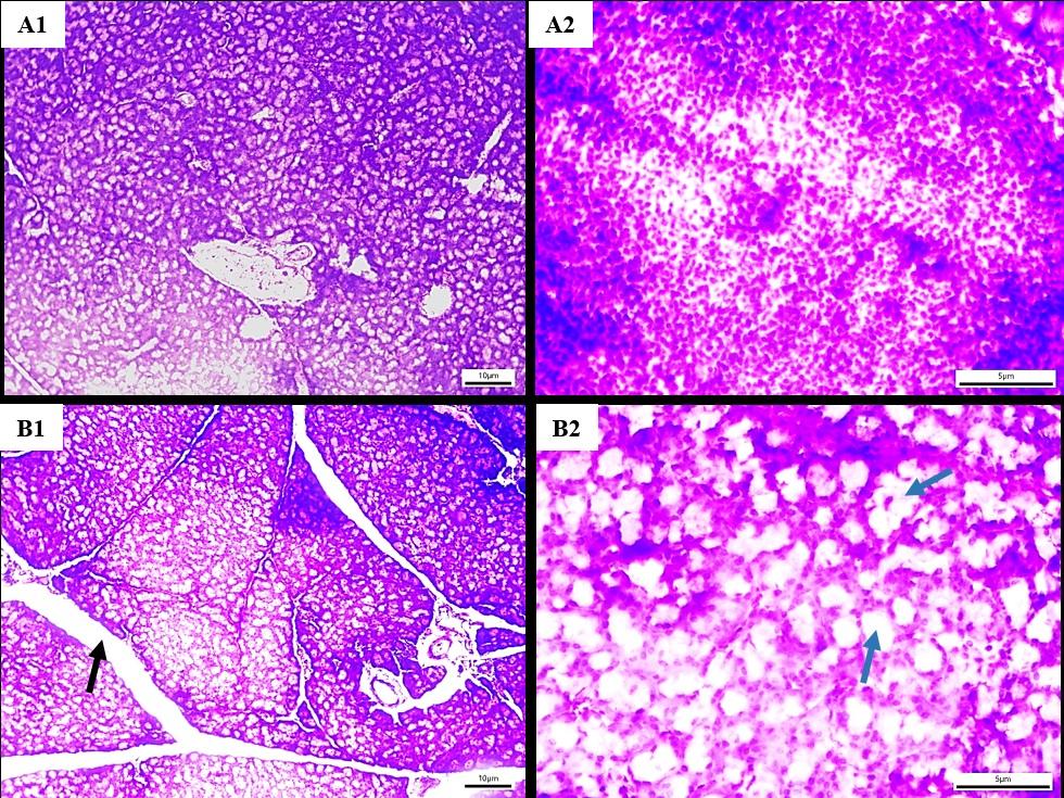 Figure 4: Histoarchitecture of the fresh (A1 & A2) and epoxy resin-immersed (B1 & B2) spleen. A1 & B1 – 10X magnification, A2 & B2 - 40X magnification, black arrows – trabeculae, blue arrows – empty spaces |
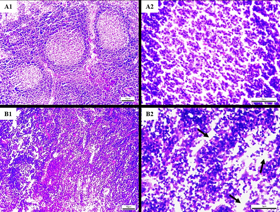 Figure 5: Histoarchitecture of the fresh (A1 & A2) and epoxy resin-immersed (B1 & B2) lymph node. A1 & B1 – 10X magnification, A2 & B2 - 40X magnification, black arrows – empty spaces due to localized loss of lymphocytes |
The findings of the current study on the gross and histomorphological characteristics of epoxy resin immersed specimens provide insights into its effectiveness and limitations as a preservation technique. These findings, based on a detailed analysis of various tissues, highlight how epoxy resin immersion preserves both the macroscopic and microscopic features of organs and tissues, while also revealing certain changes in cellular structure that are inherent to the process.
The study results show that epoxy resin immersed visceral organs retained their gross morphological features in near-natural states. The specimens were hard, dry, odorless, lightweight, and durable, with no visible alteration observed even after one month of storage at room temperature except for yellowish discoloration of the samples. This is consistent with the known advantages of plastination, which preserves the structural integrity of biological specimens by replacing water in tissues with a polymer, typically silicone or epoxy resin (von Hagens, 1986; Chisholm and Varsou, 2018).
However, a color change was noted in the epoxy resin immersed visceral organs, which appeared darker and yellowish over time. This is likely due to the formation of pigments during the fixation process, where formalin fixation can induce darkening in certain tissues (Weber & Henry, 1993). Tissue discoloration was reported by Elnady (2016) due to formalin fixation. Bernal et al. (2022) performed an additional pigmentation step using pastry or gourmet dyes to resolve this issue. However, Sora et al. (2002) who carried out the study on slices rather than whole organs, reported no change in color following resin plastination. Formalin is commonly used to fix tissues before plastination, but its interaction with certain organs can result in pigmentation or discoloration. Conversely, the musculoskeletal specimens retained their color and texture, suggesting that the epoxy resin immersion process might have less impact on tissues with more fibrous or dense structures compared to soft organs like the lymph node or spleen.
In terms of histology, the epoxy resin-immersed heart specimens showed no significant alteration in histoarchitecture, with distinctly identifiable epicardium, myocardium, and endocardium layers at the magnifications investigated. This aligns with previous studies indicating that plastination preserves the cytoplasmic and nuclear structures of cardiac tissues, which is crucial for detailed anatomical study (Cook and Al-Ali, 1997). However, while the muscle layers were well preserved, the study found that the cardiac myofibers could not be distinctly separated from each other. This might be due to the physical properties of the epoxy resin immersion process, where the immersion and curing process can cause the fibers to become more fused or aggregated, potentially reducing the fine separation between muscle fibers (Steinke, 2001).
Similar to the heart, the histological structures of the epoxy resin-immersed skeletal muscle showed minimal changes. The muscle architecture was well preserved, with clear cytoplasmic and nuclear features, but there was an increased spacing between muscle bundles and individual myofibers. Similar changes were observed for musculoskeletal specimens preserved using a cryodehydration technique (Sultana and Islam, 2023). This increased inter-fiber space may result from the shrinkage or displacement of cellular components during the dehydration and immersion steps of epoxy resin immersion, a well-documented issue (Fasel et al., 1988). Despite this, the muscle fibers retained sufficient detail to support their use in both educational and research contexts.
In contrast, the epoxy resin-immersed spleen exhibited some loss of histoarchitectural details. While the red and white pulp zones remained distinguishable, there was a loss of cellular integrity characterized by empty spaces within the tissue. This finding is consistent with the effects of plastination on softer tissues, where cellular details may be compromised due to the challenges in fully impregnating certain tissues with the resin, leading to a reduction in cellular preservation (Shahar et al., 2007). The preservation of the distinct trabecular septa in the spleen is an important feature, as it allows for the recognition of the organ's basic structural components despite the loss of finer details.
The lymph node specimens showed relatively few histological changes, with the overall structure and cellular clarity well preserved. However, localized loss of lymphocytes was observed in some areas. This suggests that while the epoxy resin immersion technique is generally effective in preserving lymphoid tissues, the delicate nature of lymphocytes may make them more susceptible to degradation or loss during the process (Sora et al., 2002). This localized cell loss may be a result of incomplete immersion or the physical stresses associated with the dehydration and sectioning steps.
The findings of this study suggest that the epoxy resin immersion technique is highly acceptable in preserving the gross and histological features of various organ systems, with notable differences between soft organs and more fibrous tissues. The preservation of morphological integrity in both visceral and musculoskeletal specimens supports the growing use of epoxy resin-immersed specimens in medical education and research. However, the study highlights certain limitations of the techniques used, particularly in preserving cellular details in softer tissues such as the spleen and lymph nodes. One key limitation of the current study is that perfusion fixation was not used, which may have contributed to the darker appearance of the samples. In addition, the study did not assess whether the current technique offers any advantage over silicone plastination or traditional formalin fixation. Therefore, further research is recommended to prepare plastinated specimens using perfusion fixation combined with vacuum pressure at low temperature to enhance immersion and minimize tissue damage, and to compare its effectiveness with silicone plastination and traditional formalin fixation.
Acknowledgments
We appreciate the technical assistance from the Department of Anatomy and Histology, Bangladesh Agricultural University, Bangladesh. Nasrin Sultana was a recipient of the research fund from the Ministry of Science and Technology (MoST), Bangladesh. The funding source had no involvement in the study design; in the collection, analysis, and interpretation of data; in the writing of the report; and in the decision to submit the article for publication.
Abas, R. 2025: Cadaver Preservation for Display in Malaysian Anatomy Museums: Methods, Challenges, and Cultural Considerations. Mal J Med Health Sci 21(3): 474-480. https://doi.org/10.47836/mjmhs.21.3.55
Bernal V, Aburto P, Pérez B, Gómez M, Gutierrez JC. 2022: Technical note of improvement of the Elnady technique for tissue preservation in veterinary anatomy. Animals 12(9): 1111.
https://doi.org/10.3390/ani12091111
Bilge O, Çelik S, Yörük MD, Koçer IB. 2018: Useful materials for cross-sectional anatomy education: silicone plastinated examples of foot and hand. Austin J Anat 5: 1080-1084.
Binawara BK, Rajnee CS, Mathur KC, Sharma H, Goyal K. 2010: Acute effect of formalin on pulmonary function tests in medical students, Pak J Physiol 6(2): 8-10. https://doi.org/10.69656/pjp.v6i2.796
Chisholm F, Varsou O. 2018: Resin‐embedded anatomical cross‐sections as a teaching adjunct for medical curricula: is this technique an alternative to potting and plastination? J Anat 233(1): 98-105. https://doi.org/10.1111/joa.12816
Cook P, Al-Ali S. 1997: Submacroscopic interpretation of human sectional anatomy using plastinated E12 Sections, J Int Soc Plast 12(2): 17-27. https://doi.org/10.56507/XICY2283
Cunningham MDF, Chuang MSK, Mackenzie LW, Easteal RA. 2008: Plastinates and Modern Learning Practises. J Int Soc Plast 23: 55.
Elnady FA. 2016: The Elnady Technique: An innovative, new method for tissue preservation. ALTEX-Alt Anim Exp 33(3): 237-242. https://doi.org/10.14573/altex.1511091.
Fasel J, Mohler R, Lehmann B. 1988: A technical note for improvement of the E12 technique. J Int Soc Plast 2(1): 4-7. https://doi.org/10.56507/LNBR6798
Haizuka Y, Nagase M, Takashino S, Kobayashi Y, Fujikura Y, Matsumura G. 2018: A new substitute for formalin: application to embalming cadavers. Clin Anat 31(1): 90-98. https://doi.org/10.1002/ca.23011
Islam R, Sultana N. 2021a: Preservation of musculoskeletal specimens of goat by the Elnady technique: an innovative low-cost alternative to plastination. J Plast 33(2): 1-12. https://doi.org/10.56507/QBZN2088
Islam R, Ayman U, Sultana N. 2021b: Histological architecture and biometric characteristics of indigenously plastinated organs of goat. Int J Morph 39(3): 759-765. http://dx.doi.org/10.4067/S0717-95022021000300759.
Jordan G, Thomasius R, Schröder H, Wulf JS, Schlüter O, Sumpf B, Lang KD. 2009: Non-invasive mobile monitoring of meat quality. J Verbr Lebensm 4: 7-14. https://doi.org/10.1007/s00003-009-0389-1
Kamruzzaman M. 2016: Formalin crime in Bangladesh: a case study. Eur J Clinic Biomed Sci 2(5): 39-44. https://doi.org/10.11648/j.ejcbs.20160205.12
Latorre R, de Jong K, Sora MC, López‐Albors O, Baptista C. 2019: E12 technique: Conventional epoxy resin sheet plastination. Anat Histol Embryol 48(6): 557-563. https://doi.org/10.1111/ahe.12507
Ottone NE, Baptista CA, Del Sol M, Ortega MM. 2020: Extraction of DNA from plastinated tissues. Forensic Sci Int 309: 110199. https://doi.org/10.1016/j.forsciint.2020.110199
Ottone NE, del Sol M, Fuentes R. 2016: Report on a sheet plastination technique using commercial epoxy resin. Int J Morph 34(3): 1039-43. http://dx.doi.org/10.4067/S0717-95022016000300036
Ottone NE. 2023: Epoxy sheet plastination technique, In: Advances in Plastination Techniques. Springer, Cham (pp. 127-176). https://doi.org/10.1007/978-3-031-45701-2_6
Pashaei S. 2010: A brief review on the history, methods and applications of plastination. Int J Morph 28(4): 1075-1079. http://dx.doi.org/10.4067/S0717-95022010000400014.
Shahar T, Pace C, Henry RW. 2007: Epoxy plastination of biological tissue: VisDocta EP73 technique. J Int Soc Plast 22: 46-49. https://doi.org/10.56507/ZDLR4482
Sora MC, Brugger PC, Strobl B. 2002: Shrinkage during E12 plastination. J Int Soc Plast 17: 23-27.
https://doi.org/10.56507/DIUH4490
Steinke H, Pfeiffer S, Spanel-Borowski K. 2001: A new plastination technique for head slices containing brain. Ann Anat 184(4): 353-358. https://doi.org/10.1016/S0940-9602(02)80055-3
Sultana N, Islam R. 2023: Efficacy of cryodehydration technique in preserving the gross and histoarchitectural details of goat visceral and musculoskeletal specimens. J Adv Vet Anim Res 10(4): 720-729.
https://doi.org/10.5455/javar.2023.j727
Sultana N, Khan MZI, Amin T, Jahan MR, Uddin I. 2019: Preservation of internal organs of goat by an alternative method to plastination. J Plast 31(1): 14-18. https://doi.org/10.56507/CMZK7885
von Hagens G. 1986: Heidelberg Plastination Folder: Collection of technical leaflets for plastination. Biodur Products, Rathausstrasse 18, Heidelberg, 69126, pp 811-12, 9/ 1-14.
Von Hagens G. 1979: Immersion of soft biological specimens with thermosetting resins and elastomers. Anat Rec 194(2): 247-255. https://doi.org/10.1002/ar.1091940206
Weber W, Henry RW. 1993: Sheet plastination of body slices - E12 technique, filling method. J Int Soc Plast 7: 16-22. https://doi.org/10.56507/EZGX2343