1Department of Anatomy, Biochemistry & Physiology, University of Hawaii School of Medicine, Honolulu, HI, USA
2Golden Honu Services of Oceania, Honolulu, HI, USA
3Department of Biological Sciences, Florida Atlantic University, Boca Raton, FL, USA
Temporospatial sharing of plastinated specimens remains a problem. The purpose of this study was to demonstrate a workflow to create and license high-resolution 3D digital models of plastinated head specimens from endangered olive ridley (Lepidochelys olivacea) and loggerhead (Caretta caretta) sea turtles. Two endangered sea turtle heads were obtained as a result of longline bycatch and drawn from a longstanding biological specimen collection at the University of Hawaii. The specimens were plastinated using the standard room temperature technique. Photogrammetry was employed to generate detailed digital 3D anatomical models. The models were uploaded to a web-based platform for publishing and licensing under a Creative Commons license, enabling exchange across agnostic hardware. Examples are provided for active engagement. Results provide direct accessibility and interactivity for educators and researchers. This combined approach offers a valuable resource, facilitating education exchange and licensing, particularly of imperiled species like the olive ridley and the loggerhead sea turtles.
olive ridley; loggerhead; sea turtles; photogrammetry of plastinates; creative commons license
Katherine Edling, Department of Anatomy, Biochemistry & Physiology, University of Hawaii School of Medicine, Honolulu, HI 96813 e-mail: kle42@georgetown.edu





The olive ridley (Lepidochelys olivacea) and loggerhead (Caretta caretta) sea turtles are threatened species classified as endangered (U.S. ESA) and vulnerable (IUCN) (Abreu-Grobois & Plotkin, 2008; Casale & Tucker, 2017). Both face numerous threats that have led to significant population declines in certain areas. Habitat loss is a considerable concern, especially in areas where coastal development and rising sea levels from climate change erode vital nesting beaches (Poloczanska et al., 2009). The warming temperatures associated with climate change also threaten both species' feeding habitats, such as coral reefs and pelagic foraging zones, some of which are degrading due to ocean acidification (Hoegh-Guldberg & Bruno, 2010). Bycatch in fisheries is one of the most pressing dangers (Cáceres-Farias et al., 2022). Olive ridley and loggerhead turtles are particularly vulnerable to longline and trawl fishing operations, which result in accidental capture (bycatch) (Lewison et al., 2004). When caught, they often suffer serious injuries, such as esophageal perforations caused by fishing hooks (Valente et al., 2007) or barotrauma (Garcia-Parraga et al., 2014). Drowning is also common among these turtles due to entanglement, which can lead to pulmonary edema and hemorrhage (Work et al., 2010). Further, synchronized nesting (arribada), with its associated low hatching success, along with frequently unmanaged egg harvest and the harvest of adult and large juvenile turtles, is taking a toll on olive ridley populations (Cáceres-Farias et al., 2022). Most of the turtles affected by bycatch are immature, since hatching habitats overlap with commercial fishing zones, hindering the survival of young turtles into reproductive adulthood. Plastic entanglement is also a significant issue, with turtles becoming trapped in discarded fishing nets or other marine debris, leading to injury or death (Schuyler et al., 2014). Overall, the combinations of habitat destruction, climate change, bycatch, harvest, and pollution pose serious threats to the survival of the olive ridley and loggerhead turtles, requiring ongoing conservation efforts to stabilize their populations and protect them from further decline.
Outreach and education are important to the conservation of endangered and threatened species because they increase societal awareness and provide incentives for decision makers to implement measures to conserve species (Rodriguez et al., 2017). Education can be enhanced with the availability of assets, but gaining access to specimens from endangered species can be challenging, due to permitting issues and other regulatory roadblocks. Having access to preserved specimens that can be shared might facilitate this goal. One such preservation technique is plastination. Plastination preserves biological specimens by replacing water and fat with curable polymers such as silicone, resulting in dry, odorless, and durable anatomical specimens that are invaluable for education and research (DeJong & Henry, 2007). This method has become indispensable in preserving specimens for museum display, facilitating public education, and broadening the impact on the audience (Jiang et al., 2022). Many museums use plastination to educate the public about both human and animal biology. For example, the Body Worlds exhibition has used plastinated human bodies to teach visitors about human anatomy (BODY WORLDS, n.d.). Similarly, plastinated animal specimens, including marine species, are preserved to ensure that rare specimens can be used for study and educational purposes over an extended period (Miller et al., 2017). Through plastination, museum attendees can interact with detailed, three-dimensional specimens of animal anatomy, fostering a greater appreciation for species, like these turtles, and their ecological importance.
Converting plastination specimens to a digital format could enable even broader dissemination for display and education. This can be accomplished through photogrammetry, a technique that produces 3D models by layering multiple photographs and extracting surface meshes comprising x, y, and z coordinates. Transforming physical plastinates into digital assets broadens access to rare or delicate specimens, extending their reach beyond the limitations of physical displays. Photogrammetry has been successfully employed in museums and research fields to preserve and share digital versions of fossils, artifacts, and wildlife, further demonstrating its potential in conservation and education (Falkingham, 2012; Bot & Irschick, 2019; Irschick et al., 2022). Creative Commons licensing is a relatively recent sharing mechanism that enables a creator who owns the copyright to a digital asset to provide public access without surrendering ownership. Under this mechanism, the creator of the work may require a user to acknowledge the creator. The creator can also specify whether the work can be used for commercial or noncommercial distribution and whether derivative versions can be created and shared. If derivations are used, then the same license must also be applied. Thus, the creator determines exactly how the work can be utilized by the public.
Incidental deaths of the olive ridley and loggerhead sea turtles, both of which are ecologically significant species and classified as vulnerable and endangered, respectively, presented a unique opportunity for preservation of turtle anatomy using plastination, enabling the preservation of intact head and dissected craniofacial regions. The purpose of this study, in part, was to test the hypothesis that endangered olive ridley and loggerhead turtle heads can be successfully preserved using standard room temperature (RT) plastination methodology. This study also determined whether a photogrammetry workflow can be applied to obtain digital models of the endangered species that can be distributed and licensed electronically. As a result of this study, 3D online models of the crania will be available to download utilizing a creative commons license.
Specimens. The specimens included one olive ridley sea turtle of unknown age and sex, and one male loggerhead sea turtle of unknown age that were necropsied as part of health surveys of longline bycatch turtles (Work et al., 2002; Work et al., 2010). Both were presumed to be adults based on generalized size and morphology. The heads were removed following necropsy, weighed 1.91 kg (olive ridley) and 3.80 kg (loggerhead), and were fixed in10% buffered formalin. The turtle heads remained immersed in the formalin solution and kept at room temperature (RT) for 3 months to allow sufficient time for the fixative to reach the deep tissues. After fixation, the specimens were rinsed in running tap water (24 hours) to remove formalin, preparing them for the subsequent dehydration process. Dissection of the olive ridley specimen was performed on the right side following Jones et al. (2012), identifying craniofacial features focusing on bone, muscle, and visceral structures. The objective was to understand functional morphology and biological variation within the context of comparative anatomy. Specifically, the dissection centered on contrasting bone topography and muscle attachments to infer jaw movements, as well as visceral structures to understand craniofacial functional activity. Head scutes were removed to expose bone and muscle features, including the superior oblique, constrictor colli, and adductor mandibularis externus and internus. This process also exposed the lacrimal and Harderian glands. Since the heads were relatively fresh, good contrast was still available for contrasting bone with other connective tissue structures.
Plastination. Specimens were plastinated using RT methods (Raoof et al., 2007). Following fixation, specimens were dehydrated by being placed in a chemically resistant bucket with a sealable lid. Specimens were immersed in fresh technical quality acetone (-25 °C, 10:1 ratio) and placed in an explosion-proof freezer (Lab-Line Frigid Cab, HI). Acetone concentration was measured weekly (manometer, HGF Scientific Stafford TX). A total of three acetone bath changes achieved an acetone concentration of 98%. Defatting (degreasing) was performed by warming the specimens to RT followed by sequential immersions in four fresh acetone baths (> 99%). The dehydrated/degreased specimens were submerged into PR10 polymer /Cr20 cross linker solution (100:8, Dow Chemical) and placed in a medium sized vacuum chamber attached to an oil-free two-stage vacuum pump (Labport, KNF, Neuberger) at 22 °C (RT). Forced impregnation was achieved by applying vacuum pressure (approximately 350 mmHg) and continued until bubble release ceased. Specimens were removed and cured by blotting dry followed by being lightly coated with the crosslinker and then wrapped and placed in an airtight plastic container to achieve additional curing.
Photogrammetry & Modeling. Photogrammetry was employed following the methods of Hashida et al. (2021) to create digital 3D models of the turtle heads. Briefly, each head was placed on a turntable, where systematic photographs were taken from three different heights, ensuring comprehensive coverage of the specimen's anatomical features. The images were captured using a DSLR camera (Canon EOS 6D, fixed 50 mm lens, Canon USA, Melville, NY) to ensure high-resolution and detailed photography. A polarizing filter and non-reflective film placed on the flash were used to minimize reflectance from surfaces. Individual specimens were placed on a turntable and rotated for a full 360° revolution on the platform (10° per rotation) with a ground signal sent to the mounted camera shutter to automate the process and eliminate external disturbances during photo-capture. A total of thirty-seven photographs were captured per revolution, and this process was repeated three times with the tripod-mounted camera positioned at three different height levels to maximize surface capture and ensure retention of important landmarks and textures.
The captured images were uploaded to Adobe Lightroom® (Adobe, Inc., Mountain View, CA, USA), where lighting adjustments were made to enhance the realism of the specimens. Following this, the images were processed in RealityCapture® (Capturing Reality s.r.o., Drienova 3, 821 01 Bratislava, SK), and 3D models were aligned. Artifacts were removed from the high-poly mesh models using ZBrush (maxon.net/en/zbrush). A low-polygon version intended for real-time rendering was created with Substance Painter (Adobe Inc., Mountain View, California, USA). This real-time rendering engine uses these data to simulate surface lighting and optimally visualize the anatomical structure. The models were published to Sketchfab (sketchfab.com) and annotated, providing an interactive platform for students and educators to explore the turtle heads from various angles and in detail. Due to limitations with the Sketchfab software, two models of the same specimen were created to accommodate 40 annotations (see Figure 3). The creative commons attribution “non-commercial-share alike” (CC BY-NC-SA) was selected for end user licensing.
Morphological Descriptions. Gross morphological features were identified and depicted in the photographs of the plastinates (Fig. 1) while specific features were annotated directly on the digital models in Figures 2 and 3. The models and annotations are accessed using the QR codes listed or the links provided in the corresponding captions.
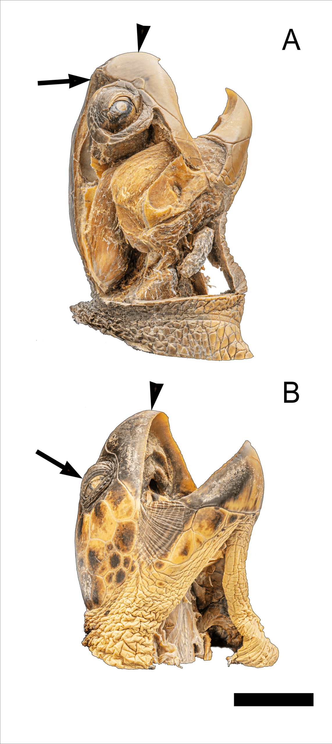
Figure 1. Plastinates of the olive ridley (A) and loggerhead (B) turtle heads for gross and generalized comparison to their corresponding models depicted in Figures 2 & 3. A) The olive ridley displayed a tapered and sharp beak (arrowhead) with two pairs of prefrontal scales (arrows). B) The logger head turtle showed a more robust beak (arrowhead) with eyes positioned dorsolateral (arrow) in the craniofacial region. Specific anatomical features are annotated in the online models (Figures 2 & 3). Scale bar = 10 cm.
Olive ridley (Lepidochelys olivacea). The olive ridley had a relatively small and streamlined head compared to other sea turtle species, contributing to its hydrodynamic profile in the water (Pritchard & Mortimer, 1999). As seen in the plastinated head (Fig. 1A), the cranium is somewhat tapered along anteriorly and proportionally smaller than that of the loggerhead (Fig. 1B). The olive ridley displays a moderately elongated neck facilitating flexible, multidirectional movement that supports prey capture (Pritchard & Mortimer, 1999).
The skull itself was relatively lightly built with less robust bone compared to the loggerhead, likely reflecting the olive ridley’s preference for soft-bodied prey such as jellyfish and invertebrates (Wyneken, 2001). The beak, or rhamphotheca, was intact and sharply defined in the plastinate. The olive ridley specimen (Fig. 1A) retained accurate proportions and spatial relationships among major structures. Natural coloration was largely preserved with skin and muscle layers distinguishable, and the head remained dry, odorless, and firm. Overall anatomy was displayed without collapse or distortion. Externally, the olive ridley exhibited two pairs of prefrontal scales characterized as the keratinized plates along the superior aspect of the snout between the eyes and posterior to the nostrils (Pritchard & Mortimer, 1999). The epidermal scales on the head were preserved in the plastinate, maintaining the natural surface texture and arrangement consistent with the species’ cranial anatomy.
Loggerhead (Caretta caretta). The loggerhead (Fig. 1B) had a robust and broad head, distinguishing it from other sea turtles. The head of loggerhead sea turtles is large and robust providing a reinforced structure suitable for their durophagous diet of hard-shelled prey (Marshall et al., 2014). Bone was more prevalent, robust and expansive in the loggerhead skull with the rhamphotheca heavier, reflecting the species’ adaptation to consuming harder, more resistant prey, including crustaceans and mollusks (Wyneken, 2001). The neck of the loggerhead was short and muscular, likely stabilizing the head, which is necessary for its powerful jaw action during crushing shells (NOAA Fisheries, 2022). The beak was strong and hooked, designed for grasping and consuming hard-bodied organisms (Wyneken, 2001). The jaw muscles of the loggerhead were well-developed and more powerful than those of the olive ridley, reflecting their specialized feeding habits (Wyneken, 2001). Loggerhead sea turtles have dorsolateral positioned eyes that support a visual field adapted to benthic foraging, enabling them to locate prey on the seafloor (Warriach et al., 2020).
The loggerhead specimen (Fig. 1B) maintained accurate anatomical proportions and the proper spatial relationships of major structures. Natural color retention was good, with differentiation between skin and muscle layers preserved. The specimen remained dry, firm, and free from unpleasant odor, effectively preserving the structural integrity of the turtle's head and neck.
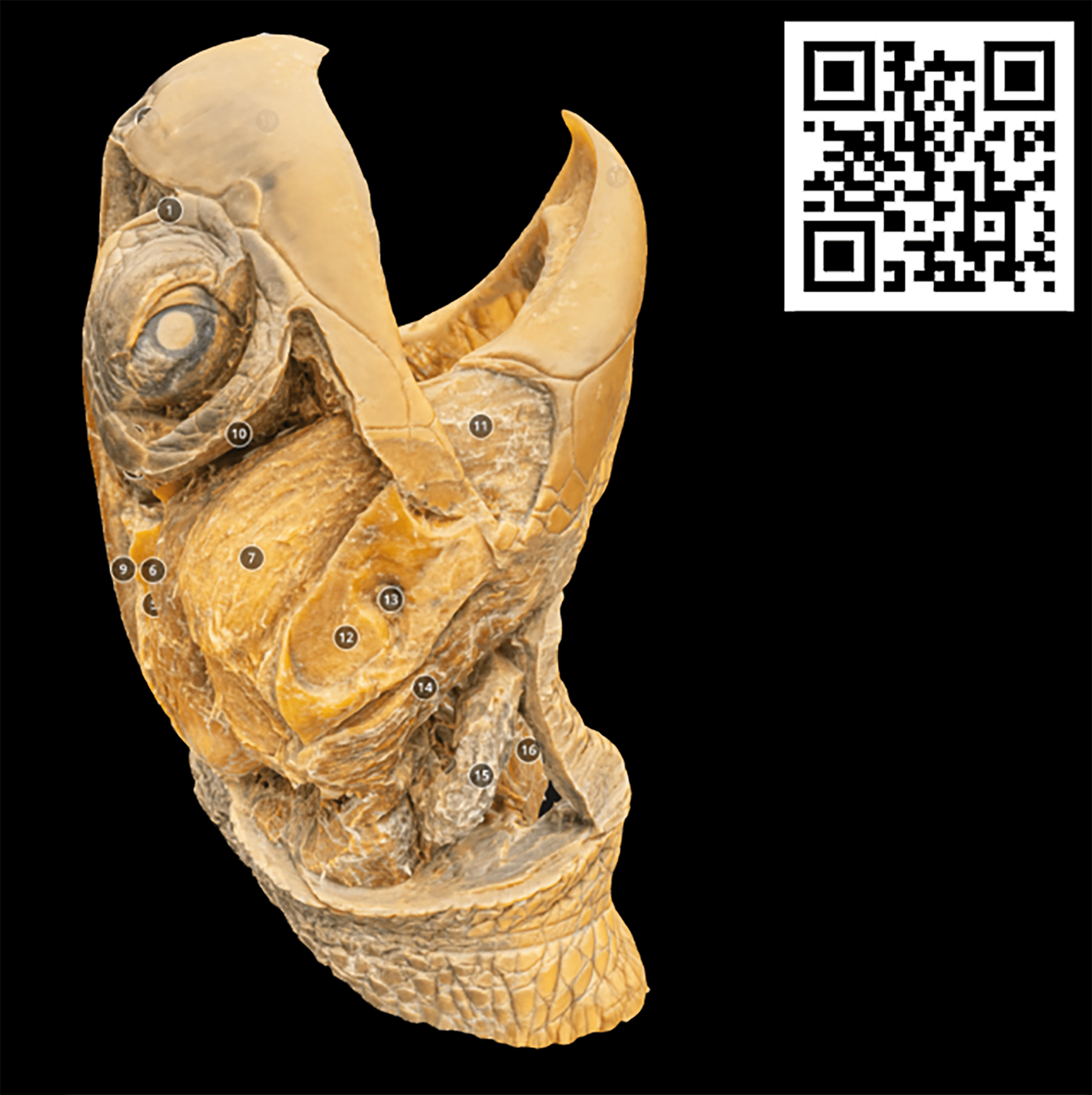
Figure 2. Interactive 3D model of the olive ridley sea turtle head corresponding to Figure 1A including annotations. This model is directly accessible using the QR code in this figure or link https://skfb.ly/pzREn
The olive ridley model includes numbered anatomical features labeled in Sketchfab and available in the QR code in Figure 2. For example, in dorsal view the nasal capsule (1) and nares (20) are seen. In the dorsal aspect of the mouth, the internal nares (17) are found caudal to the upper rhamphotheca’s (19) oral margin. Both upper (19) and lower rhamphothecae (18) maintain their keratinized structure and adhere tightly to the underlying bones. Bone structures such as the frontal (8) and a portion of the parietal bone (9) are labeled in the model.
The model also highlights major components of the cranial musculature, including the adductor mandibularis externus muscle group, with its deep (5), lateral (6), superficial (7), and medial parts (11), and the adductor mandibulae internus (4). These structures function in jaw closure and during feeding (Jones et al., 2012). The depressor mandibulae (14) and constrictor colli (15) are also visible, contributing to jaw depression and throat constriction, respectively (Jones et al., 2012). The posterior neck musculature, including the longus colli (16), is preserved and visible in the 3D model. Orbital and glandular anatomy are detailed, with clear visualization of the superior oblique muscle (2), lacrimal gland (3), and Harderian gland (10). The tympanic cavity or antrum postoticum (12) and the sole ear bone, the stapes (columella) (13), were located along the posterolateral aspect of the cranium.
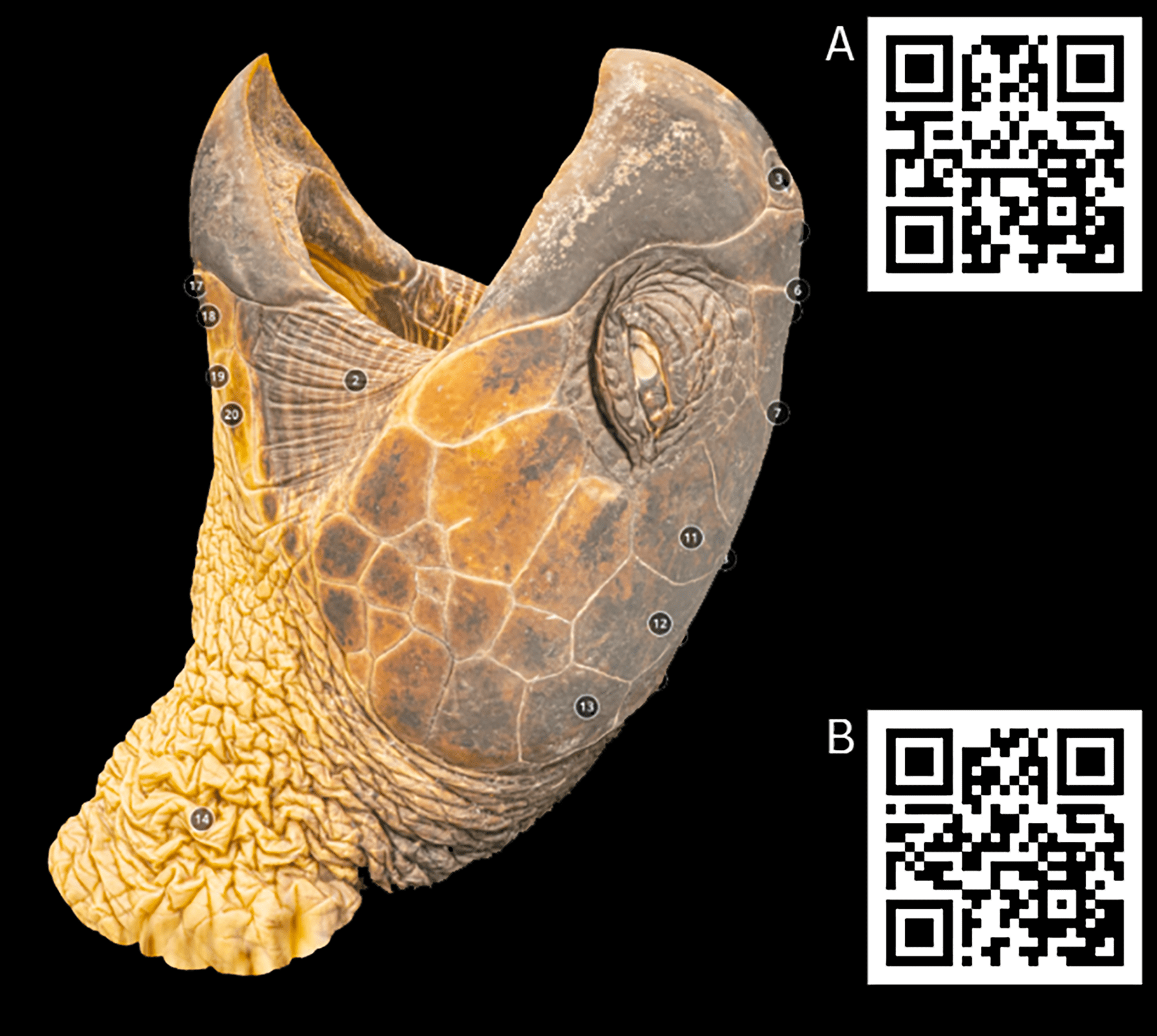
Figure 3. Interactive 3D model of the loggerhead sea turtle head corresponding to Figure 1B including annotations. This model is directly accessible using the corresponding QR code in this figure or link 3A) https://skfb.ly/pzRJF 3B) https://skfb.ly/pzRJG. Due to the large number of annotations, the loggerhead is presented as two models (3A and 3B).
The loggerhead model (Fig. 3) provides insight into the robust cranial morphology of this species. Facial and cranial scale patterns remain distinct, with identifiable prefrontal (3A, 4–5), supernumerary (3A, 6), frontal (3A, 7), frontoparietal (3A, 8), parietal (3A, 9–10), and temporal (3A, 11–13) scales. The naris (3A, 3), opening of the glottis (3A, 15), choanae (3A, 16) are preserved within the respiratory pathway; while a portion of the tongue (3A, 1), commissure of the jaw (3A, 2), and submandibular scales (3A, 17–20) are visible, helping to define some interior and exterior oral structures. Natural folding of the neck skin (3A, 14) and well-preserved dermal features are also observed.
The upper (3B, 19) and lower (3B, 20) rhamphotheca are well preserved. The mouth’s keratinized surfaces exhibit their structural specializations for durophagy. Supporting this function, large jaw and neck muscles, including the plastrosquamosus (3B, 3), branchiomandibularis visceralis (3B, 4), and coraco-hyoideus (3B, 5), are visible, emphasizing the strength behind this feeding mechanism (Wyneken, 2001). The internal carotid artery (3B, 1) and esophagus (3B, 2) are also labeled, further enhancing anatomical context. Ocular structures, including the pupil (3B, 6), lower eyelid (3B, 7), and upper eyelid (3B, 8), are intact, and the surrounding supraocular (3B, 9), tympanic (3B, 10–15), and postocular (3B, 16–18) scales help define the orbital region.
Plastination ensures the long-term preservation of physical specimens by stabilizing tissue structure and preventing decay, making it a gold standard in anatomical preservation. Photogrammetry complements plastination by creating interactive 3D models (Figs. 2,3), enhancing educational utility, and enabling detailed, remote examination of anatomical structures that might otherwise be difficult to access. Such resources may enrich the learning process by providing novel modes of interaction and aiding students with different learning preferences (Triepels et al., 2019). This dual approach expands the availability of high-quality anatomical resources beyond physical limitations, enabling inclusive and global educational opportunities (Hashida et al., 2021).
The integration of 3D models into anatomy curricula holds promising potential for enhancing understanding (Yammine & Violato, 2015) and photogrammetry of plastinations enables a unique dataset for creating online models. Plastinated specimens are well suited for photogrammetry since their rigidity ensures stability during photographic processing thus reducing blurring due to movement. Blurring and wet surface reflections are more common with dissected materials requiring extensive post processing and editing. Plastination retains color retention and definition, reducing the need for post processing and the corresponding time allotment. These features likely contributed to findings reported by Hashida et al., (2021) indicating that students preferred online models within their learning management systems generated from plastinations compared to models derived from cadaveric specimens or artistically rendered representations. With the increase of online learning, plastination combined with photogrammetric modeling should provide a unique approach for creating online learning modules and delivery of 3D anatomical educational content.
Traditionally, public use of anatomical and museum specimens involves copyright to protect intellectual property, ensuing that the owner maintained exclusive rights to reproduce and distribute their work. The length of the copyright generally extends for the author's life plus 70 years. One intent of a copyright was to encourage creativity and learning; however, this typically requires cumbersome and excessive processes for individuals to gain lawful access to the work for utilization and create derivatives. Creative Commons, an NPO, was first launched in 2002 to facilitate issuing of standard and feeless licenses enabling creators to easily share their work while retaining copyright (Plotkin, 2002). The author can easily set conditions of use for the work through standard attributions. Individuals gain read/write access to anatomy and museum electronic collections, stimulating creativity and knowledge sharing through online activities (Verzina et al., 2022). This ease-of-access is particularly meaningful where specimens that are rare or endangered can be easily accessed online by students and researchers as well as the public at large. In the case of the olive ridley and loggerhead specimens presented here, attributions were set on Sketchfab during the uploading process and these models are now widely available.
Successful plastination relies on meticulous dehydration and polymer infiltration techniques, which must often be adapted for specimens with unusual tissue compositions and organ size (von Hagens et al., 1987). For example, Jiang et al. (2022) described the plastination of a sperm whale which required unique challenges due to the immensity of the specimen, while Miller et al. (2017) confronted methodology issues to achieve successful plastination of the heart of a blue whale. In both cases, the necessity of modifying plastination protocols to accommodate species-specific anatomical features was highlighted. These adaptations of the processes offer valuable guidance for addressing similar obstacles in reptilian plastination, where unique tissue properties complicate standard techniques. Methodological variations related to the timing of dehydration, impregnation, and curing were implemented with the turtle heads described here ultimately requiring over one year to process. Although lengthy, the turtle head specimens are highly durable and should retain display qualities for many years.
The Reptilia class comprises cold-blooded and atmospheric breathing animals characterized by several anatomical specializations that vary broadly typically requiring specialized variation in plastination methodology (Ekim et al., 2017). The reptilians feature thick epidermal cornification that can limit fixation and hinder curing (Wendel et al. 2008). Significant loss of natural color in snakes as well as the need for internal filling due to tissue shrinkage has been reported as additional challenges for reptile plastination (Ekim et al., 2017). The tough, keratinized dermal layers of reptiles, such as the loggerhead and olive ridley turtles, require extensive preparation to achieve effective fixation and polymer infiltration (Silva et al., 2004). Additionally, dense and highly mineralized cranial structures complicate plastination, often necessitating prolonged processing times to ensure adequate penetration of fixatives and polymers. A faint offensive odor, reminiscent of putrefying fish, was noticed during the defatting and impregnation steps. Although less apparent following impregnation, the specimens remained in the curing phase for an extended period and the specimens were not used until the odor dispersed.
Alternate approaches involving CT or MR scanning provide excellent models of turtle anatomy (Rollot et al., 2024). These volume-based reconstruction methods have advantages since they retain both surface and internal morphological information. Additionally, volume rendering approaches typically require less processing time compared to plastination. However, volume-based models require the availability of large storage arrays limiting ease-of-exchange between viewers or embedding in educational learning management systems. The surface modeling approach used here renders models that require much less storage space enabling customization and rapid exchange between viewers (Chang et al., 2016). Surface models are uploaded to widely available platforms and are easily embedded into learning management systems. These surface models also retain surface colors and textures that typically cannot be reproduced using CT or MR approaches. Thus, surface models, such as those presented here, provide advantages for educational resource implementation that are not available otherwise.
This technique provides a scalable framework for creating digital repositories of rare or endangered species. For example, 3D models can preserve the intricate anatomical details of marine species, including rare species and those threatened by environmental changes. Miller et al. (2021) describe the plastination of the heart of an endangered southern resident killer whale (Orincus orca) that is included in a museum exhibition facilitating education and scientific observation. Similarly, Jiang et al., (2022) achieved an ambitious large scale plastination of an endangered sperm whale that is now a prominent museum exhibition. AbouHashem et al. (2015) argue that such models, whether physical or digital, can democratize access to anatomical resources, ensuring that rare or endangered specimens contribute to broader scientific and educational contexts. Additionally, plastination and display of endangered marine species impact society beyond the display. One of the last Yangtze giant soft-shell turtles (Rafetus swinhoei) was plastinated and displayed in Ngoc Son Temple, Hanoi, Viet Nam. The plastinated turtle, requiring over a year to plastinate, is called Cu Rua, or Great-Grandmother Turtle, and has great cultural significance. Westergaard & Jørgensen (2021) argue that preservation of rare animals, in particular, Cu Rua, position them in a liminal existence and become sacred cultural objects creating a sense of transcendence. Thus, the cultural and social implications of plastinations and their derivatives can be enormous.
Integrating plastination with photogrammetry extends the physical and digital essence of a biological specimen and transforms it into a versatile educational tool that is deliverable across time and space. These methods bridge gaps between traditional preservation techniques and modern digital education, promoting innovative, interactive, and practical ways to explore anatomy. Furthermore, it emphasizes the potential of such approaches to democratize access to anatomical education, making high-quality resources available to a global audience and fostering interdisciplinary learning. This study established a workflow for electronic ease-of- exchange of anatomical models derived from plastinated specimens, with a focus on the head (craniofacial) anatomy of two endangered marine turtle species, olive ridley (Caretta caretta) and loggerhead (Lepidochelys olivacea). Overall, the plastinated heads provided a clear, accurate representation of turtle craniofacial anatomy, including preserved bone positions, keratin structures, and general muscle form, with minimal distortion. These qualities make it a valuable teaching aid and anatomical reference for species-specific adaptations in sea turtles as well as serving as a basis for photogrammetry. Future efforts will aim to expand the collection by generating additional specimens as well as utilizing the specimens in an online educational setting.
LIMITATIONS
Complete removal of lipids and water was needed to ensure proper impregnation with plastic polymer. The specimens were dehydrated for over 6 weeks, allowing for the gradual replacement of water in the tissues with acetone without causing significant shrinkage or distortion. Nonetheless, this process, in addition to formalin fixation, may have caused differential shrinkage in the specimens. Additionally, quantified measurement of educational effectiveness based on these specimens was not assessed. This effort will require an extensive assessment utilizing both qualitative and quantitative methodologies (Guaraná et al., 2023; Liang et al., 2024). This study provides an approach for plastination and digital preservation of turtle heads; however, it may require additional experimentation to achieve optimal preservation across reptilian genera and species.
ACKNOWLEDGMENTS
Any use of trade, firm, or product names is for descriptive purposes only and does not imply endorsement by the U.S. Government. We thank Zarek Kon and Bashir El-Orm for their technical assistance, and Drs. T. Work, W. Joyce, and K. Tamura for their helpful comments on earlier versions of this paper.
AbouHashem Y, Dayal M, Savanah S, Štrkalj G. 2015: The application of 3D printing in anatomy education. Med Educ Online, 20(1).
https://doi.org/10.3402/meo.v20.29847
Abreu-Grobois A, & Plotkin P, (IUCN SSC Marine Turtle Specialist Group). 2008: Lepidochelys olivacea. The IUCN Red List of Threatened Species.
https://doi.org/10.2305/IUCN.UK.2008.RLTS.T11534A3292503.en
BODY WORLDS. (n.d.). BODY WORLDS & The Story of the Human Body. https://bodyworlds.com/exhibitions/human/
Bot JA, Irschick DJ. 2019: Using 3D photogrammetry to create open-access models of live animals: 2D and 3D software solutions. In: Grayburn J, Lischer-Katz Z, Golubiewski-Davis K, Ikeshoji-Orlati V. (Eds.), 3D VR in the Academic Library: Emerging Practices and Trends. pp. 54-72. Council on Library and Information Resources, Arlington, VA.
Cáceres-Farias L, Reséndiz E, Espinoza J, Fernández-Sanz H, Alfaro-Núñez A. 2022: Threats and vulnerabilities for the globally distributed olive ridley (Lepidochelys olivacea) sea turtle: A historical and current status evaluation. Animals 12(14):1837.
https://doi.org/10.3390/ani12141837
Casale P, Tucker AD. 2017: Caretta caretta (amended version of 2015 assessment). The IUCN Red List of Threatened Species
https://doi.org/10.2305/IUCN.UK.2017-2.RLTS.T3897A119333622.en
Chang CW, Atkinson G, Gandhi N, Farrell ML, Labrash S, Smith AB, Norton NS, Matsui T, Lozanoff S. 2016: Computerized 3D anatomical modeling using cone beam computed tomography (CBCT) scans of plastinated hearts. Surg Radiol Anat 38(7):843-853.
https://doi.org/10.1007/s00276-016-1645-6
DeJong K, Henry RW. 2007: Silicone plastination of biological tissue: Cold-temperature technique Biodur S10/S15 technique and products. J Int Soc Plastination 22:2-14.
https://doi.org/10.56507/ZLMJ7068
Ekim O, Hazıroğlu, RM, İnsal B, Bakici C, Akgün RO, Tunali S. 2017: A modified S10B silicone plastination method for preparation and preservation of scaled reptile specimens. Ankara Üniv Vet Fak Derg 64:155-160.
https://doi.org/10.1501/Vetfak_0000002792
Falkingham PL. 2012: Acquisition of high-resolution three-dimensional models using free, open-source, photogrammetric software. Palaeont Electron 15(2):1-15. palaeo-electronica.org/content/93-issue-1-2012-technical-articles/92-3d-photogrammetry
https://doi.org/10.26879/264
García-Párraga, D., Crespo-Picazo, J. L., de Quirós, Y. B., Cervera V, Martí Bonmati L, Díaz Delgado J, Arbelo M, Moore MJ, Jepson PD, Fernández A. 2014: Decompression sickness ("the bends") in sea turtles. Dis Aquat Org 111(3):191-205.
https://doi.org/10.3354/dao02790
Guaraná JB, Aytaç G, Müller AF, Thompson J, Freitas SH, Lee UY, Lozanoff S, Ferrante B. 2023: Extended reality veterinary medicine case studies for diagnostic veterinary imaging instruction: Assessing student perceptions and examination performance. Anat Histol Embryol 52(1):101-14.
https://doi.org/10.1111/ahe.12879
Hashida K, Shirahata K, Thompson J, Liang C, Takakusagi M, Stenger K, Lozanoff BK, Labrash S, Rettenmeier C, Gestewitz M, Lee U-Y, Doll S, & Lozanoff S. 2021: Plastinated instructional assets utilization during collaborative online head and neck anatomy in the medical educational curriculum. J Plast 33(2):28-38.
https://doi.org/10.56507/KDHJ2784
Hoegh-Guldberg O, Bruno JF. 2010: The impact of climate change on the world's marine ecosystems. Science 328(5985):1523-1528.
https://doi.org/10.1126/science.1189930
Irschick, DJ, Christiansen F, Hammerschlag N, Martin J, Madsen PT, Wyneken J, Brooks A, Gleiss A, Fossette S, Siler C, Gamble T, Fish F, Siebert U, Patel J, Xu Z, Kalogerakis E, Medina J, Mukherji A, Mandica M, Zotos X, Detweiler J, Perot B, Lauder G. 2022: 3D visualization processes for recreating and studying organismal form. iScience, 25(9) 104867.
https://doi.org/10.1016/j.isci.2022.104867
Jiang W-B, Han J, Ma X-W, Liu H, Yu S-B, Sui H-J. 2022: Plastination of a sperm whale. J Anat 240(4):669-677.
https://doi.org/10.1111/joa.13581
Jones MEH., Werneburg, I., Curtis, N, Penrose R, O'Higgins P, Fagan MJ, Evans SE. 2012: The head and neck anatomy of sea turtles (Cryptodira: Chelonioidea) and skull shape in Testudines. PLoS ONE 7(11), e47852.
https://doi.org/10.1371/journal.pone.0047852
Lewison R., Freeman SA, Crowder LB. 2004: Quantifying the effects of fisheries on threatened species: The impact of pelagic longlines on loggerhead and leatherback sea turtles. Ecol Lett 7(3):221-231.
https://doi.org/10.1111/j.1461-0248.2004.00573.x
Liang CG, Matsunaga M, Takakusagi M, DeMeo J, Thompson JD, Rettenmeier C, Aytaç G, Lee UY, Lozanoff S. 2024: A comparison of pre‐and post‐clinical education learning preferences among medical students who elected to dissect compared to those who did not during the COVID‐19 pandemic. Anat Sci Educ 17(2): 396-412.
https://doi.org/10.1002/ase.2362
Marshall CD, Wang J, Rocha-Olivares A, Godínez-Reyes C, Fisler S, Narazaki T, Sato K, Sterba-Boatwright BD. 2014: Scaling of bite performance with head and carapace morphometrics in green turtles (Chelonia mydas). J Exp Mar Bio Ecol 451:91-97.
https://doi.org/10.1016/j.jembe.2013.11.004
Miller JR, Henry RW, Nader P, Engstrom MD, Iliff S, Cheremiski V, von Hagens G. 2017: The challenges of plastinating a Blue Whale (Balaenoptera musculus) heart. J Plast 29:22-29.
https://doi.org/10.56507/SJTL1096
Miller JR, Lopez Albors O, Raverty S, Cottrell P, Engstrom MD, Latorre R. 2021: Plastination of an endangered southern resident killer whale heart (Orcinus orca). J Plast 33(1):1-8.
https://doi.org/10.56507/JWUL5276
NOAA Fisheries. (2022). Loggerhead turtle. Species Directory. https://www.fisheries.noaa.gov/species/loggerhead-turtle#overview
Plotkin H. 2002: All Hail Creative Commons: Stanford professor and author Lawrence Lessig plans a legal insurrection. SFGate. Com 11.
Poloczanska, ES, Limpus, CJ, Hays GC. 2009: Vulnerability of marine turtles to climate change. Adv Mar Biol 56: 151-211.
https://doi.org/10.1016/S0065-2881(09)56002-6
Pritchard PCH, Mortimer JA. 1999: Taxonomy, external morphology, and species identification of sea turtles. In: Eckert KL, Bjorndal KA, Abreu-Grobois FA, Donnelly M. (eds), IUCN/SSC Marine Turtle Specialist Group. https://www.widecast.org/Resources/Docs/Pritchard_and_Mortimer_1999_Sea_Turtle_Taxonomy.pdf
Raoof A, Henry RW, Reed RB. 2007: Silicone plastination of biological tissue: Room temperature technique Dow/Corcoran technique and products. J Int Soc Plastination 22:21-25.
https://doi.org/10.56507/AWAC9285
Rodriguez SL, Peterson MN, Moorman CJ. 2017: Does education influence wildlife-friendly landscaping preferences? Urban Ecosyst 20:489-496.
https://doi.org/10.1007/s11252-016-0609-2
Rollot Y, Evers SW, Ferreira GS, Girard LC, Werneburg I, Joyce WG. 2024: Skull osteology, neuroanatomy, and jaw‐related myology of the pig‐nosed turtle Carettochelys insculpta (Cryptodira, Trionychia). Anat Rec 307(9):2966-3020.
https://doi.org/10.1002/ar.25411
Schuyler Q, Hardesty BD, Wilcox C, Townsend K. 2014: Global analysis of anthropogenic debris ingestion by sea turtles. Conserv Biol 28(1):129-139.
https://doi.org/10.1111/cobi.12126
Silva MVF, Monteiro YF, Miranda RP, dos Santos AB, Bittencourt APSV, Carretta M, Menezes FV, Delpupo FVB, Bittencourt AS. 2024: From highways to biological collections: plastination of wild animal victims of roadkill in the Sooretama Biological Reserve, Brazil. Braz Arch Biol Technol 67:1-12:e24230044.
https://doi.org/10.1590/1678-4324-2024230044
Triepels CPR, Smeets CFA, Notten KJB, Kruitwagen RFPM, Futterer JJ, Vergeldt TFM, Van Kuijk SMJ. 2019: Does three-dimensional anatomy improve student understanding? Clin Anat 33(1): 25-33.
https://doi.org/10.1002/ca.23405
Valente ALS, Parga ML, Velarde R, Marco I, Lavin S, Alegre F, Cuenca R. 2007: Fishhook lesions in loggerhead sea turtles. J Wildl Dis 43(4):737-741.
https://doi.org/10.7589/0090-3558-43.4.737
Vezina B, Anas S, Craig C, Giblin R, Hollich S, Kefalea R, Keller P, Matas A, Margoni T, Petrasova K, Poritz J, Rimmer M, Terras M, Thomas, H, Zeinstra M. 2022: Towards better sharing of cultural heritage - an agenda for copyright reform: A Creative Commons Policy Paper, pp. 1-25. Creative Commons. https://creativecommons. org/wp-content/uploads/2022/04/Creative-Commons-Policy-Paper-on-GLAMs-Final. pdf. 2022.
von Hagens G, Tiedemann K, Kriz W. 1987: The current potential of plastination. Anat Embryol, 175(4), 411-421.
https://doi.org/10.1007/BF00309677
Warriach N, Wyneken J, Blume N. 2020: Feeding behavior and visual field differences in loggerhead and leatherback sea turtles may explain differences in longline fisheries interactions. Endang Species Res 41:67-77.
https://doi.org/10.3354/esr01012
Wendel H, Stark R, Plendl J. 2008: Plastination of reptiles for veterinary medicine (abstract). J Plast 23:62.
Westergaard G, Jørgensen D. 2021: Making specimens sacred: Putting the bodies of Solitario Jorge and Cụ Rùa on display. In: Animal Remains, Bezan S & McKay R (eds), pp. 68-86. Routledge Press.
https://doi.org/10.4324/9781003129806-7
Work TM, Balazs GH. 2002: Necropsy findings in sea turtles taken as by-catch in the North Pacific longline fishery. Fish Bull 100(4):876-880. https://www.researchgate.net/publication/228706862_Necropsy_findings_in_sea_turtles_taken_as_bycatch_in_the_North_Pacific_longline_fishery
Work TM, Balazs GH. 2010: Pathology and distribution of sea turtles landed as bycatch in the Hawaii-based North Pacific pelagic longline fishery. J Wildl Dis 46(2):422-432.
https://doi.org/10.7589/0090-3558-46.2.422
Wyneken J. 2001: The anatomy of sea turtles. NOAA Technical Memorandum NMFS-SEFSC-470. Pp 172. https://repository.library.noaa.gov/view/noaa/8502
Yammine K, Violato C. 2015: A meta-analysis of the educational effectiveness of three-dimensional visualization technologies in teaching anatomy. Anat Sci Educ 8(6):525-538.
https://doi.org/10.1002/ase.1510

