1Department of Anatomy and Comparative Pathology, Faculty of Veterinary, Campus de Espinardo, 30100 Murcia, Spain
2Discover-IN SL, Complejo Pleiades-Vitalis, Campus de Espinardo, 30100 Murcia, Spain
OrganKits: a new material for STEAM (science, technology, engineering, arts, and mathematics) education based on plastination was developed to create portable, classroom-ready anatomical kits for secondary education. This paper describes the technical workflow used to prepare, plastinate, assess quality, and assemble six themed multi-organ sets designed for repeated classroom use. Fresh swine, ovine, and bovine organs obtained from abattoirs were processed using organ-specific standard operating procedures (SOPs) based on silicone (S10) and epoxy and polyester plastination, with modifications introduced to preserve luminal patency, flexibility, sectional readability, and handling resistance. The workflow incorporated specimen coding at reception, stage-by-stage database monitoring, checklist-based quality assessment, microchip tagging, photographic recording, and briefcase assembly log to ensure full traceability. The final production comprised 432 specimens across 18 types, including whole organs, sectioned organs, flexible hollow viscera, a bronchial tree cast, musculoskeletal preparations, and methacrylate image sheets derived from sheet-plastinated sections or histological slides. Five additional specimens were rejected during quality control because they did not meet the checklist of standards. The completed OrganKits were then distributed to partner secondary schools for two rounds of educational implementation under the STEAM scope. This work shows that classical plastination methods can be adapted to a standardized, quality-assured production model for school-oriented STEAM approaches. Its main methodological contribution is integrating organ-specific technical modifications with traceability and quality-control measures that support reproducibility, portability, and repeated educational use.
plastination; quality assurance; STEAM education; OrganKits
Octavio López Albors, Department of Anatomy and Comparative Pathology, Faculty of Veterinary, Campus de Espinardo, 30100 Murcia, Spain
e-mail: albors@um.es





Plastination involves a group of tissue preservation techniques in which water and lipids are replaced by curable polymers such as silicone, epoxy, or polyester, thereby producing dry, durable, odorless, and nontoxic anatomical specimens that can be handled and stored with remarkable ease (Henry et al., 2019; Sora et al., 2019). Owing to these properties, plastinated materials have become well established in human and veterinary anatomical education, particularly in higher education, where they are valued for their anatomical realism, long-term stability, and suitability for repeated student handling (Latorre et al., 2007; Latorre et al., 2016; Sora et al., 2019). Moreover, previous educational studies show that students perceive plastinated specimens positively and that they may contribute to motivation and anatomy learning when incorporated appropriately into practical teaching sessions (Latorre et al., 2007; Nguyen et al., 2019).
In secondary education, however, the situation differs. Biology or science teaching at this level requires realistic, manipulable materials that help students understand organ morphology and function, but the resources most often used are textbooks, two-dimensional illustrations, artificial plastic models, or, in some cases, fresh and wet-preserved organs. Each material has limitations in anatomical realism, handling, potential toxicity, or storage requirements. In this context, plastinated organs appear as an attractive alternative because they combine real anatomy with safety and durability, making them especially suitable for the varied classroom environments found in secondary schools (López Albors et al., 2022; Delgado-Ruiz et al., 2023). Despite these potential advantages, the published evidence on the use of plastinated specimens at this level is very limited. After an extensive literature search, the only reports concerning a structured use of plastinated organs in secondary schools are largely restricted to a small number of pilot experiences related to science teaching in secondary schools (Douglass and Glover, 2003; Williams and Rae, 2019; López Albors et al., 2022; Delgado-Ruiz et al., 2023). This point matters because the technical demands of school-oriented plastinated specimens differ from those of university teaching collections. Beyond anatomical fidelity, these materials must be especially robust, easy to transport, safe for repeated manipulation, visually intuitive, and suitable for integration into interdisciplinary classroom activities (Douglass and Glover, 2003).
Recently, a project entitled “OrganKits: a new material for STEAM education based on plastination” was developed within the Erasmus+ Key Action 2 (https://erasmus-plus.ec.europa.eu/programme-guide/part-b/key-action-2) between 2022 and 2025. Coordinated by the University of Murcia and aimed at the European secondary education field, the project sought to design and validate a new educational proposal based on the use of plastinated organs through STEAM (science, technology, engineering, arts and mathematics) and project-based learning strategies. Technically, the project required producing six themed kits of plastinated specimens: CardioHealth & Emotions, NeumoHealth & Environment, NutriHealth & Wellness, MentalHealth & Mindfulness, SportsHealth & Dependence, and ReproHealth & Gender. The kits, mounted in transportable briefcases, included a collection of silicone-plastinated animal organs and supplementary methacrylate sheets with anatomical images from epoxy- or polyester-plastinated specimens or histological slides. These materials were enriched by detailed Teacher Guides, Educational Guides, and a library of digital learning objects designed specifically for each OrganKit. The project's challenge was not only educational but also technical, as it required producing classroom-ready plastinated specimens under a standardized methodology with quality controls and traceability tailored to the project's requirements.
This paper describes the methodological particularities involved in preparing and plastinating each specimen in the organ collection included in the project, and presents the procedures followed to standardize working protocols, evaluate quality, and maintain specimen traceability throughout the process.
Design and technical workflow
The technical production model was designed around the six OrganKits mentioned above. The physical contents included plastinated specimens of swine, ovine, and bovine origin, together with printed methacrylate sheets derived from epoxy- or polyester-plastinated sections or histological material. The common framework comprised specimen selection, preparation (dissection and fixation), plastination, quality assessment, and assembly into transportable briefcases.
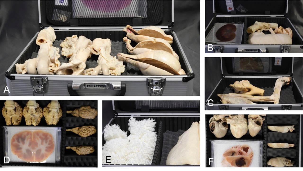
Figure 1. Composition of the six OrganKits as arranged in the customised briefcases. A, ReproHealth & Gender (three sets housed in the same briefcase); B, NutriHealth & Wellness; C, SportsHealth & Dependence; D, MentalHealth & Mindfulness (three sets housed in the same briefcase); E, NeumoHealth & Environment; F, CardioHealth & Emotions (three sets housed in the same briefcase)
The six kits were configured as follows (Fig. 1). The CardioHealth kit contained plastinated swine hearts, including one complete specimen, one sagittally sectioned half-heart, and a methacrylate lamina showing a transverse section at the base of the heart. The NeumoHealth kit contained ovine flexible lungs, a bronchial cast, and a histological lamina of lung tissue. The NutriHealth kit contained a swine stomach, small intestine, and large intestine portions, plus a lamina of a sagittal section of the kidney. The MentalHealth kit contained an entire bovine encephalon, a half sagittal encephalon, and a lamina with a transverse section at the level of the thalamus. The SportsHealth kit contained a plastinated bovine elbow, an ovine hindlimb dissection distal to the knee, and a lamina with a sagittal section of the stifle. Finally, the ReproHealth kit contained a plastinated ovine testicle and female reproductive tract, and a histological lamina of ovarian tissue.
In addition, the kits included a domestic endoscope to facilitate exploratory activities and simulate endoscopy and bronchoscopy procedures.
All organs were collected from local abattoirs after veterinary inspection and delivered fresh to the plastination laboratory of Discover-IN SL (University of Murcia). Upon arrival, the organs were processed according to standardized operational protocols (SOPs) previously created and progressively validated by the authors in accordance with ISO9001-2015 requirements. Validation involved testing and repeating several modifications of the common silicone S10 (Biodur®), as described by Henry et al. (2019). Each SOP included the dissection and fixation details for each specimen, as well as any significant modifications to the mentioned publication. Overall, these modifications aimed to better preserve morphology, especially the lumen of hollow organs, and to enhance the specimen's pedagogical value for use in secondary schools.
To ensure traceability, we labeled each organ upon arrival and recorded the reference number in a database designed to track each step of the dissection and plastination process. Once curing (polymerization) was complete, we assessed specimen quality against a predefined checklist of anatomical landmarks. Organs that did not meet the checklist requirements were discarded.
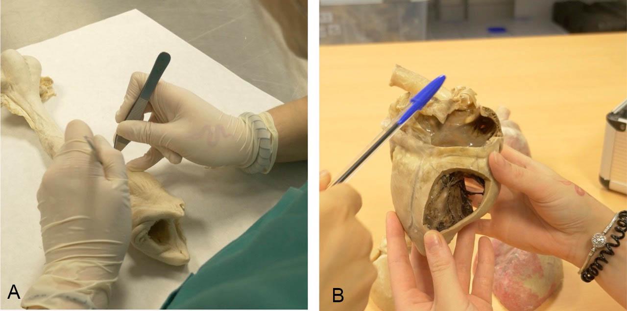
Figure 2. A, Final validation of a cured ovine uterus; B, Quality check of a pig heart prior to validation
After final manicuring and quality assessment (Fig. 2), we tagged all specimens with a microchip and packed them in aluminum briefcases labeled with the project logos. For packaging, we logged the contents of each briefcase in a document, recording the specimen’s name, microchip number, briefcase number, and destination school. A photographic record was also taken of both the individual elements and the briefcase's internal configuration.
Plastination protocols
CardioHealth
Pig hearts were received as part of a cardiopulmonary block that also included diaphragm, esophagus, and liver portions. After cleaning with tap water, the pericardium was removed and intracardiac clots were expelled by introducing water through the caudal vena cava while gently massaging the heart to simulate pumping. The main vessels were then intubated. On the right side, the caudal and cranial vena cava and the vein of the middle pulmonary lobe were prepared. On the left side, the aorta was cannulated, while the brachiocephalic trunk and left subclavian artery were closed with a tight knot. To avoid fluid circulation between the lungs and the heart, the root of each lung was closed with a ligature.
Fixation combined immersion with injection-dilatation. First, the left azygos vein was ligated to prevent reflux. Then 5% formaldehyde was introduced through the caudal vena cava until it emerged from the cranial vena cava. The cranial vena cava was sealed with a stopper, and filling continued until the right side acquired the desired volume, after which the caudal vena cava was also closed with a stopper. The left side was dilated by introducing fixative through the vein of the middle pulmonary lobe until outflow through the aorta and its branches was observed. The aorta and its branches were then sealed with rubber stoppers, and filling continued until adequate left-sided expansion had been achieved. The dilated cardiopulmonary block was then immersed in 5% formaldehyde for one week and, after fixation, the lungs and trachea were removed.
After routine dehydration, impregnation, and curing of the hearts, those destined to display the internal morphology were sagittally sectioned with a bandsaw.
NeumoHealth
Sheep lungs had to be free of parenchymal cuts or visible disease. A silicone tube was inserted into the trachea and repeated washes with running water were performed until the parenchyma became pale pink. We specifically avoided overinflation to prevent pleural rupture. After drainage, fixation was performed by perfusion-dilatation through the tracheal tube using a peristaltic pump while the lungs were immersed in 3-5% formaldehyde. Continuous pumping was maintained for 24-48 hrs, after which the lungs remained in immersion as required.
After dehydration (in cold acetone) and before forced impregnation, the lungs were filled through the trachea with silicone mixture to prevent parenchymal collapse and preserve bronchial lumen. After impregnation with S10, slow curing at room temperature was accompanied by continuous airway insufflation through the trachea so the finished specimen remained compressible and could recover its shape after manual deformation.
To complement the flexible lungs, we produced tracheobronchial tree casts. For this, we separated fresh lungs from the heart, washed them repeatedly, and connected them to an air compressor through the trachea. Controlled insufflation was maintained for at least 5-6 days to dry the lungs while preventing collapse. Room temperature vulcanizing (RTV) silicone was mixed with catalyst at 10% of the silicone weight. We generally used a total mixture volume of approximately 150-180 mL. We injected the RTV mixture very slowly through the tracheal tubing to avoid excessive pressure. After injection, the specimens cured for at least two days. A small sample of the mixture was retained as a control for polymer hardening. Corrosion was then achieved by boiling the lungs in water for about 1 hr, mechanically removing softened parenchyma with forceps, and subsequently immersing the cast in 15% hydrogen peroxide for 24 hrs. The process was repeated if residual tissue persisted.
NutriHealth
Stomachs were obtained from complete pig gastrointestinal packages extending from esophagus to rectum. The esophagus was fully preserved, and approximately 20 cm of duodenum was retained distal to the pylorus. Gastric contents were removed through esophagogastric intubation and repeated low-pressure water rinsing. This intubation was maintained until the end of plastination to preserve the esophageal lumen. The stomach was fixed by immersion in increasing concentrations of formaldehyde, starting at 5%. Prior to immersion, the duodenal end was tied, and the stomach was filled through the esophagus with 5% formaldehyde solution for approximately three weeks before dissection. Dissection consisted of removing fat and connective tissue along the greater and lesser curvatures before plastination.
The small intestine specimen corresponded specifically to the jejunum. It was isolated from the complete gastrointestinal block after identifying the duodenum and ileum. After isolation, the jejunal segment was washed internally and externally using repeated low-pressure lavage and gentle massage to avoid tears of the wall or mesentery. Fixation was then performed by dilatation-immersion. One end was ligated, the segment was filled with 5% formaldehyde to the desired volume, and then the opposite end was also ligated. The specimen was finally immersed in fixation solution for about one week.
The colon was isolated from the digestive block by first identifying the cecum and then separating the ascending, transverse, and descending colon from adjacent attachments and tissues. As with the jejunum, fixation was performed by lumen filling and immersion in 5% formaldehyde.
As with the lungs, after routine impregnation, the stomachs, jejunum, and colon were cured at room temperature with continuous insufflation of compressed air to preserve lumen size and maintain flexibility. This slow, air-assisted curing protocol yielded flexible specimens suitable for repeated handling and simulation of gastroscopic examination.
MentalHealth
We extracted bovine brains from skulls obtained at abattoirs. The mandible was disarticulated to facilitate manipulation. We made a transverse cut at the orbital level and a shallow sagittal incision in the fronto-occipital region, preserving the internal lamina of these bones. The skull was opened by controlled blows with a hammer and chisel. During brain removal, the dura mater was sectioned to gain access to the subarachnoid space. The cranial nerves were dissected and preserved, the epiphyseal recess sectioned, and the hypophysis preserved. The final extraction was performed cautiously to avoid tearing the olfactory bulbs and peduncles. Fixation was carried out at approximately 4 °C for about two months by immersion in formaldehyde solution with progressive concentration increase. To prevent flattening or deformation from contact with the container base, the brains were suspended in the solution with sutures attached to the vessels of the circle of Willis. Brains were first immersed in 2.5% formaldehyde, and the concentration was increased by 2.5% each week until it reached 10%. In the second week of fixation, the lateral, third, and fourth ventricles were injected with 5% formaldehyde until slight dilatation was observed.
After fixation, some of the brains were sagittally sectioned. Brains were dehydrated in cold acetone at -20 °C. Acetone purity was monitored every two days until a stable reading above 98.5% was maintained for five consecutive days.
SportsHealth
We isolated a bovine elbow joint from skeletal thoracic limbs obtained at abattoirs by sectioning the humerus, radius, and ulna 10 cm proximal and distal to the joint. Dissection focused on preserving the lateral and medial collateral ligaments. Before fixation and plastination, we removed bone marrow from the bone cavities. During curing, we routinely performed repeated forced flexion-extension of the joint to preserve articular movement in the final specimen.
Sheep hindlimbs were transversely sectioned just proximal to the stifle joint. Dissection was planned to display details of the stifle, distal musculature, major tendons, and the common peroneal and tibial nerves. Fixation of the distal limb was carried out by arterial injection through the femoral artery and immersion in 5% formaldehyde solution before dissection. The skin was removed only to the mid-metatarsal level. The dissection preserved the stifle ligaments and menisci, the common peroneal and tibial nerves, the common calcaneal tendon, and all leg muscles. We drilled bone perforations before dehydration to facilitate degreasing. The main technical target was not only anatomical identification but also retention of mobility and flexibility in the finished specimen; to achieve this, active flexion-extension was applied during curing.
ReproHealth
The ovine female reproductive tracts also included the rectum and urinary bladder. Dissection focused on exposing the vulva, vagina, cervix, uterine body, uterine horns, uterine tubes, ovaries, and broad ligament with minimal injury. Silicone tubing was introduced through the vulva to dilate the vagina. The rectum was emptied and trimmed and then cannulated through the anus. In the urinary portion, excess fat was removed and a second tube was introduced through the vulva into the urethra and bladder to maintain lumen visibility. Fixation was performed by immersion in 5% formaldehyde, but before immersion, the uterine horns were injected so that they remained partly dilated. This fixative solution was aspirated, and the uterine lumen was flushed with tap water before dehydration.
We prepared fresh ovine testicles by vascular injection before fixation. The testicular artery was cannulated and injected with Biodur 07®, a low-viscosity red polymer, followed by Biodur U® 10%. After vascular injection, the tunica vaginalis was punctured to reduce shrinkage, and a sagittal cut was made on the free border of the testicle, leaving the parenchyma open like a book. The specimen was then immersed in 10% formaldehyde for at least two weeks.
Methacrylate image sheets
Each kit incorporated a methacrylate lamina produced from selected images of polyester and epoxy plastinated slices or histological slices. Images were scanned at 1200 dpi, stored in TIFF format, and post-processed; if necessary, they were adjusted for brightness, contrast, and saturation. An external provider produced 15 x 25 cm methacrylate laminas from these images after quality pre-testing confirmed adequate transparency and structural readability.
We produced 432 specimens, corresponding to 24 replicas of each of the 18 specimen types (Table 1). Across the six OrganKits, these categories included pig heart and digestive specimens; sheep respiratory, hindlimb, and reproductive specimens; and cow encephalon and elbow preparations, along with associated methacrylate image sheets from epoxy- and polyester-plastinated sections and histological slides. During quality assessment, five specimens: two pig stomachs, two sheep testes, and one hindlimb dissection, were discarded because they did not comply sufficiently with the quality standards.
| OrganKit name | Organkit composition | Specimens produced (n) |
| CardioHealth & Emotions | Whole pig heart, sagittal half-heart, heart-base methacrylate sheet | 72 |
| NeumoHealth & Environment | Flexible sheep lung, bronchial cast, lung histology methacrylate sheet | 72 |
| NutriHealth & Wellness | Pig stomach, intestinal specimen(s), kidney-section methacrylate sheet | 72 |
| MentalHealth & Mindfulness | Whole bovine brain, sagittal half-brain, thalamic-section methacrylate sheet | 72 |
| SportsHealth & Dependence | Bovine elbow, ovine hindlimb dissection, sagittal stifle-section methacrylate sheet | 72 |
| ReproHealth & Gender | Ovine female reproductive tract, ovine testicle, ovary histology methacrylate sheet | 72 |
Note. Five specimens failed the quality assessment and were discarded (two pig stomachs, two sheep testicles, and one sheep hindlimb dissection).
Figure 1 shows the exact composition of each OrganKit case. Once assembled, the cases were distributed to the partner schools for two rounds of educational implementation and assessment (Fig. 3). Although the pedagogical outcomes of that phase are outside the scope of the present paper and will be published separately, the implementation provided robust evidence that OrganKits constitute a valuable, motivating, and adaptable STEAM-based resource for secondary education.
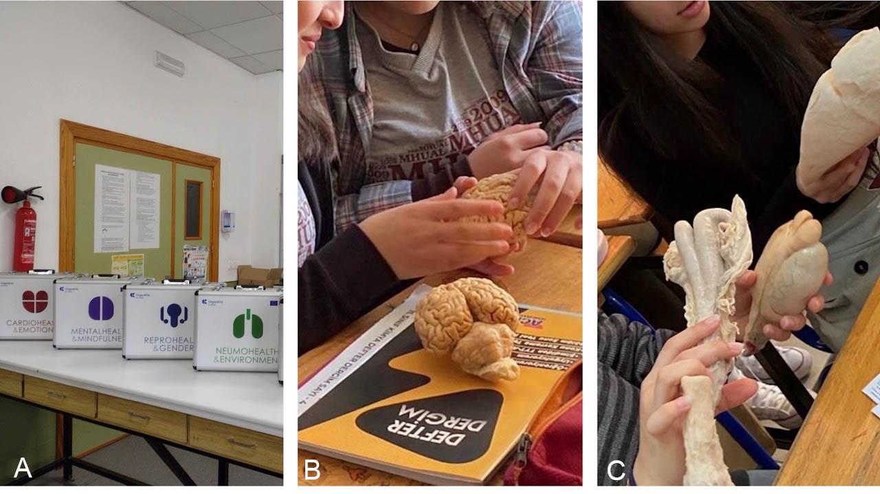
Figure 3. A, OrganKits ready for use in a secondary school setting; B,C, use of plastinated organs from different OrganKits during a learning activity
This study's main contribution is a standardized plastination workflow designed to deliver anatomical specimens optimized for secondary education. Plastination has long been understood as a set of techniques whose final quality depends not only on the polymer involved, but also on rigorous control of each of its key steps: specimen preparation, fixation, dehydration, forced impregnation, and curing (von Hagens et al., 1987; Henry et al., 2019). In our case, we integrated these principles into a comprehensive framework involving production management, quality assurance, and specimen traceability. This represents a notable advancement over conventional laboratory workflows, which typically focus on preserving the anatomical fidelity of individual specimens but not on production variables such as reproducibility and scalability. In this work, we carried out plastination according to organ-specific standard operating protocols (SOPs), each incorporating a checklist for quality assessment. Upon completion, we tagged each specimen with a microchip identifier, ensuring full traceability from raw material acquisition through final assembly. These features distinguish this methodology from most published descriptions of plastination practice, where systematic quality reporting and specimen-level traceability are rarely formalized. In this regard, the OrganKits project may serve as a methodological reference for future large-scale plastination programs subjected to rigorous quality assurance and systematic production.
The manufacturing workflow required several technical adjustments to ensure both appropriate-to-use morphological preservation and inter-specimen standardization. Those intended to preserve the morphology and flexibility of hollow viscera were particularly relevant. Previous studies have highlighted the importance of maintaining the morphology of these organs by inflation or hydrostatic distension throughout the plastination process (Henry, 1987; Holladay, 1989; Henry et al., 1997; Henry et al., 2019). As described above, hollow organs required modifications during preparation, fixation, and/or curing to preserve luminal size and retain flexibility. Specifically, the protocols for the heart, stomachs, intestines, uterus, and lungs incorporated hydrostatic dilatation during fixation. We also performed additional steps during curing; notably, stomachs and lungs underwent prolonged slow curing at room temperature under continuous airflow. Similarly, musculoskeletal specimens underwent slow curing coupled with repeated flexion-extension to preserve joint dynamics. Although none of these modifications is truly novel, this work makes a technical contribution by consolidating organ-specific adaptations into a standardized workflow that may serve as a practical reference for researchers seeking to establish plastination programs beyond the usual small-scale production of plastinates in their own laboratories.
From an educational perspective, previous literature shows that students widely accept plastinated specimens, which can substantially enhance anatomical comprehension when appropriately integrated into medical or veterinary curricula (Latorre et al., 2007; Nguyen et al., 2019; Goh et al., 2024). This evidence is particularly relevant in secondary education, where published experience with plastinated specimens remains considerably more limited than at university levels. The available literature suggests that plastinated material can enrich biology and science teaching in secondary schools by providing anatomical specimens in a safe, durable, and visually and emotionally engaging way (Douglass and Glover, 2003; Williams and Rae, 2019; Delgado-Ruiz et al., 2023). However, it also shows that this field is still in its early stages of development. Within that context, the OrganKits project represents a substantive contribution. For the first time, partner schools from four European countries and diverse national curricula served as the testing ground for a structured educational proposal in which plastinated organs were used not merely in biology lessons but as the material core of a STEAM-based, project-based learning framework. Thus, the range of disciplinary application is considerable. As documented on the project website (www.organkitsproject.com), plastinated organs were used directly, or played a central role, in a broad range of project-based learning activities in mathematics, technology, engineering, arts, physical education, and related areas. By integrating anatomical innovation and educational design, the OrganKits project extends plastination into a broad, yet still lightly explored, educational domain. Although the educational outcomes, in terms of learning gains and user experience, fall outside the scope of the present paper and will be reported separately, the OrganKits model already represents a new and significant contribution to the field of secondary-school educational research, positioning plastinated organs at the core of future innovative and interdisciplinary teaching proposals.
ACKNOWLEDGMENTS
The authors acknowledge the OrganKits consortium partners and the Erasmus+ framework that supported the project OrganKits: a new material for STEAM education based on plastination (Project No. 2022-1-ES01-KA220-SCH-000089616). The authors also acknowledge the technical staff involved in specimen preparation, plastination, quality control, and assembly of the educational briefcases.
Delgado-Ruiz MC, Bernal-Sánchez R, López-Albors O, Latorre-Reviriego R. 2023: Órganos plastinados: recursos para una propuesta de innovación educativa STEM en Educación Secundaria [Plastinated organs: resources for a proposal for STEM educational innovation in secondary education]. RiiTE Revista Interuniversitaria de Investigación en Tecnología Educativa [RiiTE Interuniversity Journal of Research in Educational Technology] 15:103-119. doi: 10.6018/riite.575831.
Douglass C, Glover R. 2003: Plastination: preservation technology enhances biology teaching. Am Biol Teach 65(7):503-510. doi: 10.1662/0002-7685(2003)065[0503:PPTEBT]2.0.CO;2.
Goh JSK, Chandrasekaran R, Sirasanagandla SR, Acharyya S, Mogali SR. 2024: Efficacy of plastinated specimens in anatomy education: A systematic review and meta-analysis. Anat Sci Educ 17(4):712-721. doi: 10.1002/ase.2424.
Henry RW. 1987: Plastination of an integral heart-lung specimen. J Int Soc Plastination 1(2):20-24. doi: 10.56507/KQKI2988.
Henry RW, Janick L, Henry C. 1997: Specimen preparation for silicone plastination. J Int Soc Plastination 12(1):13-17. doi: 10.56507/HVSK9838.
Henry RW, von Hagens G, Seamons G. 2019: Cold temperature/Biodur®/S10/von Hagens'--silicone plastination technique. Anat Histol Embryol 48(6):532-538. doi: 10.1111/ahe.12472.
Holladay SD. 1989: Plastination of inflated hollow gastrointestinal organs from large animals. J Int Soc Plastination 3(1):34-37. doi: 10.56507/UZSD4906.
Latorre RM, García-Sanz MP, Moreno M, et al. 2007: How useful is plastination in learning anatomy? J Vet Med Educ 34(2):172-176. doi: 10.3138/jvme.34.2.172.
Latorre R, Bainbridge D, Tavernor A, López-Albors O. 2016: Plastination in anatomy learning: An experience at Cambridge University. J Vet Med Educ 43(3):226-234. doi: 10.3138/jvme.0715-113R1.
López Albors O, Gil Cano F, Latorre Reviriego R. 2022: Órganos plastinados como recurso de innovación docente en biología de educación secundaria [Plastinated organs as a resource for teaching innovation in secondary school biology]. EDUCA Revista Internacional para la Calidad Educativa [EDUCA International Journal for Educational Quality] 2(1):86-99.
Nguyen VH, Pham PT, Joo K, Jeter CB. 2019: Dental students' and residents' opinions and performance of anatomy learning via cadavers or plastinated specimens. J Plastination 31(1):6-13. doi: 10.56507/KMZL8564.
OrganKits project. 2025: OrganKits - A new material for STEAM education based on plastination. URL: https://organkitsproject.eu/ [accessed May, 2026].
Savoini EH. 2018: Plastination of large mammal knees. Abstracts Presented at The 19th International Conference of the ISP, Dalian. J Plastination 30(2):40.
von Hagens G, Tiedemann K, Kriz W. 1987: The current potential of plastination. Anat Embryol (Berl) 175(4):411-421. doi: 10.1007/BF00309677.
Williams S, Rae G. 2019: Exciting New Orleans youth with plastinated anatomic specimens. FASEB J 33(S1):441.2. doi: 10.1096/fasebj.2019.33.1_supplement.441.2.

