1 Queen’s University, Department of Biomedical and Molecular Sciences, 18 Stuart Street, Kingston, Ontario, Canada, K7L 3N6 bl46@queensu.ca
2 University of Manitoba, Department of Human Anatomy and Cell Science, 745 Bannatyne Avenue, Winnipeg, Manitoba, Canada madeline.damjanovic@umanitoba.ca
3 McMaster University, Education Program in Anatomy, Department of Health Sciences, 1200 Main Street W, Hamilton, Ontario, Canada, L8N 3Z5 damjanoi@mcmaster.ca
Fresh frozen and soft-embalmed donors provide realistic and flexible tissue qualities, making them invaluable for surgical skills training and medical research; however their potential to be utilized as long-term teaching specimens is constrained by the progressive and rapid degradation of soft tissues. Because of this tissue degradation, body donors used for surgical skills training may be prepared for cremation with anatomical regions still intact. The objective of this study was to evaluate the feasibility of plastinating male genitourinary specimens using the Biodur® S 10 standard technique from soft-embalmed and fresh frozen donors following surgical skills sessions, thereby maximizing body donor usage. A hard-embalmed specimen was also plastinated for comparison purposes. Plastination produced high quality durable specimens regardless of initial preservation technique. The only discernable difference were focally dark areas within the plastinate that was obtained from a fresh frozen donor. As plastinates can be utilized for long term educational purposes, this approach increases the potential usage of human body donors that are initially soft-embalmed or fresh frozen for surgical skills.
Dissection; Education; Embalming; Pelvis; Plastination
Isabella Damjanovic, 1200 Main Street W, Hamilton, Ontario, Canada, L8N 3Z5
Email: damjanoi@mcmaster.ca

![]()


Plastination of human cadaveric material, including dissected specimens, isolated organs, and body slices, has played a fundamental role in anatomical education for several decades (Riederer, 2014). Plastination is a long-term preservation technique in which bodily fluids are replaced with a polymer, resulting in durable anatomical specimens that are easy to handle while eliminating many of the health hazards traditionally associated with cadaver-based learning (von Hagens, 1979; Atwa et al., 2021). Moreover, plastinated specimens are not confined to laboratory settings; they can be handled safely without gloves, are easily transportable, and are typically odorless and structurally rigid. These characteristics make them particularly well suited for use in lecture theatres, tutorial spaces, and smaller educational facilities (Riederer, 2014). Plastinated specimens are especially valuable for instruction in disciplines such as neuroanatomy and gross anatomy and are also highly effective for the long-term preservation of rare or unique pathological specimens (Riederer, 2014).
Fresh frozen and soft-embalmed human body donors are widely utilized in surgical skills training and clinical research because they preserve realistic tissue characteristics, with features such as color, texture, and mobility remaining largely unaltered (Balta et al., 2015). However, long-term usage is not feasible due to tissue deterioration, which also eliminates the potential for further educational use following surgical skills practice (Damjanovic et al., 2024). At Queen’s University, the pelvic organs of human body donors are rarely used for surgical simulations. As such, fresh frozen and soft-embalmed donors are typically prepared for cremation with intact genitourinary viscera. Preservation via formalin-fixation, dissection, and plastination of genitourinary organs would allow for continued educational use of surgical skills donors for a wide range of learners in a variety of disciplines.
There is minimal published literature related to the preservation (Damjanovic et al., 2024) or plastination (Hunter, 2019) of cadaveric specimens for long term utilization after donors have been used for surgical skills. The aim of this project was to describe a technique for maximizing body donor usage through the fixation and plastination of genitourinary specimens from soft-embalmed and fresh frozen donors following surgical skills sessions. Here we describe the dissection and preservation techniques, as well as the potential educational significance of the specimens produced using this method.
The genitourinary organs from three male body donors were included in this study; one donor was hard-embalmed (Table 1), one was soft-embalmed (Table 2), and one was fresh frozen at -20℃. All three cadavers were generously donated to the Human Body Donor Program at Queen’s University. Ethics approval for this study was granted by the Queen’s University Health Sciences and Affiliated Teaching Hospitals Research Ethics Board (6045572). The hard-embalmed and soft-embalmed donors were preserved by perfusing the femoral arteries using a PORTI-BOY Mark V embalming machine. The soft-embalmed donor was initially used for weekly surgical skills sessions over a duration of two months and was stored in a refrigerator at 4℃ when not in use. The fresh frozen specimen had been subjected to three freeze-thaw cycles as the donor was previously used for various surgical skills training sessions and was stored in a freezer at -20℃ when not in use.
Each specimen was initially harvested by disarticulating the hip joints and cutting through the soft tissues located between the rectum and sacrum/coccyx in a semi-circular path. The specimens were dissected immediately after procurement using standard instruments (scalpel, forceps, scissors, probe). They were stored in 10% formalin when dissection was not occurring. The basic dissection steps were repeated for each specimen. The bladder and prostate gland were sectioned in the sagittal plane and the shaft of the penis was sectioned in the coronal plane. Final cutting of the bony pelvis was done with a handheld reciprocating saw. The remaining exposed bony elements were scraped with scalpel blades to remove as much soft tissue and periosteum as possible.
After dissection was complete, the specimens were stored for 24 hours in a 3% hydrogen peroxide solution for bleaching, with the fresh frozen specimen held for an additional 24 hours in solution as it was focally darker in color. The specimens were generously rinsed with cold water after the bleaching process was complete. They were then transferred to a cold water bath where water was allowed to continually run over the specimens for 24 hours. The rinsed specimens were placed in a bucket of cold water and placed in a refrigerator for 24 hours at 4℃.
Dehydration was done in consecutive acetone baths (90%, 95%, 99%, and 100% acetone) at -20℃ for one week for each bath. Dehydration was considered complete when an acetometer read >99% at 20℃. Defatting was performed with a new batch of 100% acetone at room temperature for 24 hours. All specimens were dehydrated and defatted together for each phase of the process.
Plastination was performed using the Biodur® S 10 standard technique. The silicone liquid polymer (Biodur® S 10) was mixed with silicone hardener (Biodur® S 3) in a 100:1 ratio, respectively. Forced impregnation was performed at -20℃ with all three specimens in the same 35 litre Heidelberg plastination kettle (Biodur®). The specimens were initially placed in the chamber for 24 hours before vacuum was applied. Vacuum was gradually increased over a period of 12 days. Forced impregnation was considered complete when the pressure was steady at ~4 mm Hg for three consecutive days without any visible bubbles in the plastination chamber.
The specimens were removed from the plastination kettle and placed in a curing chamber outfitted with racks to allow for excess polymer to drip away. A small container with gas curing hardener (Biodur® S 6) was placed in the curing chamber for 21 days. After this initial curing period the specimens were examined for tacky spots. These areas were wiped with paper towels and the specimens were left on wire racks at room temperature for an additional 14 days. After curing for a total of 5 weeks, the specimens were compared to each other in terms of texture, color, and overall quality. Photographs were taken. After documenting the findings, the specimens were included in the Anatomy Museum at Queen’s University for usage in anatomy laboratory sessions for courses in a variety of programs.
Table 1: Hard-embalming solution
| Ingredient | Volume |
| Potassium acetate | 150 mL |
| Dettol | 600 mL |
| Phenol | 1 L |
| Glycerol | 1 L |
| Formalin (37% formaldehyde) | 1 L |
| Ethanol (95%) | 20 L |
Table 2: Soft-embalming solution
| Ingredient | Volume |
| Phenol | 1.8 L |
| Glycerol | 4 L |
| Ethanol (95%) | 8 L |
| Water | 8 L |
All three specimens demonstrated adequate tissue fixation after the dissection and preservation period (Fig. 1). The integrity of the tissue of the fresh frozen pelvic organs was delicate compared to the hard-embalmed and soft-embalmed specimens; however, this finding did not cause difficulty during the dissection process. Additionally, the fresh frozen specimen was focally darker in color compared to the hard-embalmed and soft-embalmed specimens (Fig. 2). This darker color was most noticeable in the superficial muscles of the perineum and erectile tissues/fascia of the penis. We speculate that this was mainly due to tissue decomposition that would have occurred while the specimen was subjected to several freezing/thawing cycles while the donor was used for surgical skills sessions. It is also possible that the specimen may be darker in color because it was fixed by simply submerging in 10% formalin, while the hard-embalmed and soft-embalmed specimens were fixed via arterial submersion with embalming fluids that contained ethanol and other chemicals.
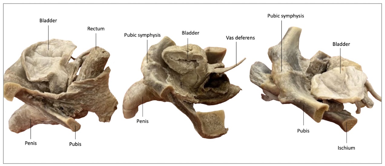 Figure 1. Lateral view of plastinated GU specimens: A, hard-embalmed; B, soft-embalmed; C, fresh frozen |
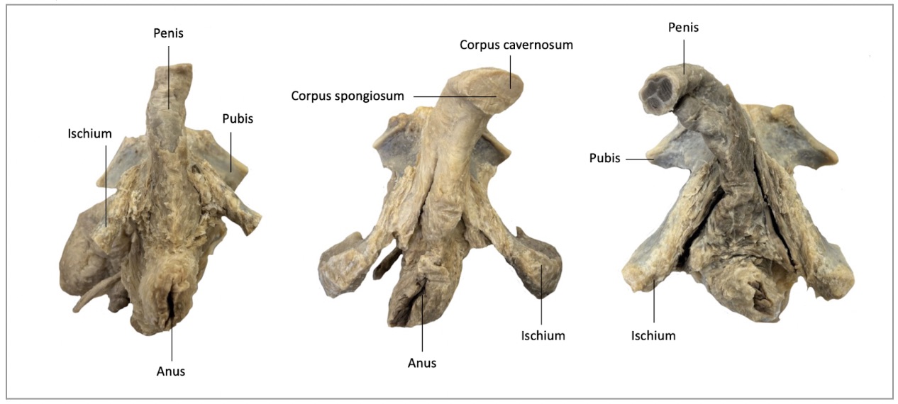 Figure 2. Inferior view of plastinated GU specimens: A, hard-embalmed; B, soft-embalmed; C, fresh frozen |
It was initially thought that the dark areas may lighten to the same degree as the other two specimens during the bleaching phase if the fresh frozen specimen was given additional time in the hydrogen peroxide bleaching solution. However, after an additional 24 hours of bleaching, it was observed that the bladder was becoming overbleached and therefore the specimens were removed from the bleaching solution. Upon completion of plastination each specimen produced a firm, durable product that is useful for teaching gross anatomy to a variety of learners. There is not a discernable textural difference between the three preservation types. We did not notice any difference in flexibility in the final plastinated specimens between the hard-embalmed and soft-embalmed examples, although a soft-embalmed cadaver has significantly more flexibility compared to a hard-embalmed whole body. The flexibility of a soft-embalmed specimen is similar to a fresh-frozen donor.
The present study demonstrates a reproducible technique for plastinating genitourinary specimens obtained from both soft‑embalmed and fresh frozen donors following surgical skills sessions which were found to be comparable to the more traditional hard-embalmed plastinated specimens. By preserving these organs following surgical skills training, this approach maximizes the educational value of each donor and provides durable, high‑fidelity specimens for long term anatomical education.
Plastination, first developed in the late 1970s, has long been recognized as a means of permanently preserving human tissues and organs as dry, odorless, and durable specimens that retain gross anatomical detail for teaching and demonstration purposes (Riederer, 2014; Torres-Palsa et al., 2025). Studies in anatomy education have documented several benefits of plastinated specimens, including enhanced visualization of spatial relationships, ease of handling compared to wet specimens, and positive learner engagement (Riederer, 2014; Chytas et al., 2019). In qualitative comparisons, students perceived plastinated specimens to be authentic and valuable for understanding complex three‑dimensional anatomy (Goh et al., 2024). Despite these favorable perceptions, systematic reviews and meta‑analyses have highlighted that the evidence-based date is limited, with few controlled studies demonstrating clear preference of plastinates over traditional teaching methods (Chytas et al., 2019; Goh et al., 2024).
The current project highlights the potential of plastination techniques to bridge traditional anatomy teaching methods with surgical skills simulation and training sessions while maximizing the use of soft-embalmed and fresh frozen donors. This technique allows the donor to be used for multiple educational purposes; first for hands-on surgical skills practice, and subsequently as a long‑term anatomical resource for a variety of learners such as high school, undergraduate, and professional graduate students. The plastinated genitourinary specimens retained structural integrity and tactile realism similar to that of hard-embalmed plastinated specimens, enabling learners to appreciate complex anatomical relationships, complementing cadaveric dissection, digital tools, and other educational learning resources. This approach may be particularly valuable in programs with limited donor availability, as learning efficiency can be enhanced through repeated utilization of surgical skills donors.
Despite these strengths, several limitations should be acknowledged. The number of donors included in this project was limited, which may restrict the generalizability of our findings. While all three plastinated specimens had similar textural properties, the genitourinary organs obtained from the fresh frozen cadaver were focally dark in color compared to the soft-embalmed and hard-embalmed specimens, even after additional bleaching time.
Furthermore, plastination requires specialized equipment, lab space, chemicals, and technical expertise, which may limit widespread implementation in some educational settings or institutions.
This study presents a practical method for plastinating genitourinary specimens following surgical skills sessions with both soft-embalmed and fresh frozen donors, enabling repeated use of each donor while preserving relevant anatomical features and maximizing educational value. By enhancing both the durability and utility of cadaveric material, this technique broadens the educational potential of donors, providing a valuable resource for both anatomical and surgical education, particularly in settings where donors and cadaveric material are limited.
ACKNOWLEDGEMENTS
The authors sincerely thank those who donate their bodies for the advancement of anatomical education and research. Results from such research have the potential to increase scientific knowledge and improve patient care. These donors and their families will always have our highest respect and gratitude.
STATEMENT OF ETHICS APPROVAL
Ethical approval for this study was granted by the Queen’s University Health Sciences and Affiliated Teaching Hospitals Research Ethics Board.
Atwa H, Dafalla S, Kamal D. 2021: Wet specimens, plastinated specimens, or plastic models in learning anatomy: perception of undergraduate medical students. Med Sci Educ 31(4): 1479-1486. https://doi.org/10.1007/s40670-021-01343-6
Balta JY, Cronin M, Cryan JF, O'Mahony SM. 2015: Human preservation techniques in anatomy: A 21st century medical education perspective. Clin Anat 28(6): 725-734. https://doi.org/10.1002/ca.22585
Chytas D, Piagkou M, Johnson EO, Tsakotos G, Mazarakis A, Babis GC, Nikolaou VS, Kaseta MK, Natsis K. 2019: Outcomes of the use of plastination in anatomy education: current evidence. Surg Radiol Anat 41(10): 1181-1186.
https://doi.org/10.1007/s00276-019-02270-3
Damjanovic IG, Damjanovic MM, Donaldson E, Bale LSW. 2024: Embalming cadaveric upper limbs after freezing and thawing: a novel technique for maximizing body donor usage through fresh frozen and formalin-fixed preservation. Euro J Anat 28(2): 215-226. https://doi.org/10.52083/SDQC3737
Goh JSK, Chandrasekaran R, Sirasanagandla SR, Acharyya S, Mogali SR. 2024: Efficacy of plastinated specimens in anatomy education: A systematic review and meta-analysis. Anat Sci Educ 17(4): 712-721.
https://doi.org/10.1002/ase.2424
Hunter A. 2019: Thiel Plastination: Improving the aesthetics and flexibility of traditional plastinated specimens. Doctoral dissertation, University of Dundee.
Riederer BM. 2014: Plastination and its importance in teaching anatomy. Critical points for long‐term preservation of human tissue. J Anat 224(3): 309-315. https://doi.org/10.1111/joa.12056
Torres-Palsa MJ, Dominguez J. 2025: Implementation of plastinated specimens in a Doctor of Physical Therapy human anatomy curriculum. Anat Sci Educ 18(7): 709-717. https://doi.org/10.1002/ase.70043
von Hagens G. 1979: Impregnation of soft biological specimens with thermosetting resins and elastomers. Anat Rec 194(2): 247-255. https://doi.org/10.1002/ar.1091940206