Department of Anatomy and Histology, Faculty of Veterinary Science, Bangladesh Agricultural University, Mymensingh-2202, Bangladesh
Cryodehydration is an efficient and cost-effective method for preserving the gross morphology of anatomical specimens. However, repeated freezing and thawing cause substantial cellular damage due to micro-ruptures of the cell membrane. Objectives: Therefore, this study investigated the role of cryoprotectants and controlled thawing protocols in optimizing the cryodehydration technique for preparing cryodehydrated specimens, with particular focus on preserving histoarchitectural integrity. Method: This study used four healthy goats, labeled G1 (control), G2 (formalin-preserved), G3 (cryodehydrated using a routine protocol), and G4 (cryodehydrated using glycerol as a cryoprotectant with controlled thawing at 4 °C). Results: Although the G4 specimens required more freeze-thaw cycles, both protocols (G3 and G4) preserved color, texture, and morphology excellently. The specimens were soft, dry, odorless, lightweight, and durable. In the histological study, G2 showed better histoarchitectural preservation resembling the control, while G3 showed massive histoarchitectural alterations. These included noticeable thinning of the heart wall, increased spaces between cardiac myofibers, decreased bronchial and bronchiolar circumference and reduced alveolar surface area in the lungs, loss of hepatocytes and widened sinusoidal spaces in the liver, damage to both renal tubules and glomeruli, loss of lymphoid tissue in the spleen, and shrinkage of skeletal myofibers. In contrast, G4 demonstrated better preservation of histoarchitecture, maintaining cellular integrity along with distinct cytoplasmic and nuclear clarity. Conclusion: Based on these findings, glycerol as a cryoprotectant, combined with controlled thawing at 4 °C, enhances the efficacy of the cryodehydration technique in preserving tissue histoarchitecture.
controlled thawing; cryodehydration; cryoprotectant; glycerol; tissue preservation
Prof. Dr Nasrin Sultana, Department of Anatomy and Histology, Faculty of Veterinary Science
Bangladesh Agricultural University, Mymensingh 2202, Bangladesh
email: nsultana.bau@gmail.com; nasrin.sultana@bau.edu.bd





Veterinary anatomy education and research rely extensively on the preservation of animal cadavers and their various organs or body parts (Nam et al., 2020). However, the preservation of anatomical specimens remains a significant challenge due to the inherent susceptibility of biological tissues to decomposition (Han and Bischof, 2004). Although several chemical-based preservation techniques have been developed and are widely used, they often raise considerable health and safety concerns (Jangde et al., 2015). Commonly employed preservatives, including formalin, ethanol, zinc chloride, glycerol, Kaiserling solution, and sodium chloride solution, are known to be toxic and have been associated with adverse health effects such as allergic reactions, skin and mucous membrane irritation, headaches, and even respiratory complications including asthma (Haizuka et al., 2018; Kumari et al., 2019; Rezoana et al., 2022). These chemical preservatives also present handling difficulties, particularly in educational settings, as they cannot be safely managed without protective equipment, thereby limiting their utility for instructional purposes (Alsharif et al., 2017). In response to these challenges, alternative preservation methods such as plastination and its various modifications have been developed (Elnady, 2016; Sora et al., 2019; Islam and Sultana, 2021; ElShafey et al., 2022). While these techniques offer safer and more durable preservation of anatomical specimens, their adoption in countries with limited resource, such as Bangladesh, is constrained by high set-up costs and the need for specialized, often patented, chemicals (Islam and Sultana, 2021). In this regard, cryodehydration is considered as a cost-effective and practical alternative for anatomical specimen preservation (Sultana and Islam, 2023).
Cryopreservation is the use of low temperature to preserve structurally intact living cells and tissues, facilitating their long-term storage and viability (Pegg, 2014). Although the modern scientific application of cryopreservation is relatively recent, the concept itself is not new. The fundamental idea of using low temperatures for preservation has historical roots dating back centuries (Whaley et al., 2021). Cryodehydration represents a critical phase in the cryopreservation process, wherein animal cadavers or anatomical specimens are dehydrated through successive cycles of freezing and thawing following initial fixation with formalin (Martins and Sakalem, 2022; Sultana and Islam, 2023). This method, classified as a dry preservation technique, results in lightweight, odorless specimens that are easy to store, transport, and handle without protective equipment (Teixeira Filho et al., 2019). During the subsequent washing and freeze-thaw cycles, residual formalin is also effectively eliminated, thereby minimizing potential health risks associated with chemical exposure (Teixeira Filho et al., 2019; Martins and Sakalem, 2022). However, microrupture of cellular membranes induced by repeated freeze–thaw cycles poses a significant challenge to maintain the histoarchitecture of tissues in a near-natural state (Teixeira Filho et al., 2019; Kilbride et al., 2021). Our previous research demonstrated significant histomorphological and histomorphometric alterations in cryodehydrated specimens (Sultana and Islam, 2023). To address the challenge of preserving tissue histoarchitecture during cryodehydration, the incorporation of cryoprotective agents like glycerol combined with controlled thawing at low temperature (0 °C to 4 °C) may enhance histoarchitectural preservation and yield better outcomes (da Silva et al., 2021; Whaley et al., 2021).
The cryoprotective properties of glycerol were initially identified by chance. It is now understood that freezing-induced cellular injury occurs through two primary mechanisms: direct mechanical disruption caused by ice crystal formation, and secondary damage resulting from the elevated concentration of solutes as water progressively freezes (Pegg, 2002; Whaley et al., 2021). Cryoprotectants mitigate these effects mainly by limiting ice formation at subzero temperatures. In addition to this, thawing at low temperature (4 °C) allows for a gradual rehydration of cells by slowing the warming process, thereby minimizing cellular osmotic stress. This low-temperature approach also inhibits the fusion of small ice crystals into larger ones, which could otherwise compromise cellular integrity during thawing (Pegg, 2014; da Silva et al., 2021). To the best of our knowledge, no studies have yet examined the comparative efficacy of a modified cryodehydration technique incorporating cryoprotectants and controlled thawing, in contrast to standard cryodehydration methods and traditional formalin preservation. While cryodehydration is a proven, cost-effective dry preservation technique, it remains a relatively novel approach in Bangladesh that requires further investigation and adoption (Sultana and Islam, 2023). Therefore, in light of the protective effects of glycerol as a cryoprotectant and the benefits of low-temperature thawing, the present study was designed to evaluate the comparative effectiveness of this modified cryodehydration method in preserving the morphological and morphometric characteristics of anatomical specimens, from both macroscopic and microscopic perspectives.
Collection of specimens
The study was conducted following the established guidelines of Animal Welfare and Experimentation Ethics Committee [Approval No.: AWEEC/BAU/2023(44)]. Four healthy adult Black Bengal goats (approximately 18 months of age and similar body weight) were procured from the local market and labelled as G1, G2, G3, and G4. Following euthanasia (Thiopental Sodium @ 70 mg/kg body weight), tissue samples, including the heart, lungs, liver, kidneys, spleen, and musculoskeletal specimens, were collected. The initial weight (g) of each specimen was recorded immediately after collection.
Fixation and freezing
All the freshly collected specimens were fixed with 10% neutral buffered formalin solution (G1 and G2 – 72 hours, G3 and G4 – one week). However, in the G4 group, 5% glycerol was added as cryoprotectant with the formalin solution. Due to its penetrating and hygroscopic properties, glycerol is expected to diffuse into the tissue and be retained even after washing. Therefore, although the washing step reduces surface and unbound substances, a proportion of glycerol likely persists within the tissue matrix and contributes to protection during subsequent freezing-thawing cycles (da Silva et al., 2021). Following 72 hours of fixation, the specimens from G1 group were processed for histological study and those in the G2 group were preserved in 10% neutral buffered formalin for three months, followed by histological investigation. The specimens from groups G3 and G4 were washed under running tap water for 45 minutes to remove the formalin before beginning the cryodehydration process.
Cryodehydration protocol
To prepare the cryodehydrated specimens, we followed the method described by Sultana and Islam (2023), with some modifications. In this study, glycerol was added to the formalin as a cryoprotectant, and controlled thawing at 4 °C was performed for the G4 group to assess its efficacy in minimizing the tissue damage. In brief, the protocol was as follows: specimens from G3 and G4 were kept in a deep freezer (LG 345L chest freezer with fast freezing, Model No: LG GCS415GQFG, South Korea) at -20 °C for approximately 22 hours to ensure complete freezing of the tissue water. G3 specimens were thawed at room temperature (25 ± 2 °C) while G4 specimens were thawed at 4 °C (refrigerator-controlled thawing). Thawing was carried out for approximately 2 hours. After thawing, surface moisture was gently absorbed with a tissue napkin. This freezing-thawing cycle was repeated every 24 hours for complete drying of the specimens. The completion of the cryodehydration process was determined based on stabilization of specimen weight across consecutive cycles. Specifically, the process was considered complete when the difference in weight between two successive freezing-thawing cycles was less than 1%, indicating minimal remaining moisture content. In addition, supportive physical characteristics such as reduced weight, lighter coloration, fibrous consistency, and spongy texture were observed to confirm the endpoint. Afterward, the final weight of the specimens was recorded, and they were coated with wood glue (Fevicol SH Synthetic Resin Adhesive, Pidilite Industries Ltd., India) to prevent moisture absorption. Finally, the specimens were stored in airtight plastic containers with silica gel. The weight loss of each specimen was calculated using the following formula:
Weight loss (%) = (Initial weight - Final weight)/(Initial weight) ×100
Histological study
To investigate the histoarchitectures of the specimens, a 0.5 cm2 sample was taken from each specimen (including fresh, formalin-preserved, and cryodehydrated samples) and processed for routine staining using H&E stain, following the method described by Islam et al., (2021). From each organ of every animal, three tissue specimens were carefully excised from three different regions. Thus, the individual tissue specimen was considered the experimental unit for histological study, rather than the animal itself. This design enabled within-animal comparisons and helped reduce the influence of inter-individual variability. For each tissue specimen, five histological sections were prepared and examined. From each section, ten microscopic fields were randomly selected for morphometric analysis to ensure representative sampling.
Microscopic images were captured using a photomicroscope (B-293, Optika, Italy). The histomorphology and histomorphometry, such as the thickness of the heart wall (µm), circumference of the bronchi and bronchioles (µm), the surface area of lung alveoli (µm2), the circumference of the hepatic central veins (µm), the circumference of the renal tubules (µm), the glomerular surface area (µm2) of the kidney, and the cross-sectional area (µm2) of the myofibers were measured using Image J software.
All morphometric measurements were performed independently by two observers in a blinded manner, without prior knowledge of the specimen group allocation, to reduce observer bias and improve measurement reliability.
Statistical analysis
SPSS (version 22) was used in this study to analyze the data. The Shapiro-Wilk test was performed to check the data for normality. To determine significant differences between the mean values, one-way ANOVA was performed, with P < 0.05 considered statistically significant. The results were presented as the mean ± standard error of the mean (SEM).
Macroscopic features of the cryodehydrated specimens
The macroscopic characteristics of cryodehydrated specimens prepared using two different protocols are presented in Figures 1 and 2. The samples in group G3 required fewer freeze-thaw cycles (approximately 20 for cavitary organs, 30 for parenchymatous organs, and 40 for musculoskeletal specimens) compared to the samples of group G4, which required approximately 25, 35, and 50 cycles for the corresponding specimen types.
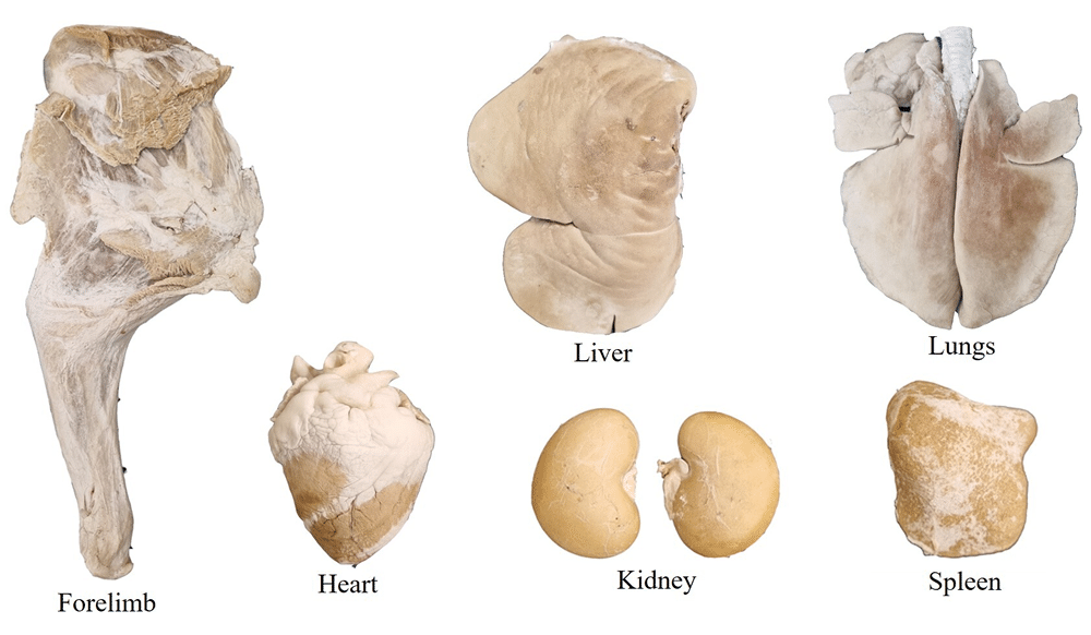 Figure 1. Cryodehydrated specimens (Group G3) prepared through repeated cycles of freezing and thawing at room temperature |
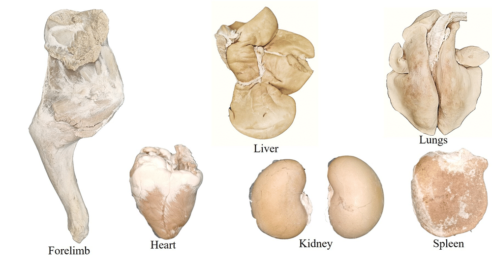 Figure 2. Cryodehydrated specimens (Group G4) prepared using glycerol as a cryoprotectant and controlled repeated freezing and thawing (at 4 °C) cycles |
Both protocols yielded cryodehydrated specimens that exhibited excellent preservation of color, texture, and morphology, closely resembling their natural appearance. The specimens were soft, dry, odorless, lightweight, and durable, maintaining their structural integrity without any visible alteration when stored at room temperature for up to three months. Notably, no significant wrinkles were observed in any of the organ surfaces, with the exception of the liver, which exhibited a few wrinkles on its parietal surface.
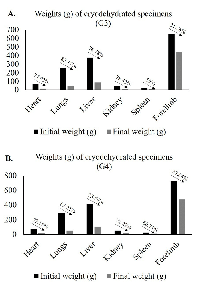
Figure 3. Weights of fresh and cryodehydrated specimens collected from goats, along with weight loss (%) following cryodehydration. A: G3 - Cryodehydrated specimens prepared by thawing at room temperature; B: G4 - Cryodehydrated specimens prepared using cryoprotectant and thawing at 4 °C
Morphometric data, including initial weight, final weight, and percentage of weight loss, are presented in Figure 3. Both protocols resulted in substantial reductions in specimen weight. The highest percentage of weight loss was observed in the lungs, whereas the musculoskeletal specimen (left forelimb) exhibited the lowest weight loss following cryodehydration. The weight loss (ranging from 31.76% to 82.21% across all specimens) made the specimens lightweight which were easy to handle.
Microscopic features of the cryodehydrated specimens
Heart
The histomorphologic and histomorphometric features of the heart are presented in Figure 4. The G2 and G4 specimens showed normal histoarchitecture like G1. The cardiac histological layers were clearly identifiable in all groups. However, the cytoplasmic clarity of the cardiac muscle was slightly lost in G3. Noticeable alteration (P < 0.05) in the heart wall thickness was found in the histomorphometric investigation of the cryodehydrated specimens (G3 and G4) irrespective of the protocols used, compared to G1 and G2. However, G4 showed significantly higher heart wall thickness compared to G3. The space between the cardiac myofibers also increased noticeably in G3 while G4 showed no such alterations.
Lungs
The histomorphologic and histomorphometric features of the lungs are presented in Figure 5. G2 and G4 exhibited relatively well-preserved histoarchitecture like G1, with only slight loss of nuclear detail of the alveolar pneumocytes. In contrast, specimens from G3 demonstrated extensive cellular damage, characterized by complete loss of nuclear and cytoplasmic clarity in the lung parenchyma, as well as in the bronchi and bronchioles.
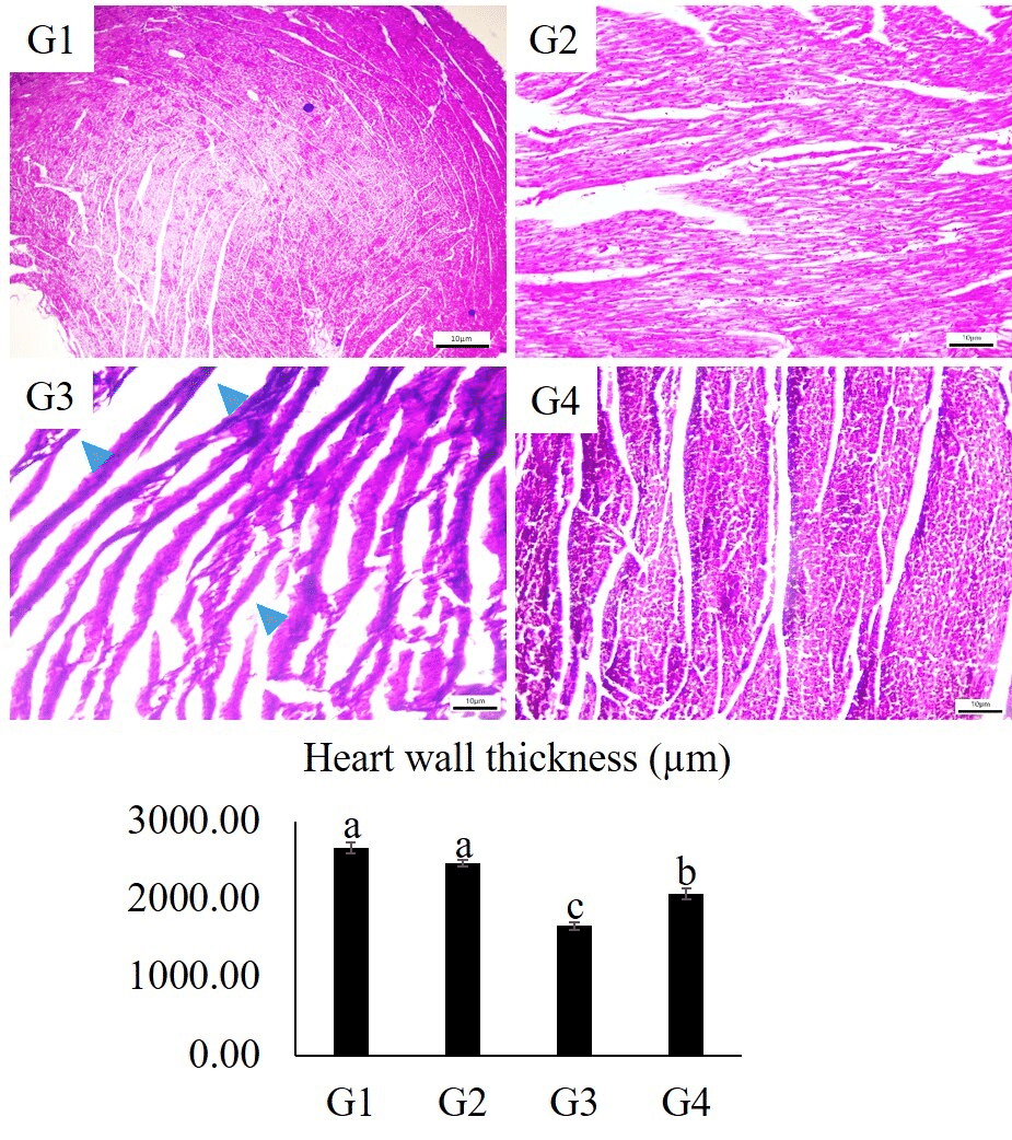 Figure 4. Morphologic and morphometric attributes (thickness of heart wall) of the heart (G1: control, G2: formalin preserved, G3: cryodehydrated (thawed at room temperature), and G4: cryodehydrated using cryoprotectant (thawed at 4 °C) of goat. Blue arrowheads indicate increased intermyofibrillar spaces resulting from extensive tissue shrinkage. Morphometric data are expressed as mean ± SEM. Statistical differences among groups were analyzed using one-way ANOVA. Values with different alphabetic superscripts are significantly different (P < 0.05). Staining method - H&E, magnification - 10 X, scale bar - 10 µm |
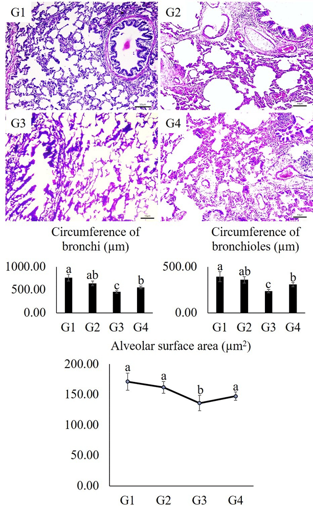 Figure 5. Morphologic and morphometric attributes of goat lungs (G1: control, G2: formalin preserved, G3: cryodehydrated (thawed at room temperature), and G4: cryodehydrated using cryoprotectant (thawed at 4 °C). Morphometric data are expressed as mean ± SEM. Statistical differences among groups were analyzed using one-way ANOVA. Values with different alphabetic superscripts are significantly different (P < 0.05). Staining method - H&E, magnification - 10 X and scale bar - 10 µm. |
Histomorphometric analysis revealed a significant reduction (P < 0.05) in bronchial and bronchiolar circumference in both G3 and G4 following cryodehydration when compared to the control group (G1). However, these attributes were significantly greater (P < 0.05) in G4 than in G3, indicating better preservation of airway structure with the G4 protocol. Furthermore, the alveolar surface area was markedly reduced (P < 0.05) in both G3 and G4 relative to G1.
Liver
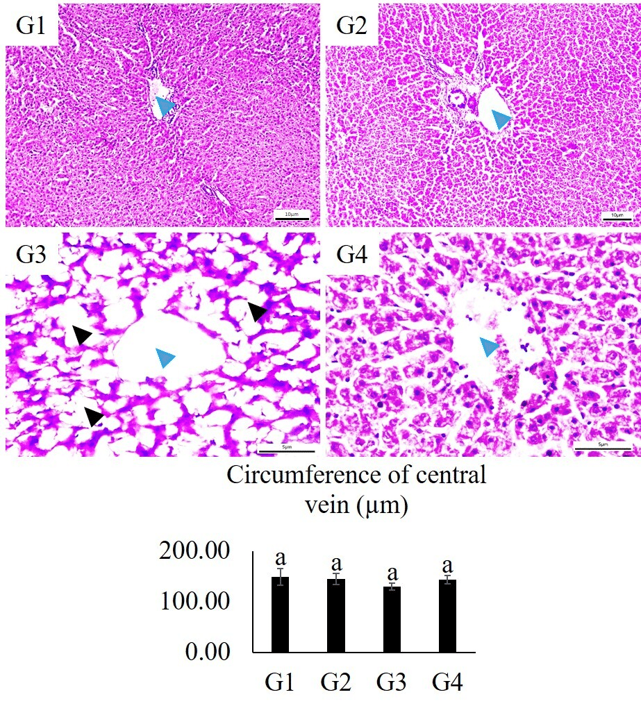
Figure 6. Morphologic and morphometric attributes of goat liver (G1: control, G2: formalin preserved, G3: cryodehydrated (thawed at room temperature), and G4: cryodehydrated using cryoprotectant (thawed at 4 °C). Blue arrowheads indicate central veins, black arrowheads indicate increased sinusoidal space between the hepatic cords. Morphometric data are expressed as mean ± SEM. Statistical differences among groups were analyzed using one-way ANOVA. Values with different alphabetic superscripts are significantly different (P < 0.05). Staining method - H&E, magnification - 40 X, scale bar - 5 µm
The histomorphologic and histomorphometric characteristics of the liver are depicted in Figure 6. G2 exhibited normal hepatic architecture similar to that of the control group (G1), with the exception of a slight increase in sinusoidal spaces between the hepatic cords. In contrast, G3 demonstrated severe structural degradation, including complete loss of cytoplasmic and nuclear detail in hepatocytes, along with markedly dilated sinusoidal spaces. Group G4 showed better preservation of tissue architecture, characterized by distinctly recognizable hepatocytic cords with intact nuclei. However, some hepatocytes showed cytoplasmic loss, and sinusoidal dilation was also seen to some extent.
Histomorphometric analysis revealed no statistically significant differences (P > 0.05) in the circumference of the central vein among the groups. Nevertheless, a trend toward decreased central vein circumference was observed in the cryodehydrated groups (G3 and G4) compared to the control (0.05 < P < 0.1), suggesting subtle structural alterations associated with the dehydration process.
Kidney
The histomorphologic and histomorphometric features of the kidney are presented in Figure 7. Histomorphologic examination revealed distinct pathological alterations in group G3, including loss of cytoplasmic detail and nuclear clarity, along with degeneration of renal tubules and glomeruli in certain areas. These observations were further confirmed by histomorphometric analysis, which showed a significant reduction (P < 0.05) in renal glomerular size in both G3 and G4 compared to the G1 and G2.
Spleen
The histomorphologic features of the spleen are shown in Figure 8. In G1, the splenic architecture appeared normal with well-organized white and red pulp regions. Lymphoid follicles were distinct, and cellular density was high with intact nuclei and cytoplasmic detail. In G2, mild disruption of splenic architecture was observed, characterized by reduced lymphoid follicle definition and slight loss of cellular density, although nuclear clarity was still relatively preserved. On the other hand, marked histopathological alterations were evident in G3. The splenic architecture was severely disrupted, with extensive loss of lymphoid tissue, reduced cellular density, and vacuolation. Nuclear detail was also significantly diminished, indicating degeneration. In G4, the splenic architecture was comparatively better preserved than in G3, with clear cytoplasmic and nuclear details. However, some loss of lymphoid tissue with slightly reduced cellular density was still observed.
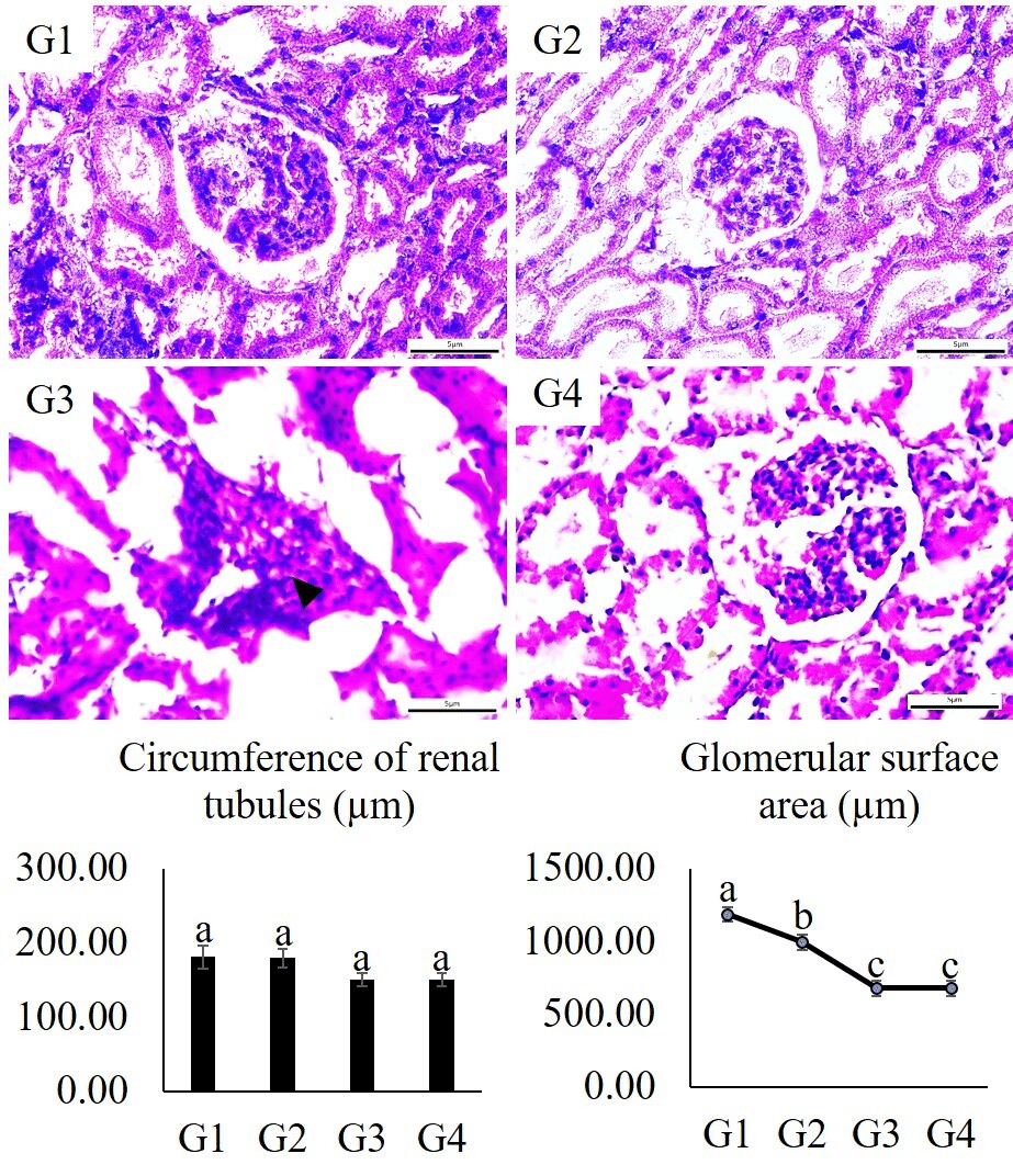 Figure 7. Morphologic and morphometric attributes of goat kidney (G1: control, G2: formalin preserved, G3: cryodehydrated (thawed at room temperature), and G4: cryodehydrated using cryoprotectant (thawed at 4 °C). Black arrowhead (G3) indicates degenerated and shrunken glomerulus. Morphometric data are expressed as mean ± SEM. Statistical differences among groups were analyzed using one-way ANOVA. Values with different alphabetic superscripts are significantly different (P < 0.05). Staining method - H&E, magnification - 40 X, scale bar - 5 µm |
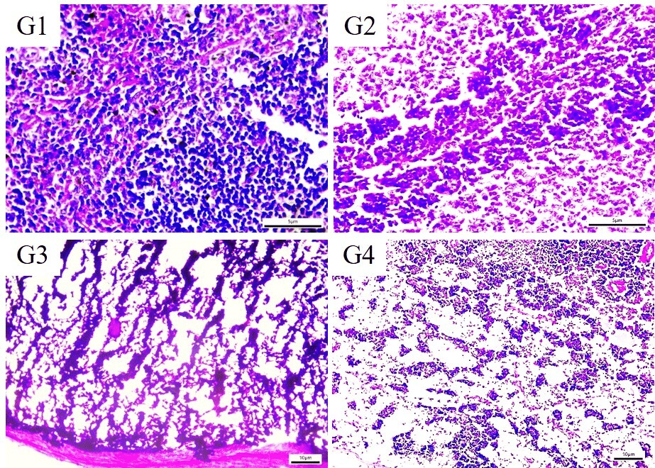 Figure 8. Morphologic and morphometric attributes of goat spleen (G1: control, G2: formalin preserved, G3: cryodehydrated (thawed at room temperature), and G4: cryodehydrated using cryoprotectant (thawed at 4 °C). Morphometric data are expressed as mean ± SEM. Statistical differences among groups were analyzed using one-way ANOVA. Values with different alphabetic superscripts are significantly different (P < 0.05). Staining method - H&E, magnification - 10 X, scale bar - 10 µm |
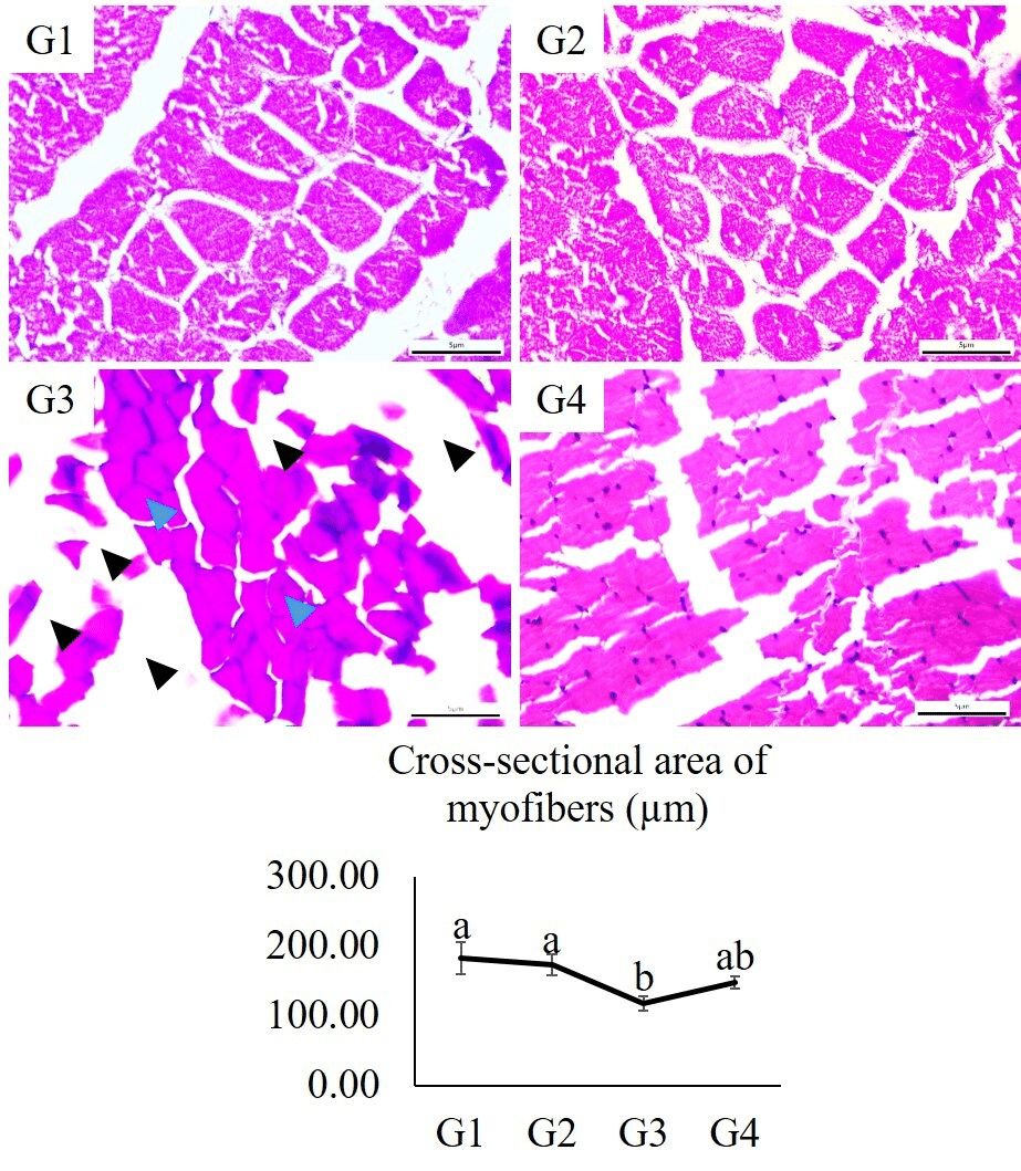 Figure 9. Morphologic and morphometric attributes of goat skeletal muscle (G1: control, G2: formalin preserved, G3: cryodehydrated (thawed at room temperature), and G4: cryodehydrated using cryoprotectant (thawed at 4 °C). Blue arrows indicate myofibers and black arrows indicate increased space between the myofibers and muscle bundles. Morphometric data are expressed as mean ± SEM. Statistical differences among groups were analyzed using one-way ANOVA. Values with different alphabetic superscripts are significantly different (P < 0.05). Staining method - H&E, magnification - 40 X, scale bar - 5 µm |
Skeletal Muscle
The histomorphologic and histomorphometric characteristics of skeletal muscle are illustrated in Figure 9. No notable histoarchitectural alterations were observed in groups G1 and G2. In contrast, G3 exhibited evident shrinkage and significant loss of myofibers, characterized by empty spaces and loss of nuclear clarity. In G4, both cytoplasmic and nuclear clarity were well preserved, although loss of myofibers remained still apparent. Histomorphometric analysis revealed no significant differences (P > 0.05) in the cross-sectional area of myofibers among G1, G2, and G4; however, a marked reduction was observed in G3.
Various techniques have long been used to preserve biological specimens for educational, research, and diagnostic purpose, yet researchers are looking for a more effective, safer, and cost-efficient alternative (Hammer et al., 2012; Hayat et al., 2018; Islam and Sultana, 2021; Islam et al., 2021; Sultana and Islam, 2023). Cryodehydration is being used to preserve organ samples or anatomical segments, even whole-body sections, and cadavers (Teixeira Filho et al., 2019; Islam and Sultana, 2021; Martins and Sakalem, 2022). In this study, we utilized a modified cryodehydration technique (use of glycerol as cryoprotectant and controlled thawing at 4 °C) to compare its efficacy in tissue preservation with routine cryodehydration (Sultana and Islam, 2023) and formalin preservation.
Macroscopic features of the cryodehydrated specimens
Preserving the gross morphology of the specimens is one of the main goals of using any kind of preservation techniques like cryodehydration. Cryodehydration is a time-consuming process influenced by the size and type of specimens (Sultana and Islam, 2023). In this study, the modified protocol (G4) required more freeze-thaw cycles and consequently more time than the routine protocol (G3). It is likely due to the higher viscosity of glycerol, which can impede water movement within tissues, slow the dehydration process, and inhibit ice crystal formation, thereby reducing the efficiency of freezing in facilitating water removal (da Silva et al., 2021). However, cavitary organs reached their final stage with fewer freeze-thaw cycles than parenchymatous organs which is in line with Martins and Sakalem, (2022) and Sultana and Islam (2023). This could be attributed to their hollow structure and lower tissue thickness, which may promote quicker water removal and thereby reduce the number of cycles needed to attain the final form (Sultana and Islam 2023).
In the current study, both routine and modified protocols produced excellent color, texture, and morphology while remaining soft, dry, odorless, lightweight, and durable, with no visible structural alterations during three months of room temperature storage, except for minor wrinkling observed on the parietal surface of the liver. These findings are consistent with previous studies in which cryodehydration of parenchymatous organs and body sections resulted in dry, lightweight, and odor-free specimens (Teixeira Filho et al., 2019; Martins and Sakalem, 2022; Sultana and Islam, 2023). These specimens can be preserved for an extended period, potentially lasting up to 20 years (Martins and Sakalem, 2022). A key feature of the cryodehydration technique is the significant weight loss due to water removal, which is essential for ensuring long-term specimen preservation by preventing tissue autolysis and microbial damage (Teixeira Filho et al., 2019). The water loss also leads to a reduction in specimen weight, making the preserved samples lightweight and easier to handle (Sultana and Islam, 2023). In the present study, cryodehydrated specimens exhibited substantial weight loss during the dehydration phase, with the least reduction observed in musculoskeletal specimens, likely due to the presence of mineral-dense bone alongside muscle tissue (Sultana and Islam, 2023).
Microscopic features of the cryodehydrated specimens
Maintaining the histoarchitectural integrity of the tissue specimens is a key requirement when selecting any preservative or preservation method (Gugic et al., 2007; Balta et al., 2015; Islam et al., 2021). However, research focused on evaluating the effectiveness of cryodehydration in preserving the histoarchitectural characteristics of various organs is very limited. Our previous study indicated significant changes in the tissue histoarchitecture after cryodehydration, where no cryoprotectant was applied and thawing occurred at room temperature; 25 °C (Sultana and Islam, 2023). To minimize tissue damage, this study utilized a modified cryodehydration protocol where glycerol was used as a cryoprotectant, and thawing was carried out at a controlled temperature of 4 °C as suggested in earlier studies (Best, 2025; Kilbride et al., 2021).
In the routine cryodehydration protocol, several changes were seen in tissue structure. These included a noticeable thinning of the heart wall, wider gaps between heart muscle fibers, and unclear cell details in the lungs. The lungs also showed smaller bronchial and bronchiolar openings and reduced alveolar surface area. In the liver, liver cells lost their clear structure, and the spaces between blood vessels were greatly widened. The kidneys showed damage to both the tubules and glomeruli. The spleen had a loss of lymphoid tissue, and the skeletal muscles had smaller muscle fibers. Similar changes were also reported in the earlier study by Sultana and Islam (2023). However, in the modified cryodehydration protocol, various tissue specimens exhibited minimal histoarchitectural alterations and tissue damage, likely due to the protective effects of the cryoprotectant and the controlled thawing process (Best, 2015; Kilbride et al., 2021).
Cryodehydration using repeated freezing-thawing cycles dehydrates and preserves tissue by forming and melting ice crystals that gradually disrupt cellular water content, allowing water molecules to escape tissue and thus maintaining tissue structure and function (Pegg, 2002; Tavares Schäfer et al., 2023). The tissue damage during cryodehydration mostly happens due to the microrupture of cells or tissues (Teixeira Filho et al., 2019; Kilbride et al., 2021). This process facilitates the subsequent phase of dehydration by allowing the slow release of fluids from within and between cells, while also helping to minimize excessive tissue shrinkage (ur Rahman et al., 2018). However, these microruptures caused by ice crystal formation within the cells and intracellular spaces are the main culprit for tissue damage, cytoplasmic drain and loss of nuclear clarity (Pegg, 2002; Sultana and Islam, 2023). Microruptures or microfractures can also be caused by sudden re-expansion of tissues resulting from rapid warming at room temperature.
Using cryoprotectant such as glycerol lowers the freezing point of water and replaces some of the intracellular water and thus reduce the likelihood of large ice crystals formation leading to minimal tissue damage (Pegg, 2002; Bhattacharya, 2018; Whaley et al., 2021; Yang et al., 2022). A similar effect is seen during low-temperature thawing which prevents small ice crystals from merging into larger ones, which could otherwise damage the cell structure during thawing (da Silva et al., 2021). Controlled thawing at temperatures below room temperature also helps to prevent tissue shock and thus limits the formation of excessive osmotic gradients (Kilbride et al., 2021). Thawing at 4 °C slows this process, giving cells time to rehydrate gradually. Glycerol also helps to maintain membrane integrity during freezing by reducing osmotic stress, which occurs as water leaves cells during freezing (da Silva et al., 2021). In addition, glycerol facilitates a more controlled dehydration process, reducing abrupt shifts in water content that can tear or rupture cells (Pegg, 2002; da Silva et al., 2021; Zhang et al., 2022). Thus, cryodehydration using cryoprotectant like glycerol and controlled thawing can preserve the histoarchitectures of anatomical specimens more efficiently by significantly minimizing tissue damage. While the present findings demonstrate promising outcomes for the modified cryodehydration protocol, we acknowledge that the study was conducted using a limited number of animals. Although multiple tissue specimens were analyzed to reduce intra-animal variability, the relatively small sample size may limit the generalizability of the results. Therefore, the findings of this study should be considered preliminary, and further investigations involving a larger number of animals are warranted to validate and extend these observations.
In addition, the interpretation of these findings should also take into account that the study was conducted exclusively on isolated organ samples, with histological evaluation performed immediately following cryodehydration; thus, outcomes may differ in the context of whole-body preservation or extended storage durations.
The routine cryodehydration technique is effective for preserving the gross morphology of anatomical specimens; however, significant tissue damage limits its utility in maintaining histoarchitectural integrity. Notably, slight modifications, such as the use of glycerol as a cryoprotectant and controlled thawing at 4 °C, enhance the technique's effectiveness in preserving both gross morphology and histoarchitecture. However, given the limited number of experimental animals, these findings should be interpreted with caution. Further studies with larger sample sizes and more robust experimental designs are necessary to confirm the effectiveness and reproducibility of this approach. We also recommend a systematic evaluation of cryoprotectants over extended storage periods to further assess the efficacy of this modified cryodehydration technique for whole-body preservation.
Acknowledgments
We gratefully acknowledge the Department of Anatomy and Histology at Bangladesh Agricultural University, Mymensingh-2202, Bangladesh, for providing the necessary research facilities. We also extend our appreciation to the Bangladesh Agricultural University Research System (BAURES) for supporting this study through research funding. The funding body had no role in the design of the study, data collection, analysis, or interpretation, nor in the writing of the manuscript or the decision to submit it for publication.
Funding
This research was supported by a grant from the Bangladesh Agricultural University Research System (BAURES), Bangladesh. The grant was awarded to RI as the Principal Investigator (Grant No.: 2023/1847/BAU).
Author contribution
NS and RI conceptualized the study. RI analyzed and interpreted the data, prepared all the figures, and drafted the manuscript. ASJ collected the samples and prepared the cryodehydrated specimens. RI and ASJ performed the histological study. Finally, NS finalized the manuscript.
Alsharif MHK, Musthafa M, Elamin AY, Ibnouf EO, Taha KM, Alfaki MA, et al. 2017: A brief review on the principles of human cadaver preservation and monitoring of microbial degradation. Forensic Med Anat Res 5(3):19. https://doi.org/10.4236/fmar.2017.53003
Balta JY, Cronin M, Cryan JF, O’Mahony SM. 2015: Human preservation techniques in anatomy: A 21st century medical education perspective. Clin Anat 28(6):725–734. https://doi.org/10.1002/ca.22585
Best BP. 2015: Cryoprotectant toxicity: facts, issues, and questions. Rejuvenation Res 18(5):422–436. https://doi.org/10.1089/rej.2014.1656
Bhattacharya S. 2018: Cryoprotectants and their usage in cryopreservation. Cryopreserv Biotechnol Biomed Biol Sci 7:1–12. https://doi.org/10.5772/intechopen.73413
da Silva AM, Pereira AG, Brasil AV, Macedo LB, Souza-Junior J, de Moura CEB, et al. 2021: Influence of freezing techniques and glycerol-based cryoprotectant combinations on the survival of testicular tissues from adult collared peccaries. Theriogenology 167:111–119. https://doi.org/10.1016/j.theriogenology.2021.03.013
Elnady FA. 2016: The Elnady technique: An innovative, new method for tissue preservation. ALTEX 33(3):237–242. https://doi.org/10.14573/altex.1511091
ElShafey A, Magdy Y, Hamad AMAAR, Ahmed O. 2022: Modified form of the Elnady technique for tissues preservation. Benha Vet Med J 41(2):56–60. https://doi.org/10.21608/bvmj.2021.95641.1470
Gugic D, Nassiri M, Nadji M, Morales A, Vincek V. 2007: Novel tissue preservative and tissue fixative for comparative pathology and animal research. J Exp Anim Sci 43(4):271–281. https://doi.org/10.1016/j.jeas.2006.09.002
Haizuka Y, Nagase M, Takashino S, Kobayashi Y, Fujikura Y, Matsumura G. 2018: A new substitute for formalin: application to embalming cadavers. Clin Anat 31(1):90–98. https://doi.org/10.1002/ca.23011
Hammer N, Löffler S, Feja C, Sandrock M, Schmidt W, Bechmann I, et al. 2012: Ethanol-glycerin fixation with thymol conservation: A potential alternative to formaldehyde and phenol embalming. Anat Sci Educ 5(4):225–233. https://doi.org/10.1002/ase.1270
Han B, Bischof JC. 2004: Engineering challenges in tissue preservation. Cell Preserv Technol 2(2):91–112. https://doi.org/10.1089/153834404774101945
Hayat K, Qureshi AS, Rehan S, Rehman T. 2018: Plastination—An innovative preservative technique in anatomy. Trends Anat Physiol 1(10):2–5. https://doi.org/10.24966/TAP-7752/100003
Islam R, Sultana N. 2021: Preservation of musculoskeletal specimens of goat by the Elnady technique: an innovative low-cost alternative to plastination. J Plast 33(2):1–13. https://doi.org/10.56507/QBZN2088
Islam R, Ayman U, Sultana N. 2021: Histological architectures and biometric characteristics of indigenously plastinated organs of goat. Int J Morphol 39(3):759–765. https://doi.org/10.4067/S0717-95022021000300759
Jangde S, Arya R, Basan K, Kumar N. 2015: Preventive measures and suggested guidelines to reduce the chemical health hazards on medical students and educators. Res J Pharmacol Pharmacodyn 7(3):147–150. https://doi.org/10.5958/2321-5836.2015.00028.2
Kilbride P, Meneghel J, Fonseca F, Morris J. 2021: The transfer temperature from slow cooling to cryogenic storage is critical for optimal recovery of cryopreserved mammalian cells. PLoS One 16(11):e0259571. https://doi.org/10.1371/journal.pone.0259571
Kumari PK, Akhila S, Rao YS, Devi BR. 2019: Alternative to artificial preservatives. Syst Rev Pharm 10:99–102. https://doi.org/10.5530/srp.2019.1.17
Martins LL, Sakalem ME. 2022: Cryodehydration protocol to obtain high-quality permanent anatomical material. J Anat 241(2):545–551. https://doi.org/10.1111/joa.13669
Nam SM, Moon JS, Yoon HY, Chang BJ, Nahm SS. 2020: Comparative evaluation of canine cadaver embalming methods for veterinary anatomy education. Anat Sci Int 95:498–507. https://doi.org/10.1007/s12565-020-00547-x
Pegg DE. 2002: The history and principles of cryopreservation. Semin Reprod Med 20(1):5–14. https://doi.org/10.1055/s-2002-23515
Pegg DE. 2014: Principles of cryopreservation. Cryopreserv Freeze-Drying Protoc 3–19. https://doi.org/10.1007/978-1-4939-2193-5_1
Rezoana R, Akter L, Islam R, Bhakta S, Ayman U, Karim MR, et al. 2022: The hazardous effects of formalin and alcoholic fixative in mice: A public health perspective study. Saudi J Biol Sci 29(5):3366–3371. https://doi.org/10.1016/j.sjbs.2022.02.019
Sora MC, Latorre R, Baptista C, López-Albors O. 2019: Plastination—A scientific method for teaching and research. Anat Histol Embryol 48(6):526–531. https://doi.org/10.1111/ahe.12493
Sultana N, Islam R. 2023: Efficacy of cryodehydration technique in preserving the gross and histoarchitectural details of goat visceral and musculoskeletal specimens. J Adv Vet Anim Res 10(4):720–729. https://doi.org/10.5455/javar.2023.j727
Tavares Schäfer B, de Oliveira Pureza Pinto PL, Teixeira Filho A. 2023: Cryodehydration: Quality and durability of specimens subjected to vacuum-forced impregnation with wood glue. J Morphol Sci 40:379–384. https://doi.org/10.51929/jms.40.379.2023
Teixeira Filho A, Schäfer BT, Vives PS. 2019: Cryodehydration technique applied to anatomical segments. J Morphol Sci 36(4):219–222. https://doi.org/10.1055/s-0039-3400482
ur Rahman U, Sahar A, Ishaq A, Aadil RM, Zahoor T, Ahmad MH. 2018: Advanced meat preservation methods: A mini review. J Food Saf 38(4):e12467. https://doi.org/10.1111/jfs.12467
Whaley D, Damyar K, Witek RP, Mendoza A, Alexander M, Lakey JR. 2021: Cryopreservation: an overview of principles and cell-specific considerations. Cell Transplant 30:0963689721999617. https://doi.org/10.1177/0963689721999617
Yang J, Xu Y, Luo S, Dang H, Cao M. 2022: Effect of cryoprotectants on rat kidney decellularization by freeze-thaw process. Cryobiology 105:71–82. https://doi.org/10.1016/j.cryobiol.2021.11.180
Zhang PQ, Tan PC, Gao YM, Zhang XJ, Xie Y, Zheng DN, et al. 2022: The effect of glycerol as a cryoprotective agent in the cryopreservation of adipose tissue. Stem Cell Res Ther 13(1):152. https://doi.org/10.1186/s13287-022-02817-z

