1 University Hospital, University of São Paulo, Brazil
2 Department of Anatomy, Institute of Biomedical Sciences (ICB), University of São Paulo, Brazil
3 Department of Medical Education, College of Medicine, University of Toledo, Toledo, Ohio, USA
In anatomical and clinical practice, comparison between plastinated specimens and Computed Tomography (CT) and Magnetic Resonance (MR) has been often reported in literature. However, few studies performed imaging scans of plastinated specimens. This study was made to evaluate how plastination affects the radiological properties of the tissues, specially concerning CT. A Toshiba Aquilion 64-multdetector CT scan was utilized, at The Radiology Department of the Heart Institute, University of São Paulo, Brazil, to evaluate a heart and a diencephalon-brain stem specimen, plastinated 34 years ago, using the Biodur Silicone S10 technique. Cross-sectional images of specimens were obtained from 0.5 mm thick slices. Tridimensional images were generated by the volume rendering technique using TeraRecon Aquarius Net Viewer Workstation. The X-ray attenuation tissue coefficients by CT (in Hounsfield units) of the specimen images were measured and compared to CT images obtained from living subjects from the archives of The Heart Institute Department of Radiology. The internal and external structures were preserved both for heart and diencephalon-brain stem images. Both plastinated specimens showed increased X-Ray attenuation coefficients when compared to the living subject images. This phenomenon may be linked to the physical properties of the silicone resin. MRI scanned specimens, as expected, did not produce any signal. The CT 3D scan is an excellent method for examining plastinated specimens, especially to evaluate either inner and outer surfaces.
plastination, computer tomography, silicone; magnetic resonance imaging, imaging
Carlos A. C. Baptista, Department of Medical Education, College of Medicine, University of Toledo, Toledo, Ohio USA 43614, Email: carlos.baptista@utoledo.edu

![]()


Since the dawn of the age of computed tomography/magnetic resonance imaging techniques, and the advent of plastination technology, efforts have been made to use both techniques with plastinated specimens as useful teaching tools. Sectional anatomy has been incorporated in the curricula of medical, veterinary and dental schools in order to facilitate learning. The understanding of sectional anatomy is crucial for professionals such as radiologists and neurosurgeons.
Most studies in the literature compare sheet plastinated specimens with images from computed tomography (CT) scanning and magnetic resonance (MRI) imaging. The source of the images may be from living subjects or cadaveric specimens (Cook, 1997; Entius et al., 1997; Reina de la Torre et al., 2015; Sora et al., 2019). Some studies compared plastinated slices to CT images obtained prior to plastination, from the same specimen (Latorre et al., 2006, 2003; Rodriguez et al., 2008, 2010; Parraga et al. 2013; Ottone et al., 2016). A few studies, including Shianti et al. (2015) showed the relationship between CT in pre- and post-plastinated specimens, emphasizing the behavior of X-Ray attenuation coefficient numbers.
The majority of studies showing 3D reconstruction of images of plastinated specimens are based on computerized models (Sora et al., 2007; Tunali et al., 2008; Latorre et al., 2008; Cerqueira et al., 2008 a, b; Rodriguez et. al, 2008; Arredondo et al., 2008 a, b; Sora et al., 2013). In 2012, Tiwari et al. performed a 3D reconstruction of a plastinated 24-week fetus directly from the 16 slice-CT scan machine.
Even though MRI is also important for diagnostic purpose, Baptista et al. (1990) showed that MRI scanning of plastinated specimens produced no signals, for reasons which are discussed later in this paper.
The objective of this study was to analyze the internal and external morphological aspects and integrity of silicone plastinated specimens (Biodur® S10) using tridimensional CT (3D-CT). The radiological aspects of images were studied by comparing the X-ray tissue attenuation coefficients of plastinated specimens by CT, to the coefficients of the same tissue in CT images made from living subjects.
A human heart (Fig. 1a) and a diencephalon-brain stem (D-BS) (Fig. 1b), plastinated with silicone 34 years ago using the Biodur® S10 silicone standard technique, described by von Hagens (1979, 1985, 1986), were the subjects of this study. For 3D-CT scanning, a 64-multidetector Toshiba Aquilion CT Scanner (Fig. 2) was used and belonged to the Heart Institute of the College of Medicine of the University of São Paulo, Brazil. The radiological parameters of the images acquired are shown in Table 1. The scanner obtained 434 transverse cross-section images from the plastinated heart, and 317 images from the plastinated diencephalon-encephalic trunk (Figs. 3a, 3b).
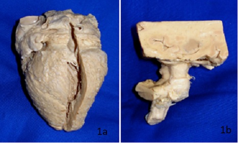 Figure 1. Silicone plastinates: a: human heart, |
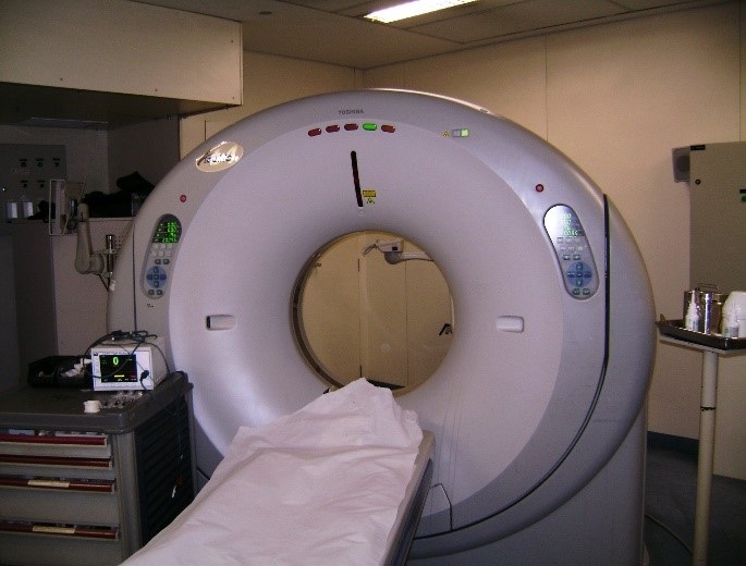 Figure 2. Toshiba Aquilion 64-multidetector CT scan from Radiology Department of Heart Institute – University of São Paulo, Brazil. |
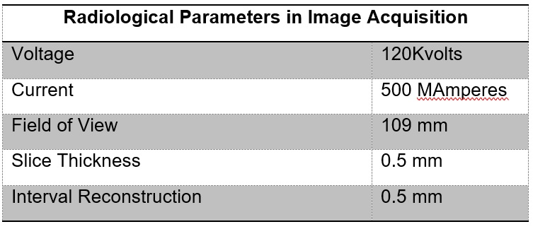 Table 1 - Radiological Parameters in Image Acquisition |
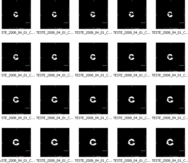 Figure 3a. Set of CT scans of plastinated human heart cross-sections. |
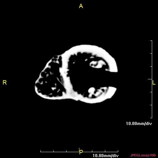 Figure 3b. A single slice CT cross-section of a plastinated human heart. |
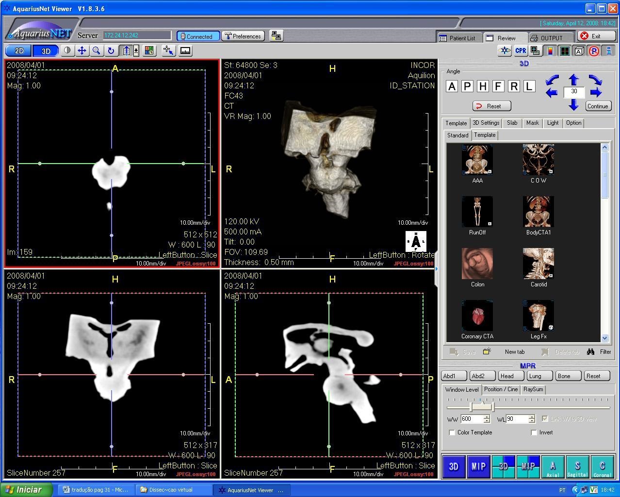 Figure 4. Workstation interface utilized to reconstruct the transverse sections into tridimensional images. |
The cross-sectional images were reconstructed on a Terarecon® Aquarius Net Viewer Workstation by a technique known as Volume Rendering, producing tridimensional images, which can be rotated in all directions (Fig. 4).
The X-ray attenuation coefficients (mean and standard deviation) were obtained from transverse images of the plastinates, measured in Hounsfield units (HU), and compared to the attenuation coefficients obtained from archived images of the same region obtained in CT scans of living subjects (Archives of the Department of Radiology, Heart Institute, University of São Paulo) (Figs. 5a, 5b, 6a, 6b, 6c).
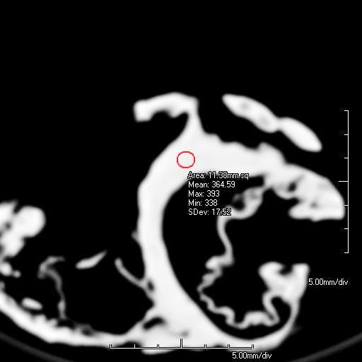 Figure 5a. Area of myocardial X-ray attenuation |
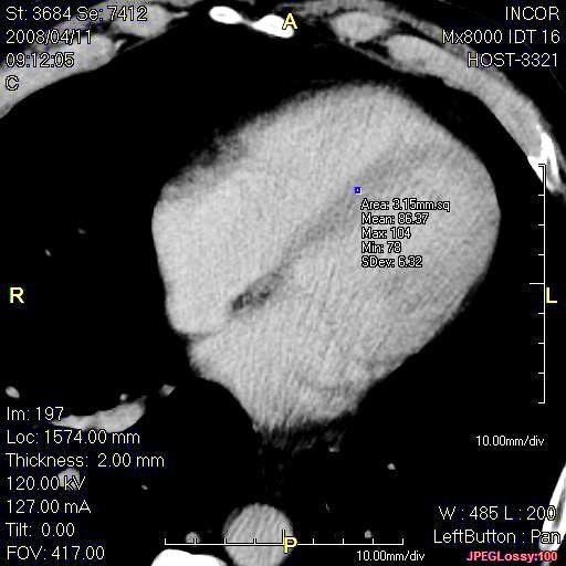 Figure 5b. Area of myocardial X-ray attenuation coefficients measurements: Living human – Blue Dot |
|
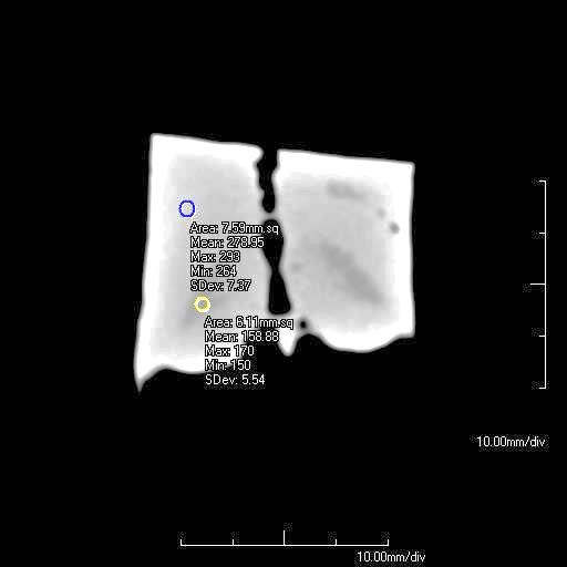 Figure 6a. Area of X-Ray attenuation coefficients |
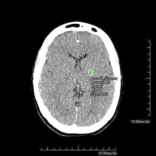 Figure 6b. Area of X-Ray attenuation coefficients measurements of D-BS, gray and white matter: Living subject – green circle, Indicates area of gray matter |
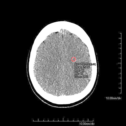 Figure 6c. Area of X-Ray attenuation coefficients measurements of D-BS, gray and white matter: Living subject –Red circle, Indicates area of white matter |
 Table 2. X-Ray tissue attenuation coefficients ( Preston R. Chapter1. Imaging the Head and Brain. In Diagnostic Imaging for the Emergency Physician. 2011, Pages 1-45.) |
X-Ray attenuation in CT indicates the following: presence of fat or air inside the tissue, air reaching values about -1000 HU (shown as black on image); 0 HU is the attenuation of water (shown as medium gray on image); soft tissue other than fat is between 0 and +100 HU; hemorrhage, and especially bone and other calcified tissues are above +100 HU, bone reaching values about+1000 HU (shown as white on the image) (Table 2).
The specimens were submitted to a 1.5 T MRI scan to evaluate their behavior in a strong magnetic field.
For the scanned plastinated heart, this study found the following:
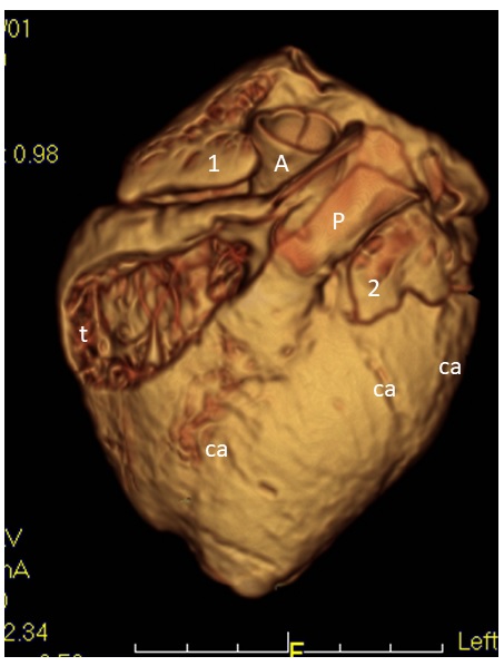 Figure 7. 3D CT reconstruction of the plastinated heart (anterior view). (1- Right atrial appendage; 2- Left atrial appendage; A – Aorta; P - Pulmonary trunk; ca - Coronary artery; t - Trabeculae carneae) |
 Figure 8. Longitudinal section of heart with internal detail, 3D CT (slab image). Inner structure: AV - Aortic valve; M - Mitral valve; CT - Chordae tendineae; S - Interventricular septum; P - Papillary muscle; TV - Tricuspid valve) |
For the D-BS plastinate, this study found the following:
The MRI scanning of the plastinates did not yield any images. There was no evidence of signal generation in the scan.
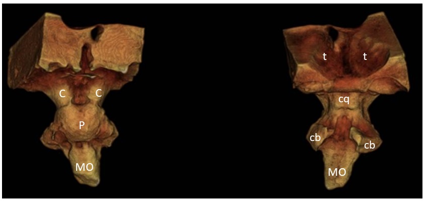 Figure 9. Anterior (left) and posterior (right) views of 3D CT reconstruction of diencephalon-brain stem plastinate. C- Cerebral peduncle; P- Pons; MO - Medulla oblongata; cb - Cerebellar peduncle’ cq - Corpora quadrigemina; t –talamus |
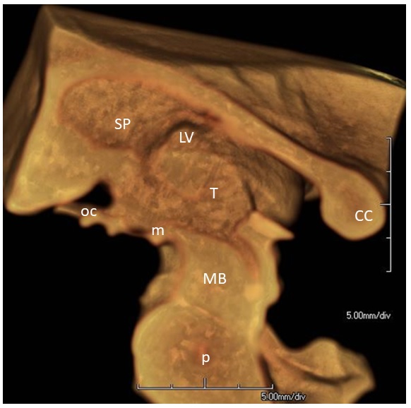 Figure 10. Sagittal section of D-BS plastinate, 3D CT (slab image). Cc – Corpus callosum (splenium); SP- Septum pellucidum; LV- Lateral ventricle; T- Thalamus MB- Midbrain; P – Pons; oc – Optic chiasm; m – Mammillary body) |
The internal and external morphology of the plastinated specimens was well characterized for both the heart and D-BS. Tiavri et al. (2012) concluded, after performing a CT scan of a 24-week plastinated fetus, that the three-dimensional morphology of the organs, soft tissue and muscle attachments was preserved.
There was an increase in the X-ray attenuation values of both the heart and D-BS CT images. Shianti et al. (2015) measured the attenuation coefficients of several organs (including liver, heart, lungs, and kidneys) before and after standard S10 plastination. Their study showed a significant increase in the coefficients of all organs studied, with minor differences occurring in bones and air-filled organs. The impregnated silicone seems to absorb more X-ray, making the specimen denser than the non-plastinated tissue, therefore raising the X-Ray attenuation values. These changes in attenuation values may be used to evaluate the degree of silicone impregnation. Once liquid is replaced by silicone, the changes in attenuation values may serve as an indicator of a complete or incomplete silicone impregnation. This phenomenon can explain the areas of low attenuation found inside the D-BS CT images. This finding is not related to the differences found in gray and white matter.
The MRI scans of the plastinated organs, as expected, produced no signal because of the absence of water and fat in the specimens. Baptista et al. (1990), in their study of MRI scans of plastinated specimens, concluded that the quality of the MRI image obtained from plastinated specimens had an indirect relationship to the time of hardening of the specimen. The specimens used for this study reported here were plastinated 34 years ago; consequently, there is no evidence of signals generated in the scan. Since MR images are generated from “vibrations” of hydrogen protons, the hardening process eliminates the possibility of such vibration.
We conclude that CT scans are an excellent method for examining plastinated specimens, especially inner or outer surfaces. Even though good images were obtained, anatomy of the wall architecture could not be determined. The impregnation of the specimens with Biodur S10 reduces the CT attenuation rates of the specimens.
ACKNOWLEDGEMENTS
We thank Professor Claudio Campi de Castro, MD, PhD, Chief Radiologist in the Magnetic Resonance sector at The Heart Institute, University of São Paulo, Brazil, for the use of the CT and MRI devices for this study.
Arredondo J, MD Ayala, O López-Albors, A Agut, JM Vázquez, F Asensio and R Latorre. 2008: Three-dimensional reconstruction of the temporomandibular joint of a feline model by means of epoxy plastinated sections. Abstract presented at The 14th International Conference on Plastination -Heidelberg and Guben, Germany, July 20-26, 2008. Int Soc Plastination 23: 61
Arredondo J, O López-Albors, A Agut, F Gil, M Soler , MJ Rodriguez and R Latorre. 2008: Epoxy plastinated slices of the temporomandibular joint of the cat are used to assess high resolution computed tomography. Abstract presented at The 14th International Conference on Plastination -Heidelberg and Guben, Germany, July 20-26, 2008. Int Soc Plastination 23: 61
Baptista CAC, Henry RW, Williams G, Brinker R. 1990: Magnetic resonance imaging of plastinated specimens: study of tissue characteristics and evaluation of the hardening process of the S10 technique. Abstract presented at The 5th International Conference on Plastination, Faculty of Medicine, University of Heidelberg, Germany July 1990. J Int Soc Plastination 4: 3.
Cerqueira EP, CAC Baptista, CC Campi, AF Silva. 2008: 3D multidetector CT reconstructions of a heart, plastinated with Biodur using the standard S10 technique. Abstract presented at The 14th International Conference on Plastination -Heidelberg and Guben, Germany, July 20-26, 2008. Int Soc Plastination 23: 51
Cerqueira EP, CAC Baptista, CC Campi, AF Silva. 2008: 3D multidetector CT reconstructions of a diencephalon and brain stem, plastinated with Biodur using the standard S10 technique. Abstract presented at The 14th International Conference on Plastination -Heidelberg and Guben, Germany, July 20-26, 2008. Int Soc Plastination 23: 51
Cook P. 1997: Sheet plastination as a clinically based teaching aid at the University of Auckland. Acta Anat 158:33-36.
https://doi.org/10.1159/000147907
Entius CAC, van Rijn RRB, Holstege JC, Stoeckart R, Zwamborn AW. 1997: Correlating sheet plastinated slices, computed tomography images and magnetic resonance images of the pelvic girdle: a teaching tool. Acta Anat 158:44-47
https://doi.org/10.1159/000147909
Latorre R, Arencibia A, Gil F, Rivero M, Ramírez G, Váquez-Autón JM, Henry RW 2003: P-40 and S10 Plastinated Slices: An Aid to Interpreting MR Images of the Equine Tarsus. J Int Soc Plastination 18: 14-22.
https://doi.org/10.56507/KDJG6154
Latorre R, F SUN, O López-Albors; MD Ayala, F Gil; S Losilla, M Orenes, RW Henry. 2008: 3D fluoroscopy reconstruction of plastinated specimens. Abstract presented at The 14th International Conference on Plastination -Heidelberg and Guben, Germany, July 20-26, 2008. Int Soc Plastination 23: 50
Latorre, R., Arencibia, A., Gil, F., Rivero, M., Henry, R. W., Ramirez, G., & Vaquez, J. M. 2006: Correlation of magnetic resonance images with anatomic features of the equine tarsus. American Journal of Veterinary Research, 67(5), 756-761.
https://doi.org/10.2460/ajvr.67.5.756
Ottone NE, Del Sol M, Fuentes R. 2016: Report on a sheet plastination technique using commercial epoxy resin. Int J Morphol 34(3):1039-1043.
https://doi.org/10.4067/S0717-95022016000300036
Párraga, E., López-Albors, O., Sánchez-Margallo, F., Moyano-Cuevas, J. L., & Latorre, R. 2013: Effects of pneumoperitoneum and body position on the morphology of the caudal cava vein analyzed by MRI and plastinated sections. Surgical Endoscopy, 27(3), 880-887.
https://doi.org/10.1007/s00464-012-2528-5
Preston R. 2011: Imaging the head and brain. In: Broder JS, editor. Diagnostic imaging for the emergency physician, 1st ed. Philadelphia PA: Elsevier Saunders, p 1-45.
https://doi.org/10.1016/B978-1-4160-6113-7.10001-8
Reina de la Torre F, Carrera-Burgaya A, Bataller-Jordà F, Pedraza S, Puig-Camps A, San-Molina J. 2015: Correlation between thin sheet plastinated slices and CT and MRI images of the central nervous system. Poster No. C-2490 Congress: ECR 2015.
Rodríguez MJ, A Agut, O López-Albors, J Arredondo , JM Vazquez , G Ramirez, R Latorre. 2008: Computed tomography imaging of the equine temporormandibular joint: a sheet plastination study. Abstract presented at The 14th International Conference on Plastination -Heidelberg and Guben, Germany, July 20-26, 2008. Int Soc Plastination 23: 60
Rodriguez, M. J., Agut, A., Soler, M., Lopez-Albors, O., Arredondo, J., Querol, M., & Latorre, R. 2010: Magnetic resonance imaging of the equine temporomandibular joint anatomy. Equine Veterinary Journal, 42(3), 200-207.
https://doi.org/10.1111/j.2042-3306.2010.00030.x
Rodriguez, M. J., Latorre, R., Lopez-Albors, O., Soler, M., Aguirre, C., Vazquez, J. M., Agut, A. 2008: Computed tomographic anatomy of the temporomandibular joint in the young horse. Equine Veterinary Journal, 40(6), 566-571.
https://doi.org/10.2746/042516408X322166
Shanthi P, Singh RR, Gibikote S, Rabi S. 2015: Comparison of CT numbers of organs before and after plastination using standard S-10 technique. Clin Anat 28(4):431-5.
https://doi.org/10.1002/ca.22514
Sora MC, Latorre R, Baptista CAC, Lopez-Arbors O. 2019: Plastination: a scientific method for teaching and research. Anat Histol Embryol 48:526-531.
https://doi.org/10.1111/ahe.12493
Tiwari S, Nandlal B, Shama Sundar N M. 2012: Plastinated fetus: 3D CT scan (VRT) evaluation. Indian J Dent Res 23:686-8.
https://doi.org/10.4103/0970-9290.107411
Tunali S, M Farrell, S Labrash, BK Lozanoff, S Doll, S Lozanoff. 2008: Computerized 3D anatomical modeling using plastinated anatomical material. Abstract presented at The 14th International Conference on Plastination -Heidelberg and Guben, Germany, July 20-26, 2008. J Int Soc Plastination 23: 50
von Hagens G, Tiedemann K, Kriz W. 1986: The current potential of plastination. Anat Embryol 175(4):411-421.
https://doi.org/10.1007/BF00309677
von Hagens G. 1979: Impregnation of soft biological specimens with thermosetting resins and elastomers. Anat Rec 194:247-255
https://doi.org/10.1002/ar.1091940206
von Hagens G. 1985: Heidelberg Plastination Folder. Collection of technical leaflets for plastination. Heidelberg: Anatomiches Institut 1, Universität Heidelberg.