1- Otago University School of Physiotherapy, Dunedin, New Zealand
2- Department of Anatomy and Structural Biology, Otago Medical School, Dunedin, New Zealand
A comparative study of spinal connective tissue and its structural arrangement was carried out using tissue preserved in epoxy resin plastinated slices and paraffin embedded sections. The 2.5mm horizontal plastinated slices provide a more complete overview of connective tissue arrangement in the spine, including fibre arrangement, bony attachment sites and, continuity with neighbouring structures when compared with corresponding 7um paraffin embedded standard histology sections. Use of plastinated slices allows distinctive macroscopic definition of connective tissue fibre arrangement of spinal tendons, ligaments and fascia. The quality of the information obtained regarding connective tissue arrangement suggests that, for spinal research on selected dense connective tissue structure, use of epoxy plastinated slices presents an attractive alternative to conventional histology.
Spinal connective tissue, Structure, Neck
G Johnson, University of Otago School of Physiotherapy, PO Box 56, Dunedin, New Zealand. Telephone: 64 3 479 5424 / Fax: 64 3 479 8414. Email: gjohnson@gandalf.otago.ac.nz

![]()


Examination of ligaments, tendons and fascia is difficult, particularly in the case of the vertebral column, where they are often closely intertwined and blended together. The orientation of collagen within these structures determines the ultimate stress and load bearing behaviour of connective tissue thus the geometric arrangement of their fibres warrants careful consideration (Behrsin and Briggs, 1988). To date, information regarding connective tissue organization within spinal structures has been derived from anatomic dissection, histology and to a lesser extent, x-ray detraction techniques. Epoxy resin plastinated slices offer an alternative strategy to the study of connective tissue organization. The successful application of this technique has been demonstrated in other body regions such as the pelvis, where the connective tissue arrangement is highly complicated (Fritsch and Hotzinger, 1995), but it has largely been overlooked in the area of spinal research. The primary aim was the visualization of spinal connective tissue structure rather than histochemical analysis and therefore the epoxy resin sheet plastination method was compared to conventional histology, in order to evaluate technical issues and quality of information obtained.
Serial cross-sectional slices were taken from a female cadaver (aged 65 years) which was obtained within 24 hours of death. Care was taken to maintain a horizontal orientation of the slices while cutting. Forty four cross-sectional horizontal slices (2.5mm thick) were taken from the region, extending 20mm above the external occipital protuberance on the occiput through the entire cervical spine to the first thoracic vertebra. The slices were processed according to the E12 sheet plastination technique (Weber and Henry, 1993) with the exception that they were cast between 0.25mm
A.P.E.T. (Amorphous Polyethylene Terephthalate, Progressive Plastic Ltd, 31-37 Fraytt St, Dunedin, New Zealand) plastic sheet. This approach was similar to the "drainage method" (von Hagens et al., 1987) as detailedby An and Zhang (1999). The connective tissue arrangement of muscle tendon and ligamentous tissue in the posterior region of the cervical spine in the plastinated slices was examined at low and high magnifications using a stereomicroscope (Wild MZ8; Leica: Heerburgg).
Tissue from a female cadaver (aged 78 years), fixed in 20% formaldehyde, was used for the histology sections. Three tissue sections from the posterior region of the cervical spine (corresponding to the upper (C1-C3), middle (C3-C5) and lower (C5-C7) regions) were detached from the underlying vertebrae using a dissecting scalpel. The blocks of tissue were dehydrated prior to embedding in paraffin. Horizontal sections 7microm thick) from each block were stained with Verhoeff's haematoxylin then counterstained with van Gieson's stain (Culling, 1957) in order to differentiate collagen, elastin and muscle fibres.
Table 1 compares spinal tissue preparation and processing, quality of the image and connective tissue differentiation in conventional histology sections and epoxy resin plastinated slices, quality of information and respective application to spinal research.
| Histology Sections | Plastinated Slices | |
| Tissue preparation | Decalcification or detachment of soft tissue from bony attachments. | No decalcification. Adequate freezer space required. |
| Tissue processing | Standard histology tissue fixation, embedding and staining 5-7 days. | Processing time approximately 6-8 weeks. |
| Image resolution | Microscopic view of connective tissue. | Macroscopic and gross anatomical view of connective tissue structure. |
| Connective tissue differentiation | Connective tissue elements and fibre orientation. | Structural organization of connective tissue within muscle tendons, ligaments and deep and superficial fascia. |
| Quality of information | Regional variation in connective tissue types and fibre orientation visualized. Tissue fragmentation around tendons and isolated calcified deposits. | Minimal tissue disruption or connective tissue damage with 2.5mm slices. Detection of fibre orientation dependent on plane of sectioning and density of connective tissue. |
| Application in spinal research | Morphological basis for biomechanical properties and magnetic resonant imaging (MRI) investigations of the spine. Structural changes occuring with age and spinal disease | Permits the study of intact connective tissue architecture and functional relationships in delicate and hard to access regions of the spine |
The plastinated slices in the horizontal plane prove highly satisfactory for visualization of selected connective tissue structures of the spine. The dense connective tissue of muscle tendons is clearly seen and their origin from the neighbouring muscles can be determined (figure 1). The cross-sectional
geometric dimensions of width and depth of major ligaments, such as the posterior longitudinal ligament, are distinguishable from bony vertebrae, loose connective and adipose tissue. The details of bony attachment sites of muscle aponeuroses are well defined, where their orientation lies in the plane of sectioning (figure 1). At the macroscopic level (under the dissecting microscope), fibre orientation within the dense connective tissues belonging to muscle tendons is visible, along with changes in their direction at different levels of the cervical spine (figure 2). Fine connective tissue septae belonging to the cervical fascia are detected at this level.
The delicate connective tissue of the atlanto-occipital and atlanto-axial membranes are not visible in the plastinated slices, nor are the cervical intervertebral disc connective tissue components of either the nucleus pulposus or annulus fibrosus.
On the histology sections collagen bundles, elastin and muscle fibres are distinguishable in various proportions in the dorsal region of the cervical spine, indicating a regional variation in connective tissue elements (figure 3). In those sections with higher proportions of loose connective and adipose tissue, the isolated collagen fibres appear disrupted with no pattern of fibre orientation apparent (figure 3). Connective tissue fibres in the lower cervical spine tissue are arranged into regular horizontal lamellae (figure 4).
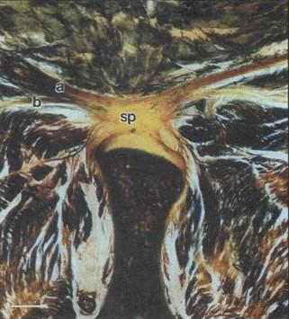 Figure 1. Macroscopic view in an epoxy resin plastinated slice of the connective tissue arrangement of bilaminar muscle tendons joining together at the midline (C6 level). The tendons of (a) trapezius and (b) semispinalis capitis are clearly distinguishable from the surrounding loose connective and adipose tissue. The direct attachment onto the C6 spinous process (sp) is evident. Bar scale = 3.6mm. |
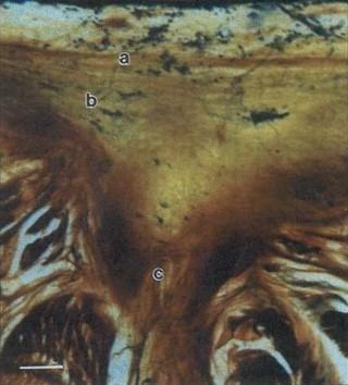 Figure 2. Macroscopic view in an epoxy resin plastinated slice of connective tissue arrangement formed by muscle tendons at the midline (C3 level). Horizontal fibres (a), oblique fibres (b) and vertical fibres (c) with the connective tissue are visible. Bar scale = 1.2mm |
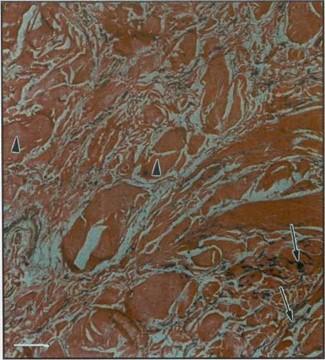 Figure 3. A horizontal histology section (7nm) stained with Verhoeff's solution and van Gieson's stain from the middle (C3-C5) cervical region showing the bundles of collagen (arrow heads), elastin (arrows) and loose connective tissue. Bar scale = 50µm. |
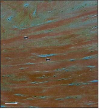 Figure 4. A horizontal histology section (7|im) stained with Verhoeff's solution and van Gieson's stain from the lower (C6- |
The results of this study demonstrate some of the advantages of epoxy resin plastinated slices over paraffin embedded histological sections in the study of connective tissue organization in the spine. The first advantage is that plastinated slices preserve the relationships of neighboring structures. For instance, in figure 1 the bands of connective tissue meeting in the midline can be identified as the tendons of trapezius and semispinalis capitis muscles. The nature of their combined attachment onto the underlying spinous process is also visible. Such views provide valuable and hitherto previously inaccessible information regarding the structural arrangement of different connective tissue components of the spine, such as the ligamentum nuchae (Johnson et al., 2000). Secondly, the relative thickness of plastinated slices compared with that of the semithin histological section prevents even the most delicate connective tissue, such as the cervical fascia, from being disrupted. The section thickness of the plastinated slices circumvents the requirements of conventional histology for decalcification of mineralized tissue or detachment of the tissue from bony attachments. Finally, although the dimensions of plastinated slices limit the degree of resolution possible compared with standard histology sections, dense connective tissue can be clearly distinguished from surrounding loose connective and adipose tissue.
The 2.5mm thick slices permitted excellent visualization of muscle tendons, fascia and ligaments which were the focus of interest for this particular study. However further work study is required to determine the optimal section thickness of plastinated sheet for both the very fine connective tissue membranes and at the other end of the spectrum, the dense connective intervertebral disc structures of the spine.
Acknowledgements
The authors thank Elaine Coory and John Howes, Department of Anatomy & Structural Biology, University of Otago for their assistance with preparation of the histology sections and, the Dean's Fund, University of Otago Medical School, Dunedin, New Zealand for purchase of the stereomicroscope.
An PC, Zhang M: A Technique for Preserving the Subarachnoid Space and its Contents in a Natural State with Different Colours. J Int. Soc Plastination. 14 (1): 12-17,1999.
https://doi.org/10.56507/CQUW3856
Behrsin F, Briggs C: Ligaments of the lumbar spine: a review. Surg Radiol Anat 10: 211-219, 1988.
https://doi.org/10.1007/BF02115239
Culling C. Handbook of Histopathological Techniques. London, Butterworth. 1957.
Fritsch H, Hotzinger H: Tomographical Anatomy of the Pelvis, Visceral Pelvic Connective Tissue, and its Compartments. Clin Anat 8: 17-24,1995.
https://doi.org/10.1002/ca.980080103
Johnson GM, Zhang M, Jones DG: The Fine Connective Tissue Architecture of the Human Ligamentum Nuchae. Spine 25 (1): 5-9, 2000.
https://doi.org/10.1097/00007632-200001010-00003
von Hagens G, Tiedemann K, Kriz W: The current potential of plastination. Anat Embryol 175 (4): 411-421,1987.
https://doi.org/10.1007/BF00309677
Weber W, Henry RW: Sheet Plastination of Body Slices - E12 Technique, Filling Method. J Int Soc Plastination 7 (1): 16-22,1993.
https://doi.org/10.56507/EZGX2343