1 Institute of Biological and Health Sciences, Federal University of Viçosa, campus Rio Paranaíba, Brazil.
2 Department of Veterinary Medicine, Federal University of Viçosa, Brazil.
Objectives - The objective of this study was the evaluation of different protocols to obtain histological slides from silicone plastinated specimens.
Materials and Methods - Samples of pig aorta, heart, and kidney were used. Four treatments for light microscopy (LM) were compared. Treatment 1 (control): fixation in 10% formalin for 48 h at room temperature; tissue samples were then processed for LM histology. Treatment 2: plastinated fragments were directly embedded in paraffin wax. Treatment 3: plastinated tissue samples were de-plastinated by immersion in 99% ethyl alcohol for 24 hours, then in methylbenzene for 48 hours; samples were then processed for LM histology. Treatment 4: plastinated samples were de-plastinated in 1,4-dimethylbenzene for 36 hours, and then processed for LM histology.
Results - The renal capsule was preserved intact in all treatments. The renal cortex showed some damage, and the epithelium of the renal tubule had some shrinkage in treatments 3 and 4. Changes in the structure of the myocardium were visible in treatments 2, 3 and 4. It was not possible to visualize the vasa vasorum in the tunica adventitia of the aorta of treatments 2, 3 and 4. All treatments showed elastic lamellae relatively well organized following Verhoeff staining.
Conclusions - We found that de-plastination with 1,4-dimethylbenzene produced a material similar in quality to de-plastination with methylbenzene, and plastinated tissue without de-plastination produced histological material similar to de-plastinated specimens.
de-plastination; plastination; histological architecture
Moema Lopes Ramos, Institute of Biological and Health Sciences, Federal University of Viçosa, campus Rio Paranaíba, Brazil, Rodovia MG-230 – Km 7, Rio Paranaíba – MG, CEP: 38810-000. Caixa Postal 22. Tel +55 34 3855 9368; E-mail: moema.lopes@ufv.br

![]()


Formaldehyde is the main fixative solution employed worldwide as a preserving solution in anatomy. It acts on biological tissues, preventing their degradation (Hambeli et al., 2010). However, the use of formaldehyde for the preservation of bodies and parts for gross anatomy study has been discouraged. In 2004, the International Agency for Research on Cancer (IARC) of the World Health Organization (WHO), classified formaldehyde as carcinogenic (group 1), tumorigenic, and teratogenic to humans (INCA, 2005).
Thus, it has become a matter of great importance to find an alternative to formaldehyde for specimen preservation in anatomy teaching. Plastination is an option to prevent the exposure of students and anatomy staff to formaldehyde (von Hagens, 1987; Latorre et al., 2007). Plastination is based on the replacement of body fluids and fats by a curable polymer. According to Ravi and Bhat (2011), one of the most interesting, important, and potentially useful qualities of silicone plastinated tissue is that its microscopic structure remains intact. This implies that the specimen can be preserved, almost indefinitely, in a form that is easily stored, while still retaining the full potential for histological examination. To access the histological structure of plastinated specimens, authors have described de-plastination with sodium methoxide (Walker et al., 1988), and methylbenzene, methylene or dichloroacetone (Ripani et al., 1996). However, the most effective substances for de-plastination, sodium methoxide and methylbenzene, are very toxic. The aim of this work, then, was to evaluate different protocols to obtain histological sections from silicone plastinated specimens.
For this study, samples of aorta, heart, and kidney of pigs were used. Four different treatments were used to investigate protocols for light microscopy (LM). Treatment 1: the fragments were fixed in 10% formalin for 48 hours at room temperature. For treatments 2, 3 and 4, samples from specimens plastinated with Biodur® S10/S3 were used. Treatment 2: plastinated fragments were directly embedded in paraffin wax without previous de-plastination. Treatment 3: plastinated fragments were de-plastinated by immersion in 99% ethyl alcohol for 24 hours, and then in methylbenzene for 48 hours. Treatment 4: plastinated samples were de-plastinated in 1,4-dimethylbenzene for 36 hours.
The samples were processed for routine histological light microscopy (LM) in a tissue processor (Leica TP 1020), including dehydration with ethyl alcohol in increasing concentrations from 70 to 100%; clearing in xylene; paraffin wax impregnation at 58° C, and embedding. Serial 5 μm sections were cut with a microtome, mounted on glass slides, and stained with hematoxylin-eosin (H & E) or Verhoeff’s stain.
The plastinated fragments used in this study came from complete pig heart, aorta and kidney, plastinated using the standardized Biodur® S10/S3 silicone method, with forced impregnation at -25oC. The heart and aorta were plastinated at the Veterinary Anatomy Laboratory, Department of Anatomy and Comparative Pathological Anatomy, Faculty of Veterinary Medicine, University of Murcia, Spain. The kidney was plastinated at the Anatomy Laboratory, Department of Morphology, Federal University of Espírito Santo, Brazil.
The comparative and descriptive analysis of the histological sections was performed under a conventional light microscope using a Motic® BA 410 microscope, observing any changes in the morphological structure of the treatments compared to the control (treatment 1). The histological images, (magnification 40X), were captured as digital images using the program Motic Images Plus 2.0 ML, in the laboratory of Reproduction of Small Animals and Wild Animals (REPAAS), at the morphology sector of the Veterinary Department, Federal University of Viçosa, Brazil.
It was found that it is possible to make histological sections of plastinated material, although some difficulties were encountered, due to the rigidity of the plastinated material, depending on the treatment employed.
Results are presented through the histology of each organ in turn. Sections obtained from treatments 2, 3 and 4 were compared with the control, treatment 1. In treatment 1, constituent elements of the renal, cardiac and aortic tissues preserved their morphological characteristics and histoarchitecture.
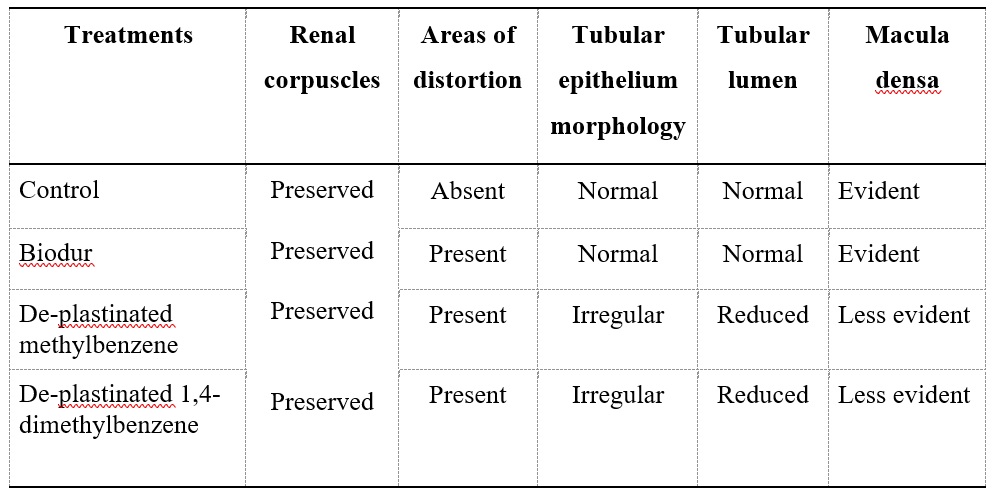 Table 1. Treatments and main histological characteristics of the kidneys |
Table 1 lists the main changes observed in the kidney histoarchitecture in all four treatments. In treatment 1, histological staining of renal tissue with H & E showed normal kidney structure, with renal corpuscles with a knot of capillaries (the glomerulus), surrounded by Bowman’s capsule (Fig 1). The tubules were observed with oval luminal morphology, and the epithelial cells showed eosinophilic cytoplasm, and central rounded nuclei (Fig. 2). The macula densa (MD) was observed in close proximity to the vascular pole of the renal corpuscle.
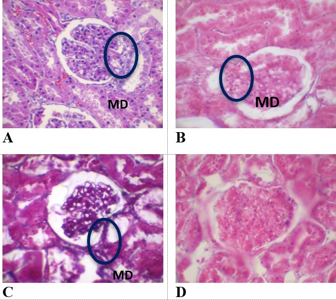 Figure 1: Photomicrographs of the cortical region of pig kidneys showing preserved renal corpuscles in all treatments and macula densa (MD). A, Control; B, Plastinated; C, De-plastinated with methylbenzene; D, De-plastinated with 1,4-dimethylbenzene. H&E staining. 400X |
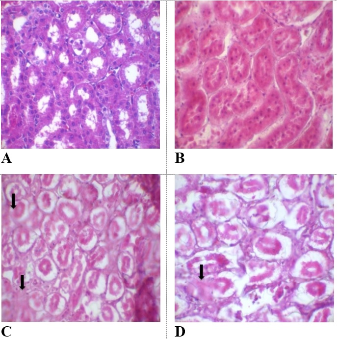 Figure 2: Figure 2. Photomicrographs of the cortical region of pig kidney showing tubular cells presenting irregularities in B and D that sometimes have loose epithelium in tubular lumen. A, Control; B, Plastinated; C, De-plastinated with methylbenzene; D, De-plastinated with 1,4-dimethylbenzene. H&E staining. 400X. Arrows indicate areas of distortion. |
Treatments 2 and 3 revealed renal corpuscles with preserved Bowman’s space, with some distortion areas presenting silicone. Epithelial cells from the parietal layer of Bowman’s capsule were evident (Fig. 1). In treatment 2 the MD was observed in close proximity to the vascular pole, with tubular architecture preserved. In treatment 3, some renal tubules revealed reduced lumen, and irregular morphology of the tubular epithelium. In treatments 3 and 4, histological changes, such as some cells of the renal tubules with morphological alterations, reduction of the tubular lumen, or capsular contour with distortion areas were also seen (Fig. 2).
The three experimental treatments showed preservation of the renal capsule, low affinity for hematoxylin staining, and small fragmented areas of tissue, just beneath the renal capsule.
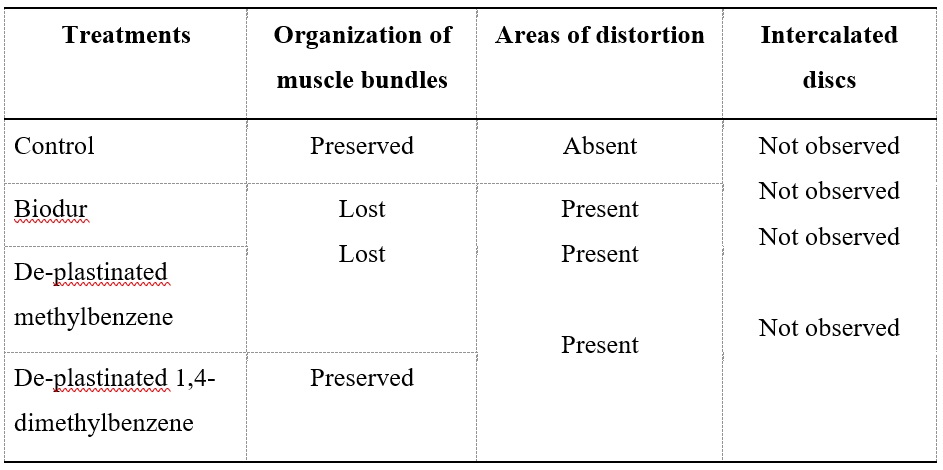 Table 2. Treatments and main histological characteristics of the heart |
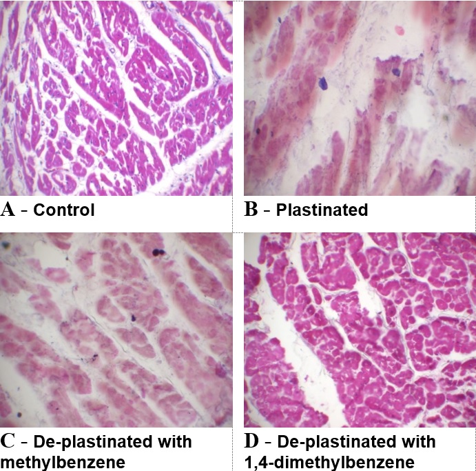 Figure 3. Photomicrographs of left ventricular myocardium of pig, showing low affinity for H&E staining with plastinated fragments in B, and fragments after de-plastination with methylbenzene in C. A and D show standard staining with H&E. 200X. |
Table 2 lists the main changes observed in the histoarchitecture of the heart in all treatments. Histological examination of the heart sections showed clear differences between treatment 1, the control, and the experimental treatments. Analysis of the myocardium in treatment 1 showed cells with a single, centrally placed nucleus, well stained by hematoxylin, surrounded by myofibrils, with blood vessels in the connective tissue. In treatments 2 and 4, the central nuclei were evident in myocardial cells. Muscle bundles showed preserved architecture. Treatments 2 and 3 revealed partial loss of organization of the muscle bundles, and low affinity for H & E staining. In treatments 2, 3 and 4, the blood vessels were not evident in the interstitium. In all of the treatments, transverse striations and intercalated discs were seen (Fig. 3). In treatments 2, 3 and 4, areas of distortion were seen in the slides. Slides from treatments 1 and 4 had similar staining with H & E, with both showing greater affinity for eosin.
Aorta
Table 3 lists the main changes observed in the histoarchitecture of the aorta in all treatments. The histological structure of transverse sections in treatment 1 demonstrates the three layers that constitute the wall of the aorta. The tunica intima showed endothelium well stained by H & E (Fig. 4). The internal elastic membrane, however, was less obvious. The tunica media showed several layers of elastic membranes and smooth muscle cells. The elastic material was well evidenced by Verhoeff's stain (Fig. 5). The tunica adventitia appeared thinner than the tunica media, and the presence of vasa vasorum in connective tissue was observed at random intervals.
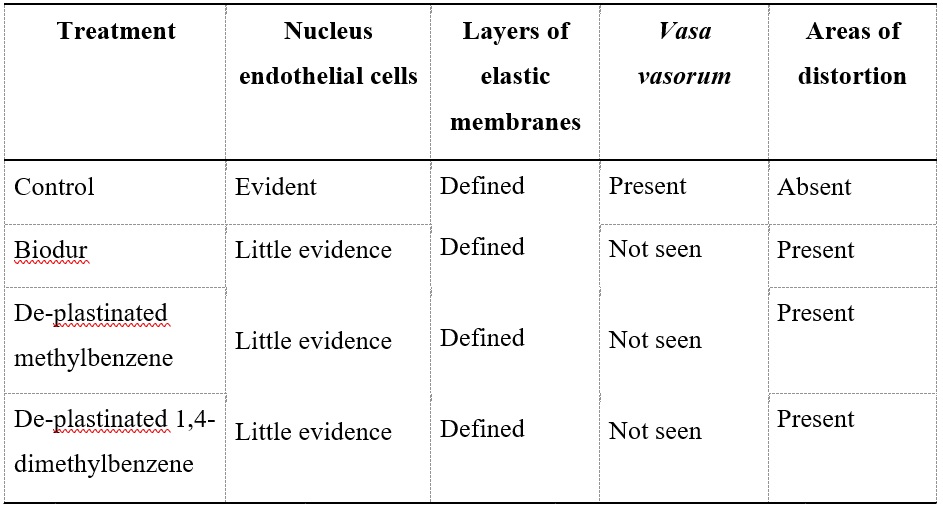 Table 3. Treatments and main histological characteristics of the aorta |
||
 Figure 4: Photomicrographs of tunica adventitia of the pig aorta showing vasa vasorum (vv) in A. |
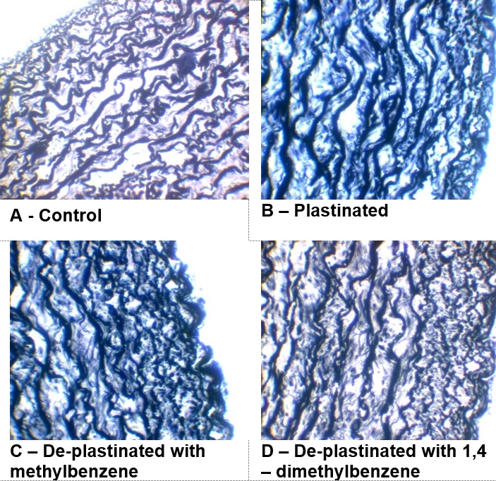 Figure 5: Photomicrographs of tunica intima of pig aorta with Verhoeff staining. 400X. |
|
In Treatments 2, 3 and 4, the tunica intima was observed, however, the endothelial nuclei were not distinct; in the tunica adventitia it was not possible to visualize the vasa vasorum (Fig. 5). In Treatments 2 and 4, areas of distortion in the endothelium were observed. In Treatment 4, spaces between the elastic membranes in the tunica media were observed. In all treatments, the elastic laminae were preserved in the tunica media, with few spaces between them. This was observed in both the H & E and Verhoeff stains.
In this study, we analyzed by light microscopy how the histoarchitecture of different organs (kidney, heart and aorta) was preserved after plastination. As in previous published studies, we used a control (Treatment 1) to compare and validate the different methods used to process the plastinated tissue samples (Treatments 2, 3 and 4) (Dellmann and Brown, 1982; Young and Heath, 2000, Gartner and Hiatt, 2003, Junqueira and Carneiro, 2008, Ross and Pawlina, 2008).
Manjunatha et al. (2014) compared histological sections of pig organs (liver, spleen and kidneys) embedded in paraffin wax, with histological sections of uncured silicone-plastinated tissues. Both were stained with H & E. Using light microscopy, they found that the tissue structure was maintained, without shrinkage, in the plastinated specimens. López-Albors et al. (2004) plastinated tissue fragments with and without the curing process, and used de-plastination with sodium methoxide, according to the protocol of Walker et al. (1988). They found that the curing process influenced tissue preservation. However, the results we report here from cured samples, showed no disruption of the tissue architecture.
Walker et al. (1988), describe de-plastination with sodium methoxide, which achieved good quality results. Others authors report the use of methylbenzene, methylene, and bichloride acetone for the same process (Ripani et al., 1988). However, our results in plastinated kidney samples without de-plastination (Treatment 2) showed a well-preserved histological structure of tubules and renal corpuscles. Moreover, the cells of the macula densa had a similar appearance to those from the control treatment (Treatment 1). Surprisingly, the results from de-plastinated kidney samples had renal tubules with reduced lumen and thin macular densa cells (Treatment 3), and the capsular space was increased in some renal corpuscles, probably due to shrinkage of the glomerular capillaries (Treatment 4). These findings for Treatments 3 and 4 agree with the results of Ripani et al. (1996), who reported lesions in the renal tubule epithelium and Bowman's capsule, after de-plastination with methylbenzene. In this study, Treatments 2, 3 and 4 showed some areas of distortion in the heart specimens, probably due to the rigidity of the material (we used fragments of the left ventricle), and also due to the hardening effect of curing, which may have caused difficulty in obtaining regular histological sections, resulting in differences in the thickness of the sections, and areas of distortion, in all three treatments.
Microscopic examination of the heart in Treatment 3 showed areas that were not well preserved, with loss of organization of muscle bundles. This problem probably occurred due to the rigidity of the cured material. It was not possible visualize blood capillaries and transverse striations in Treatments 2, 3, and 4. In addition, the slides had several areas of distortion, and the sections were not uniform. It is likely that this occurred due to the resistance of the tissue during sectioning. Our findings corroborate the results presented by Patil et al. (2016), in which clear striations in Biodur-infiltrated cardiac tissue could not be seen after de-plastination with methylbenzene.
In our findings, the tunica intima and endothelium of the aorta were mostly preserved in Treatments 2, 3 and 4. The nuclei showed poor hematoxylin affinity. The tunica media, in Treatments 2, 3 and 4, was composed of smooth muscle cells interspersed with the large amount of elastic fibers. The outer elastic limiting membrane could not be identified and, in Treatment 4, gaps between the elastic membranes were observed. This could have been due to resistance to paraffin infiltration. In the tunica adventitia of all three treatments, the non-visualization of the vasa vasorum may have been due to the loss of the surrounding connective tissue during the plastination process. The areas of refractivity in Treatment 2 and 4 probably occurred due the presence of silicone in the tissue.
According to Ravi and Bhat (2011), to achieve good results with standard staining, slides of de-plastinated samples need a slightly extended immersion time, compared to unplastinated samples. In our results, we observed a similar coloration in slides of both the de-plastinated and plastinated treatments, when stained with H & E. Both showed less evident hematoxylin staining compared to the control. This may be due to the plastination process changing the electronegativity of the nucleus, and the basophilic pattern. However, for the Verhoeff staining, the same pattern for the elastic membranes was observed in all treatments, indicating that the plastination process did not alter the uptake of the stain.
In conclusion, our experiments showed that histological slides can be made directly from Biodur® silicone plastinated specimens, and embedded directly in paraffin. This protocol presented satisfactory results, similar to those found in specimens de-plastinated with 1,4-dimethylbenzene and methylbenzene, which resulted in incomplete removal of the Biodur® silicone from the samples.
Dellmann H, Brown EM. 1982: Histologia Veterinária. Rio de Janeiro: Guanabara Koogan, 397 p.
Gartner LP, Hiatt, JL. 2003: Tratado de Histologia em Cores. 2a ed. Rio de Janeiro: Guanabara Koogan, 458 p.
Hambeli AT, Lombardi M, Prochownik M. 2010: Técnicas de conservación de piezas cadavéricas. Tercera Epoca: Rev Científica de la Facultad de Ciencias Médicas 2: 1-2.
IARC - International Agency for Research on Cancer - Summaries & Evaluations, Formaldehyde, 1995. In : http://www.iarc.fr/en/media-centre/pr/2004/pr153.html.
INCA - INSTITUTO NACIONAL DO CÂNCER - Formol ou Formaldeído 2005. In: http://www1.inca.gov.br/conteudo_view.asp?ID=795.
Junqueira, LC; Carneiro, J. 2008: Histologia básica. 11a ed. Rio de Janeiro: Guanabara Koogan, 514p.
Latorre RM, García-Sanz MP, Moreno M, Hernández F, Gil F, López O, Ayala MD, Ramírez G, Vázquez JM, Arencibia A, Henry RW. 2007: How useful is plastination in learning anatomy? J Vet Med Educ 34: 172-176.
https://doi.org/10.3138/jvme.34.2.172
López-Albors O, Gil F, Orenes M, Ayala MªD, Abellán H, Henry R, Latorre R. 2004: Curing influences the tissue preservation of silicone plastinated organs. 12th Int Conf Plast, Murcia, Spain, 2004. Abstract in J Int Soc Plastination 19: 49-50.
Manjunatha K, Prasad RV, Jamuna KV, Placid ED, Suguna R, Ramkrishna V. 2014: Comparison of histological architecture of paraffin embedded and indigenously plastinated tissues. Indian J Vet Anat 26: 132-133.
Patil SK, Jamuna, KV, Badami, S, Ramkrishna, V. 2016: Comparison of histology of cardiac Muscle using different infiltrating media. Indian J Nat Sci 6: 10558-10563.
Ravi SB, Bhat VM. 2011: Plastination: a novel, innovative teaching adjunct in oral pathology. J Oral Maxillofac Pathol 15: 133-137.
https://doi.org/10.4103/0973-029X.84475
Ripani M, Boccia L, Cervone P Macciucca DV. 1996: Light microscopy of plastinated tissue. Can plastinated organs be considered viable for structural observation? J Int Soc Plastination 11: 28-30.
https://doi.org/10.56507/PMEQ9992
Ross MH, Pawlina W. 2008: Histologia: Texto e Atlas em correlação com biologia celular e molecular. 5a ed. Rio de Janeiro: Guanabara Koogan. 908 p.
von Hagens G, Tiedemann K, Kriz W. 1987: The current potential of plastination. Anat Embryol (Berl) 175: 411-421.
https://doi.org/10.1007/BF00309677
Walker AN, Jackson RL, Powell, S. 1988: Technical communication: routine microscopy of deplastinated tissue. J Int Soc Plastination 2: 40-42.
https://doi.org/10.56507/ODUA6186
Young B, Heath J. 2000: Histologia Funcional: texto e atlas em cores. 4a ed. Rio de Janeiro: Guanabara Koogan. 415 p.