Department Anatomy & Comparative Pathology, Veterinary Faculty, University of Murcia, Murcia, Spain
In this study, plastinated specimens were used for creating a learning model to assist students’ understanding of a common cardiac pathology of dogs. Dilated cardiomyopathy is a myocardial disease, causing a progressive dilatation of all four cardiac chambers, with concomitant changes in systolic function. Two plastinated sections of canine hearts were used, one from a healthy dog, and the other belonging to a dog affected by dilated cardiomyopathy. Plastinated sections were photographed and digitized, and then a static image animation program was used to simulate the heartbeat, in a reliable manner. The digital materials were used to create a video tutorial, in which the structures of the heart were simultaneously highlighted in both anatomical and ultrasound images. The video tutorial was accessed via QR code, which was given to a set of students of the Veterinary Degree Program with previous basic training in ultrasound diagnosis. A satisfaction survey was used to monitor each student's perception of the importance of clinical anatomy and the usage and learning experience with the video tutorial. The results reported that most students considered anatomy as a fundamental subject for their professional career, although their knowledge about it at the end of the degree was not too extensive. The overall score of the video tutorial was very high, as it apparently facilitated the understanding of ultrasound imaging related to dilated cardiomyopathy. It is concluded that the combined use of plastinated material with e-learning improved the subjective perception of learning and understanding of the dilated cardiomyopathy by the students.
dilated cardiomyopathy; dog; education; heart; plastination; ultrasound
Octavio López Albors, Department Anatomy & Comparative Pathology, Veterinary Faculty, University of Murcia, 30100, Murcia, Spain email: albors@um.es

![]()


Interpretation of ultrasonic images is an advanced skill which involves repetitive exposure to obtain proficiency (Wichtel et al., 2021). In pathology where ultrasound diagnosis is required, in addition to understanding the normal anatomy of the ultrasonic image, pre- and post-graduate students should become familiar with the altered anatomy characteristic of each specific pathology. This is well represented in heart diseases where ultrasound is necessary for an accurate diagnosis (Vitarelli et. al., 2003). In fact, ultrasound exploration is the most sensitive test and the one that provides the definitive diagnosis of many heart pathologies, such as dilated cardiomyopathy (Ettinger, et al, 2017).
Dilated cardiomyopathy of the dog is a myocardial disease, characterized by a progressive and irreversible dilatation of the four cardiac chambers, with severe alteration of the systolic function (Smith et al., 2022). As for the symptoms related to this disease, they are quite unspecific. Dogs may be asymptomatic or present with a cough, dyspnea, ascites, weight loss, weak and irregular pulse, exercise intolerance, syncope, or even sudden death (Grady and Sullivan, 2004). Therefore, achieving an accurate early diagnosis is vital, as it can help to improve the patient's future quality of life. A systematic ultrasound examination is the recommended test to confirm or dismiss this pathology. In diagnosing dilated cardiomyopathy, two strategic ultrasound sections from the right parasternal approach are always required. One is the long axis view of the left ventricle outflow tract, and the second one the short axis view, transaortic.
Plastinated specimens have been widely used as innovative educational resources in anatomy and other clinical disciplines (Latorre et al., 2007, 2016; Chytas et al., 2019; Sora et al., 2019). Plastinated specimens are dry, non-toxic, durable, and suitable for digital processing. These properties facilitate the possibility of creating learning aids such as video tutorials, 3D rendered anatomical objects, images for augmented and extended reality, etc. (Garas et al. 2018; Mikami et al., 2022). Also, the use of videos is an effective strategy to promote active learning in anatomy courses (Senos et al. 2023) and embedding digitized and animated (moving) images of anatomical and pathological) plastinated specimens into video tutorials may facilitate understanding the normal and altered anatomy.
In this work we aimed to define a novel use of plastination to enhance anatomical learning within a clinical context. Anatomical and pathological plastinated specimens were used to create animated digital images embedded within a video tutorial, to train pre-graduate veterinary students in the diagnosis of dilated cardiomyopathy of the dog. The students’ perception of the pedagogical value of this combined plastinated-digital resource was evaluated by means of a randomized survey.
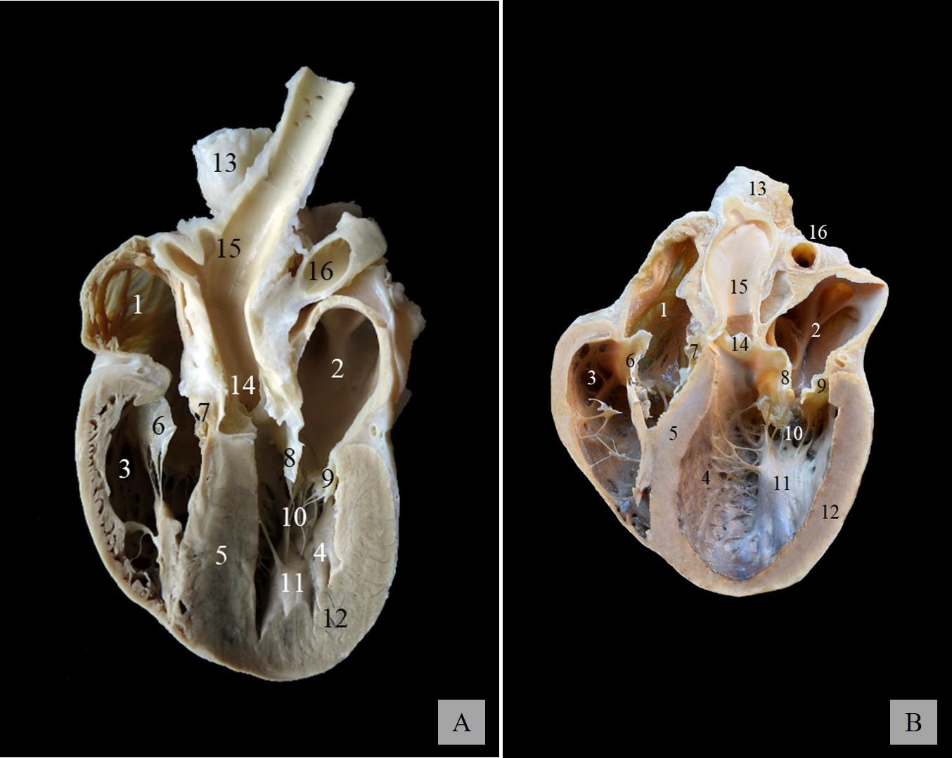
Figure 1. Plastinated dog hearts transected along the left ventricular out flow tract, to mimic right parasternal echocardiorgraphic long axis view. A; Healthy heart, B; Heart with dilated cardiomyopathy. Approximate five-chamber section: 1, Right atrium; 2, Left atrium; 3, Right ventricle; 4, Left ventricle; 5, Interventricular septum; 6, Parietal cusp of the right atrioventricular valve (tricuspid valve); 7, Angular cusp of the right atrioventricular valve (tricuspid valve). 8, Septal cusp of the left atrioventricular valve (mitral valve); 9, Parietal cusp of the left atrioventricular valve (mitral valve) 10, Tendinous cords; 11, Papillary muscles; 12, Left ventricular free wall; 13, Cranial vena cava; 14, Aortic valve; 15, Aorta; 16, Right pulmonary artery
For this study, two canine hearts were used. One heart was from a dog unaffected by heart disorders, and the other from a dog that suffered dilated cardiomyopathy. Both cadavers were donated to the Faculty of Veterinary Medicine (University of Murcia) after scheduled euthanasia for reasons other than the purpose of this study. Cadavers were donated according to the requirements of the Body Donation Program of the Faculty (as approved on 28th May 2019, modified on the 24th October 2019) .
The hearts were fixed by dilation of the chambers with 10% formalin infused with a peristaltic pump (Watson Marlow®) and subsequent immersion in 10% formalin. After fixation (3 weeks), the hearts were immersed and flushed in running tap water for at least 24 h. Silicone plastination was carried out according to the protocol of cold S-10 (Biodur®) technique (Henry et al., 2019.). The average duration of the dehydration, impregnation, and curing steps was 4, 3, and 6 weeks, respectively. Finally, the plastinated hearts were sectioned with a sharp knife so as to obtain representative slices, corresponding to the long-axis view of the left ventricle outflow tract, as defined by Thomas (1993) (Fig. 1).
The plastinated slices were photographed with a professional camera (Canon PowerShot G1X®). Selected pictures were then edited to obtain optimal images suitable for animation with the program Adobe After Effects 2022®. This software allows the collocation of reference pins over the images to deform the shape up to defined distances, hence simulating the physiological changes in morphology produced by the movement of the organ. Different layers are created to define which parts of the image should be deformed (moved), which ones are kept immobile, and those which remain in the most superficial layer when the movement is created. (Fig. 2).
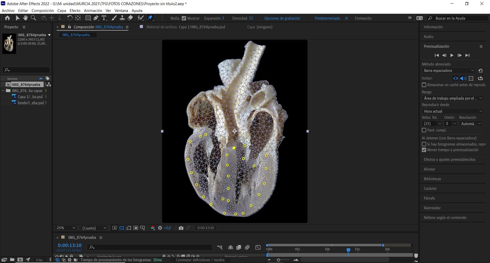
Figure 2. Using the puppet position tool to animate the image of a healthy heart. Reference points are placed to simulate the movement by creating deformations
To make the animation more realistic, the duration of each phase of the cardiac cycle and heartbeat sound were included with the Sony Vegas Pro 18.0®. Systole was defined for a duration of ⅜ of the cardiac cycle, and diastole for ⅝ of it (Salazar, 2012). These features were defined similarly in the two hearts, with the exception of the sound added to the heart with dilated cardiomyopathy, whose heartbeat corresponded to a mitral murmur, recorded and stored in a smart device with a 3M™ Littmann® CORE Digital Stethoscope.
The video tutorial was created with the Camtasia 2018R® software according to the following sequence. First, a detailed review of the heart anatomy and cardiac cycle. Then, the concept of dilated cardiomyopathy and a description of the altered anatomy in the pathological heart. Finally, a coupled identification of structures in the animated image of the pathological heart and the corresponding ultrasound video from a case of dilated cardiomyopathy (courtesy of Dr. J. Talavera, from the Hospital Veterinario Universidad de Murcia). Two versions of the video (Spanish and English) were produced and uploaded into the University of Murcia repository of multimedia objects www.tv.um.es. Access to the videos is gained by QR codes (Fig. 3) or at the following web sites, Spanish https://tv.um.es/video?id=146365&cod=a1, and English, https://tv.um.es/video?id=146596).

Figure 3. QR codes for the Spanish version (A) and English version (B)
For the evaluation of the learning product, students of the last year of the Veterinary degree were invited to participate in the whole learning experience by means of smartphones and tablets. This involved an initial phase of interaction with the plastinated hearts (handling object) followed by watching the video tutorial (digital object). A total of 30 students participated in the study.
Just after having used the learning product, students were given a link (https://encuestas.um.es/encuestas/MzczNDg.c) to a short anonymous, pre-coded on-line survey created with the “encuestas” facility integrated within the University of Murcia learning environment (Fig. 4). Data assessment was based on simple random sampling, and semi-quantitative evaluation based on a 5-level Likert scale: Strong Disagreement (1), Disagreement (2), Neutral (3), Agreement (4) and Strong Agreement (5). Results were automatically generated by the pooling system, providing values for the mean, and relative percentages. To validate the survey the Cronbach statistic was calculated with the SPSS® package (IBM).
Figure 4. Survey for students used in this study
The external and internal anatomical details of the hearts were clearly depicted in the two plastinated specimens (Fig. 1). In the normal physiological heart, dilation of the atria and ventricles was optimum, with no signs of over-dilation or deformities in the valves, papillary muscles, and tendinous cords. In the pathological specimen, the characteristic morphological features of this disease were well depicted. The left ventricular lumen was wider than normal (hyper-dilated), the interventricular septum and wall of the left ventricle were thinner, and the left atrioventricular foramen and the mitral valve wider and incongruent. Student comments on the plastinated hearts were very positive, especially the easy handling (compared to formalin fixed specimens), the resistance to manipulation and the high representation of anatomical details.
The animated images of the plastinated hearts showed a clear representation of the heartbeat in both healthy and pathological conditions. In the healthy heart, movement of the atria and ventricles was synchronous to the opening and closing of the valves, hence mimicking a physiological rate of heart beating. In the pathological heart, the animation displayed reduced mobility of the ventricles, and incomplete closure of the atrioventricular valves, so as to illustrate the potential mitral (left) and tricuspid (right) reflux.
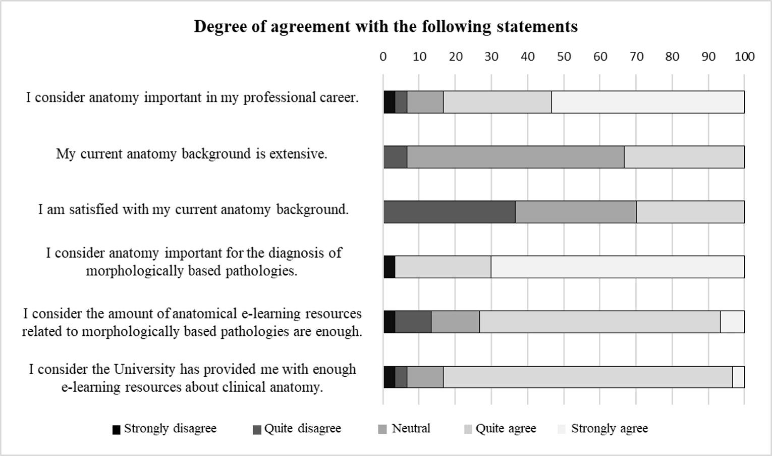
Figure 5. Results of the survey with regard to the anatomical knowledge of participants and their perception about the resources available for learning pathology concomitant with altered anatomy
Survey results showed that the average age of the respondents was 23.7 years, and the sex mostly female (83.3%). With regards to the anatomical knowledge of participants and their perception about the resources available for learning pathology concomitant with altered anatomy (Fig. 5), the majority of students considered that anatomical knowledge was relevant for their professional future (around 80%), 60% did not regard their factual anatomical knowledge as extensive, and only 30% were satisfied with it (Cronbach a=0.59 with confidence interval 0.33-0.78). On the other hand, 96% of students agreed on the importance of mastering anatomy for the clinical diagnosis of pathologies concomitant with altered anatomy, 73% considered there are enough resources available for learning this type of pathology, and 83% that the University of Murcia provided them with such resources.
When focused on the evaluation of the video tutorial, all students positively considered it helpful for learning this pathology (37% agreed and 63% strongly agreed, Cronbach a=0.72, confidence interval 0.52-0.85). Hence, on a 10-point scale they rated it at 9.5.
In this paper plastinated hearts from healthy and unhealthy dogs were used to enhance anatomical learning within a clinical context. The combination of plastinated material with an anatomical digital resource (video tutorial) was unanimously perceived by a cohort of students from the final year of the Veterinary Degree as a helpful or very helpful asset for learning the dilated cardiomyopathy of the dog. In fact, the digital resource was scored with 9.5 points out of 10. Although the number of students included in this study was only approximately 35% of the total number of the cohort, the sample can be considered as a valid representation of the total group. Firstly, the sex distribution of the participants in the survey (84% females vs 16% males) mimicked the current ratio of the group of students in the 5th Year. Also, it was very close to the figures of the whole Veterinary Degree in our Faculty (76% of females on average, data supplied by Dean’s office), and similar to the figures normally found in many European and North American Veterinary Schools (Lofstedt, 2003). Moreover, when students were asked about the working area which was of their most interest (professional profile), 60% of them chose small animal clinics, 15% large animal clinics, 12% food hygiene, 9% animal husbandry, and 4% others. These figures were also in line with the usual distribution of professional profiles among undergraduate students in our institution. Hence, with regards to the relevance of the results obtained, it might be concluded that the very positive perception of the students towards the learning asset evaluated here was not biased by different sex distribution from the original population of students, or the expected professional profile of the students.
Many previous studies have demonstrated the usefulness of video tutorials and other digital objects for learning anatomy (Morton & Colbert-Getz, 2016; Oberoi et al, 2018). Compared to live lectures, video tutorials have a great advantage, as they make it easier for students to adjust their learning process (Foertsch et al., 2002). Video tutorials offer the possibility of stopping the video when desired and being able to repeat the parts of interest as many times as needed, which makes taking notes, learning technical terms, understanding functional concepts, etc. much more flexible. On the other hand, plastinated specimens have been demonstrated as valid materials for effective learning of anatomy (Latorre et al., 2007). In the present work, both sources of knowledge have been merged (plastinated specimens plus video tutorial) into a unique innovative learning asset that takes advantage from both resources, and has a very positive effect on the student perception of learning.
Another interesting characteristic of our study was an indirect evaluation of the students’ perception on the so-called anatomical retention. Loss of knowledge during the medical curriculum is a well-known phenomenon and is a cause for concern in students and educators, with retention levels dropping down to 15-20% by the end of the degree (Doomernick et al., 2016). Such levels might be aligned with the so-called permastore concept defined by Bahrick (1984). Permastore is the state where, after a significant period of time, loss of knowledge does not decrease further and enters a state of permanent store. In our study, all students were in the last year of their degree, which in terms of curriculum was seven semesters beyond their last anatomy course. Thus, although 83% of students agreed that anatomy was an important discipline for their future, 60% of them did not consider their factual anatomical knowledge to be extensive. As mentioned above, this was surely showing the aforementioned self-evident loss of knowledge experienced by students over time. Interestingly, this phenomenon was found to be greater in traditional, non-clinically integrated curricula (such as the one of the Veterinary Faculty of University of Murcia), than in problem-oriented horizontal and vertical integrated medical curricula (Doomernick et al. 2016). It is also worthy of mention that, despite the self-perception of loss of anatomical knowledge by the students, such situation was not considered as serious, since a mean value of 2.9 out of 5 was obtained on a Likert scale regarding the degree of satisfaction with the anatomical knowledge at the moment the survey was done.
In conclusion, this paper describes a novel use of plastinated specimens in the veterinary clinic. The combined use of plastinated heart slices with an explanatory video tutorial improved the students’ perception of understanding a relevant cardiomyopathy in the dog, hence giving further evidence on the potential of plastinates to define innovative educational experiences when combined with digital technologies. The use of specialized software, such as Adobe After Effects@ was essential to create animated images representing the changes in morphology due to heartbeat. Limitations of this study were the lack of student pre-test and post-test, which would help to better assess the educational effectiveness of the learning product, and the small number of questionnaires, which likely contributed to a Cronbach's alpha measure of less than 0.7 (0.78 upper confidence limit) with regard to anatomical knowledge, and of 0.72 (0.85 upper confidence limit) with regard to the evaluation of the tutorial.
Acknowledgements.
The authors are grateful to Dr Jesús Talavera and Maria Josefa Sanz Alarcón for their advice in obtaining the ultrasound images.
Bahrick HP. 1984: Semantic memory content in permastore: fifty years of memory for Spanish learned in school. J Exp Psychol: Gen 113(1): 1-29.
https://doi.org/10.1037/0096-3445.113.1.1
Chytas D, Piagkou M, Johnson EO et al. 2019: Outcomes of the use of plastination in anatomy education: current evidence. Surg Radiol Anat 41(10): 1181-1186.
https://doi.org/10.1007/s00276-019-02270-3
Doomernik D E, van Goor H, Kooloos J G, Ten Broek RP. 2017: Longitudinal retention of anatomical knowledge in second‐year medical students. Anat Sci Ed 10(3): 242-248.
https://doi.org/10.1002/ase.1656
Ettinger S, Feldman E, Cote E, 2017: Textbook of Veterinary Internal Medicine (8th ed.). St. Louis, Missouri, US: Elsevier, p 1125-1138, 3071-3074.
Foertsch J, Moses G, Strikwerda J, Litzkow M. 2002: Reversing the lecture/homework paradigm using eTEACH® web‐based streaming video software. J Eng Educ 91(3): 267-274.
https://doi.org/10.1002/j.2168-9830.2002.tb00703.x
Garas M, Vaccarezza M, Newland G, McVay-Doornbusch K, Hasani J. 2018: 3D-Printed specimens as a valuable tool in anatomy education: A pilot study. Anat Anz 219: 57-64
https://doi.org/10.1016/j.aanat.2018.05.006
Henry RW, von Hagens G, Seamans G. 2019: Cold temperature/Biodur®/S10/von Hagens'-Silicone plastination technique. Anat Histol Embryol 48(6): 532-538.
https://doi.org/10.1111/ahe.12472
Latorre RM, García-Sanz MP, Moreno M, et al. 2007: How useful is plastination in learning anatomy? J Vet Med Ed 34(2): 172-176.
https://doi.org/10.3138/jvme.34.2.172
Latorre R, Bainbridge D, Tavernor A, López Albors O. 2016: Plastination in anatomy learning: an experience at Cambridge University. J Vet Med Ed 43(3): 226-234.
https://doi.org/10.3138/jvme.0715-113R1
Lofstedt, J. 2003: Gender and veterinary medicine. Can Vet J 44: 533-535.
Mikami BS, Hynd TE, Lee UY, et al. 2022: Extended reality visualization of medical museum specimens: Online presentation of conjoined twins curated by Dr. Jacob Henle between 1844-1852. Trans Res Anat 27: 100171.
https://doi.org/10.1016/j.tria.2022.100171
Morton DA, Colbert‐Getz JM. 2017: Measuring the impact of the flipped anatomy classroom: The importance of categorizing an assessment by Bloom's taxonomy. Anat Sci Educ 10(2): 170-175.
https://doi.org/10.1002/ase.1635
O'Grady MR, O'Sullivan ML. 2004: Dilated cardiomyopathy: an update. Vet Clin Small An Pract 34(5): 1187-1207.
https://doi.org/10.1016/j.cvsm.2004.05.009
Oberoi V, Hosseini F, Doroudi M, Vo L. 2018: Anatomy in a new curriculum: Using digital media to facilitate the learning of anatomy in the medical curriculum. FASEB J 32: 635-34.
https://doi.org/10.1096/fasebj.2018.32.1_supplement.635.34
Salazar A, Alvarado C, Lozano F. 2012: System of heart and lung sounds separation for store-and-forward telemedicine applications. Revista Facultad de Ingeniería Universidad de Antioquia 64: 175-181. ISSN 0120-6230.
Senos R, Leite CAR, dos Santos Tolezano F, Roberto‐Rodrigues M, Pérez W. 2023: Using videos in active learning: An experience in veterinary anatomy. Anat Histol Embryol 52(1): 50-54.
https://doi.org/10.1111/ahe.12839
Smith F, Tilley L, Oyama M, Sleeper M. 2022: Manual de Cardiología Canina y Felina [Manual of Canine and Feline Cardiology] Multimédica Ediciones Veterinarias.
Sora MC, Latorre R, Baptista C, López-Albors O. 2019: Plastination. A scientific method for teaching and research. Anat Histol Embryol 48(6): 526-531.
https://doi.org/10.1111/ahe.12493
Thomas WP, Gaber CE, Jacobs GJ et al. 1993: Recommendations for standards in transthoracic two‐dimensional echocardiography in the dog and cat. J Vet Int Med 7(4): 247-252.
https://doi.org/10.1111/j.1939-1676.1993.tb01015.x
Vitarelli A, Tiukinhoy S, Di Luzio, Zampino M, Gheorghiade M. 2003: The role of echocardiography in the diagnosis and management of heart failure. Heart Fail Rev 8: 181-189.
https://doi.org/10.1023/A:1023001104207
Wichtel J, Zur Linden A, Khosa D, Singh A, Sears, W, Phillips J. 2022: Validation of a novel ultrasound simulation model for teaching foundation-level ultrasonography skills to veterinary students. J Vet Med Ed 49(4): 473-483.
https://doi.org/10.3138/jvme-2020-0123