Department of Anatomy, Dalian Medical University, Dalian, 11602 7, China
A cape dolphin cadaver was used to evaluate a new polyester sheet plastination technique in this experiment. The dolphin was divided into two regions, the head and the body. After freezing the tissue at -7°C, the head was cut into forty-three 3.0mm thick sagittal slices with a high-speed band saw. The body of the dolphin was cut into 348 transverse slices of the same thickness. All slices were fixed using 10% formaldehyde and bleached using 5% dioxogen. The slices were dehydrated in a cold acetone bath and degreased in room temperature acetone. The slices were impregnated with Hoffen polyester (P45, China) and cast between two glass plates. Tissue slices were cured in a heated water bath instead of UV-light. The finished polyester slices cured properly and exhibited detailed anatomical information.
plastination; polyester; sheet; curing; dolphin; Hoffen
Telephone: 86-411-8472-0621; Fax: 86-411-8475-4558; E-mail: Suihj@hotmail.com

![]()


The rapid development of imaging technology used for medical examination and diagnosis such as magnetic resonance imaging, computed tomography, and ultrasonography have required an in depth study of sectional anatomy. Sheet plastination of specimens, a technology for tissue preservation, provides material for the study of sectional anatomical structure (von Hagens et al., 1987). This technology has been improved by researchers to produce an improved final product (Weiglein et al., 1993; deBoer-van et al., 1993; Weiglein, 1996; Cook, 1997). In our experiment, a new polyester polymer (Hoffen polyester P45, Dalian Hoffen Bio-technique Co. Ltd, room 301, number 32, Lixian Street, Hi-Tech Zone, Dalian, China) and a new curing method of sheet plastination were applied for the preservation of tissue slices for a cape dolphin (Delphinus capensis). The purpose of the present study was to find an easier and more cost effective method for sheet plastination.
Slicing
A female cape dolphin, approximately two meters in length, was found dead on a beach and was collected by a natural history museum (Fig. 1). The dolphin was frozen at -70°C for two weeks. The dolphin was cut into two sections with a hand saw at a point 55cm from the tip of the beak. In order to facilitate the cutting of the trunk of the specimen along predetermined lines, the specimen was placed in a wooden box, positioned appropriately and embedded in polyurethane foam. The head of the dolphin was cut into forty-three 3.0mm thick sagittal slices with a high-speed band saw. The tissue lost between adjacent slices due to the cutting process was approximately l.Omm in thickness. The trunk was cut into 348 transverse slices which were also 3.0mm in thickness (Fig. 2). The slices were placed in anatomic order on polyethylene grids with a piece of wire screen between the slice and the grid. The saw dust was removed with a small stream of running water. The grids with screen and slice were then placed in small stacks and tied with twine to hold each stack as a unit. All units were labeled and placed into square polyethylene containers for fixation and bleaching.
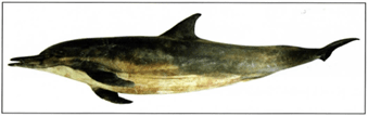 Figure 1. Cape dolphin used for sheet plastination. |
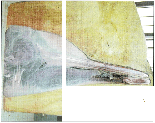 Figure 2. Sagittal section of dolphin head embedded in polyurethane foam. |
Fixation and bleaching
The slices were fixed in 10% formaldehyde for two weeks at room temperature. The polyurethane was easily removed from the tissue slices following submersion in the formaldehyde solution. After fixation, the tissue slices were rinsed in cold running water overnight to remove excess formaldehyde. Slices were then immersed in 5% dioxogen overnight to improve tissue color and transparency.
Dehydration
After bleaching, the slices were dehydrated using the freeze substitution method (von Hagens, 1985). The slices were precooled to 5°C to avoid ice crystal formation and to minimize shrinkage upon placement into cold acetone. Tissue slices were placed in the first bath of 100% acetone at -2°C they remained for seven days. Slices were then transferred into a second bath of 100% acetone at -1°C for ten days. Finally, they were put into 100% acetone for degreasing at room temperature for one week. Following final degreasing of the tissue, the slices were finally submerged in fresh 100% acetone at room temperature where they were held for one week awaiting impregnation. The concentration of acetone was monitored with an acetonometer each day during dehydration and degreasing. Once the concentration of acetone remained unchanged for three consecutive days, the slices were moved to the next dehydrating solution.
Casting and forced impregnation
The casting chamber was prepared from two plates of 5.0mm tempered glass, flexible 4.0mm latex tubing, and several large fold back clamps. This has classically been referred to as a flat chamber (Weber and Henry, 1992; Weber and Henry, 1993) (Fig. 3). The slices were removed from the final acetone bath and placed into the chambers. The chambers were filled with P45 polyester resin mixture (Hoffen polyester, China) via a funnel. The components of Hoffen polyester P45 were mixed at 1000 ml of polyester P45 resin to lOg of P45a to 30ml of P45b to Sg of P45c. The P45a and P45c were used as plasticizer and the P45b is the hardener for sheet plastination. The resin reaction-mixture is mixed immediately prior to casting since it thickens over time. Refrigeration of the reaction-mixture will retard this thickening.
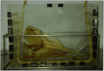 Figure 3. Chamber used for casting with tissue in place. |
Once the casting chambers were filled, large air bubbles were removed from the casting chambers manually using a piece of l.Omm stainless steel wire. The chambers were then placed upright into a vacuum chamber for impregnation at room temperature. The absolute pressure in the vacuum chamber was slowly decreased to 20.0mm Hg, 10.0mm Hg, 5.0mm Hg, and finally to O.Omm Hg according to the slow release of bubbles from the tissue slices. A pressure of O.Omm Hg was maintained until all bubbling ceased. The duration of impregnation was slightly greater than eight hours.
Curing
After the vacuum had been released, the casting chambers were rechecked for the presence of any air bubbles which would need to be removed prior to curing. The alignment of the tissue slices was also checked and repositioneded with the steel wire if necessary. Lastly, the latex tubing was closed across the top of the casting chamber and held in place by clamps.
The sheets were cured in a vertical position in a heated water bath at 40°C for 3 days (Fig. 4). A small circulatory pump was used to circulate the water within the bath to equilibrate the temperature of the water at all points within the chamber.
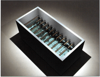 Figure 4. Representation of water bath used for curing. |
Cutting and sanding sheets
After curing, the sheets were removed from the bath and cooled to room temperature on a rack. The casting chambers were dismantled and the slices removed and covered with plastic wrap for protection. A band saw was used to cut and trim the polyester along the outside edges of the tissue slices at a distance of approximately l.Omm from the tissue. A wool sander was then used to remove the sharp edges of the slices. Following sanding, the plastic wrap was removed and the slices were placed into protective plastic bags to avoid scratching. The sheets were then ready for use or storage.
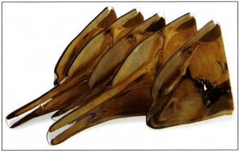 Figure 5. Sheet plastinated dolphin head |
The tissue sections produced using the new polyester polymer and new curing method yielded specimens which exhibit clear delineation between different tissue types (Fig. 5). The sections provide visually clear information about the morphology of dolphin. The polyester sheets and impregnated specimens cured completely resulting in clear, cured tissue slices within a protective polyester sheet.
The traditional P35 and P40 plastination techniques are unique means for tissue slice preservation. The P40 technique was designed as an improvement over the P35 technique as the former has a shorter processing time and uses less resin. Specimens produced using either P35 or P40 show excellent distinction between gray and white matter for brain specimens (von Hagens et al., 1987, 1994; Barnett, 2005). With the polyester technique, sections of specimens were more detailed, durable, and easier to handle than previous technique (Weber and Henry, 1992). Furthermore, these slices were also used for interpretation of medical diagnostic images such as MRI and CT scans and offered excellent reference material.
However, the curing process for the P35 and P40 techniques uses UV-light (von Hagens, 1994) which is an added expense. The process requires close monitoring of the exothermic reaction during curing and ventilators are used to keep temperatures from rising too high during curing.
In the present experiment, the sheet plastination of the dolphin was completed with the Hoffen polyester P45 technique. The slices were cured with a water bath instead of UV-light. With this new polyester for sheet plastination and new curing method, the vertical positioning of the casting chambers reduced the space required for curing specimens while the water bath provided equal dispersion of temperature during curing. The water bath keeps temperatures from rising too high as well as keeping temperatures uniform throughout the slices. The water bath also provides a more cost efficient and safer method for each specimen produced when the effects of UV-light production are taken into account.
The benefits provided by the Hoffen polyester P45 technique can effectively circumvent the disadvantages associated with the P35 and P45 plastination techniques. It is believed that the new Hoffen polyester P45 and the new curing method would facilitate sectional anatomy study and provided a good approach for sheet plastination.
The slices of this dolphin are on public display at Dalian Natural Science Museum, Dalian, China.
Barnett R, Burland G, Duxson M. 2005 : Plastination of coronal slices of brains from cadavers using the P35 technique . J Int Soc Plastination 20:16-19.
https://doi.org/10.56507/JUIT4201
Cook P. 1997: Sheet plastination as a clinically based teaching aid at the University of Auckland. Acta Anat 158:33-36.
https://doi.org/10.1159/000147907
de Boer-van Huizen, Cornelissen CJ, ten Donkelaar HJ. 1993: The P35 technique for sheet plastination of the human head . Abstract presented at The 6th International Conference on Plastination -Kingston, Ontario, Canada - July 1992. J Int Soc Plastination 7:36.
von Hagens G. 1985: Heidelberg Plastination Folder: Anatomisches Institut, Universitat Heidelberg, Germany.
von Hagens G, Tiedemann K, Kriz W. 1987: The current potential of plastination. Anat Embryo} 175:411-421.
https://doi.org/10.1007/BF00309677
von Hagens G. 1994: Plastination of brain slices according to the P40 procedure, a step-by-step description, December.
Weber W, Henry RW . 1992: Sheet plastination of the brain-P35 technique, filling method. J Int Soc Plastination 6:29-33 .
https://doi.org/10.56507/KWGD3312
Weber W, Henry RW . 1993: Sheet plastination of body slices-E l2 technique, filling method. J Int Soc Plastination 7: 16-22.
https://doi.org/10.56507/EZGX2343
Weiglein AH. 1993: Plastinated brain-specimens in the anatomical curriculum at Graz University. J Int Soc Plastination 7:3-7.
https://doi.org/10.56507/EHRX7749
Weiglein AH. 1996: Preparing and using SIO and P-35 brain slices. J Int Soc Plastination 10:22-25.
https://doi.org/10.56507/IXGV4189