Department of Anatomy1, Department of Radiology2, ERASMUS University Rotterdam, The NETHERLANDS
A new positioning technique i s presented, based on MR- and CT imaging with surface markers applied to the skin of the specimen. With this technique, the r e q u i r ed ana to mi c secti o n for cryosectioning can be defined accurately Plastinated slices, of 2 mm thickness, with the correlating MR-, CT-, and US images of double-oblique coronal imaging of the shoulder are shown. The method is equally applicable to all parts of the body and makes comparison of structures in correlative anatomic studies easier and more accurate.
CT; MRI; Cross sections
Cornelis A.C. Entius; Department of Anatomy, Room Ee 1200; Faculty of Medicine, ERASMUS University Rotterdam; P.O. Box 1738, 3000 DR Rotterdam, The NETHERLANDS Phone: 3110-4087309, FAX: 3110-4365780. E-mail: kuiper@rdiag.fgg.eur.nl

![]()


In MR imaging, with its capability of double-oblique imaging, it is relatively straight forward to obtain any orientation of the image plane one desires. This results in reproducible images of complicated structures such as joints, the brain, the heart, etc. It is, however, not always evident from surface anatomy how to cryosection a cadaver specimen to show certain structures (muscles, tendons, and bones) in the most advantageous way and how to obtain exactly matching MRI, CT, and anatomic slices for correlative anatomic studies. Cryosectioning is particularly problematic if double-oblique orientations along the axes of a complicated joint (shoulder or knee) are required.
The described procedure consists of MR- and CT imaging with surface markers applied to the skin of the specimen along the plane of the image. It results in three points on the skin of the specimen which define the required anatomic plane for cryosectioning. An example of double-oblique coronal imaging of the shoulder is presented, but the method is equally 1987 applicable to all parts of the body.
MRI was performed using a T5 Gyroscan MRI scanner (Philips) with regular software. CT images were obtained on a Somatom Plus scanner (Siemens). Both modalities yielded 2 mm thick slices, equal to the thickness of the anatomic preparations.
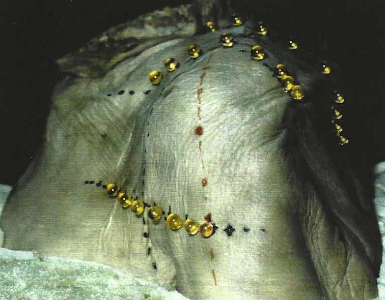
Figure 1. Overview of complete shoulder specimen with markers and support.
The left shoulder of a fixed (phenol, formaldehyde) human cadaver was sawed off in the medio-sagittal and the transverse plane 5 cm below the scapula. The specimen was then fixed on a wooden support using a polyurethane construction foam (Fig. 1). Sagittal, coronal, and transverse lines were drawn on the skin using the reference crosses projected by light beams of the MRI unit.
Oil-containing spherical capsules, glued to the skin with roughly equal spacing, were used as markers (Fig. 1). These rows of capsules lie within the primary planes of the MR scanner and are imaged without angulation. Images of the markers (not shown) were used to determine the intersections of the middle slice of the shoulder MR scan with the lines of the markers. This was done on the monitor of t h e MR un it . T he three points of intersection were marked on the specimen and a line connecting these points was drawn on the skin (the red line in Fig. 1). The sheet plastinated slices (Fig. 2) were made parallel to this line which represents the intersection of the middle slice of the shoulder MR scan (Fig. 3) with the skin of the specimen.
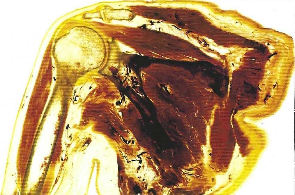 Figure 2a. Sheet plastinated slices of the shoulder made parallel to the red line in Figure1. |
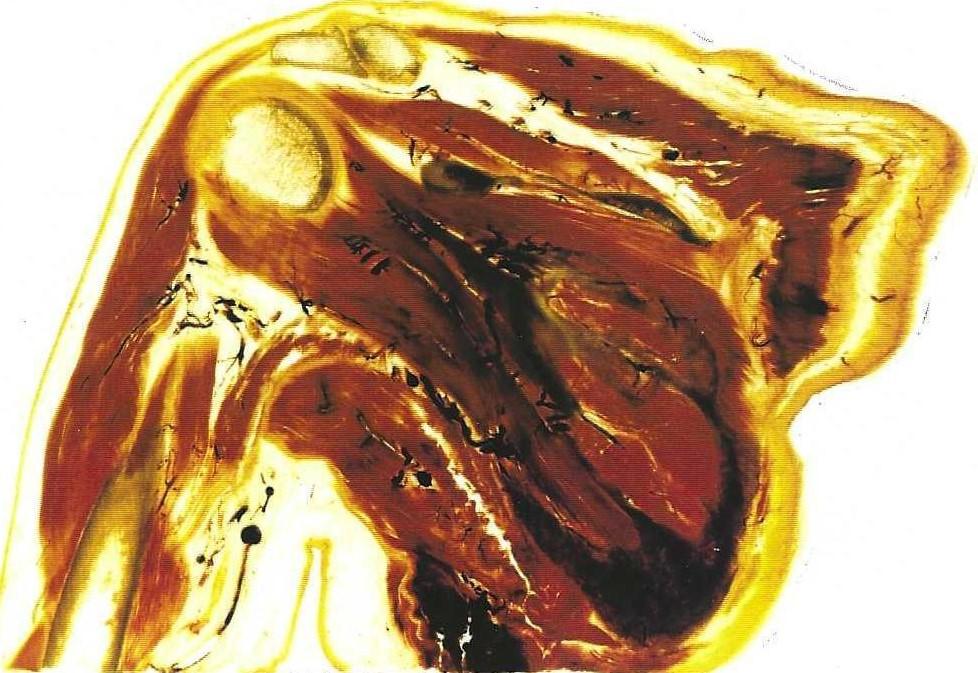 Figure 2b. Sheet plastinated slice of the shoulder made parallel to the red line in Figure 1 and 1 cm posterior to slice in Figure 2a. |
Only vertical cryosectioning using an immobile bandsaw was possible at our institution. Therefore, the specimen with the wooden support was mounted on a second horizontal support in a tilted position ensuring that the plane of sectioning had a vertical orientation. This was done visually by aligning the red line with the reference cross of the imager. CT images (Fig. 4) were also acquired in the same plane by simply aligning the red line with the reference cross of the CT scanner. The ultrasound images (Fig. 5) were acquired in a plane parallel to the red line. In addition, 3-dimensional CT reconstructions of the specimen were made to show the bone structures (Fig. 6). After the MRI, CT and Ultrasound images were obtained the specimen was frozen to minus 80 Celsius. The specimen was then sectioned in slices of 2 mm thickness using an immobile bandsaw (Reich). These slices (Fig. 2) were plastinated using the E12 technique (von Hagens et al.).
 Figure 3. Magnetic resonance images of the shoulder which correspond to the sheet plastinated slices in figure 2. |
 Figure 4. Computed tomography images of shoulder which correspond to the sheet plastinated slices in figure 2. |
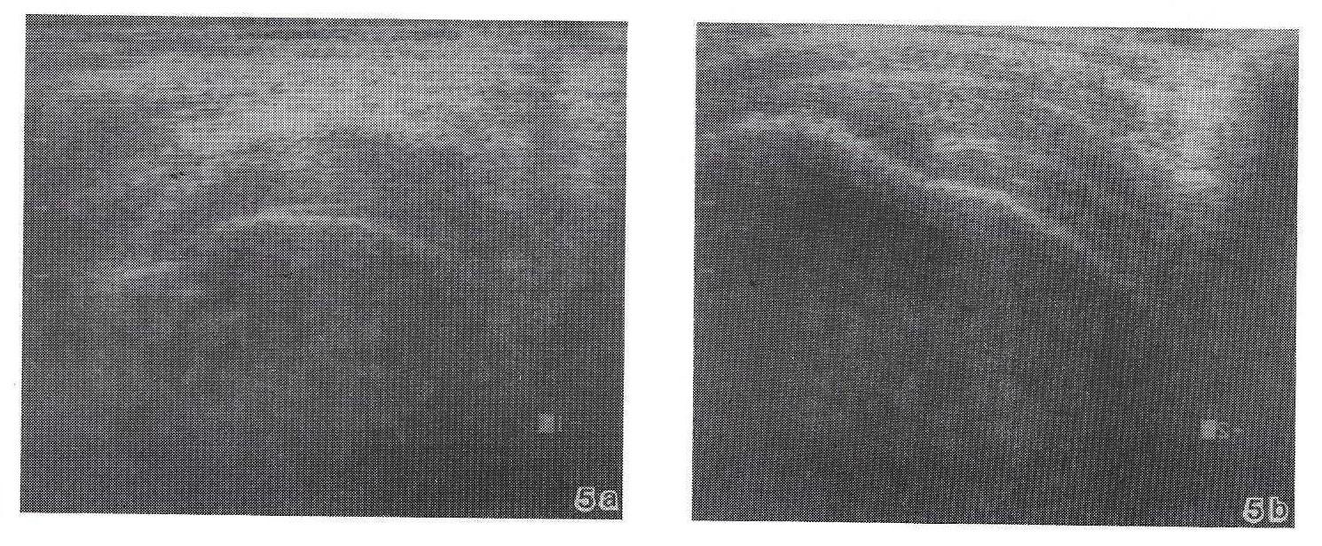 Figure 5. Ultrasound Images of shoulder which correspond to the sheet plastinated slices in figure 2. |
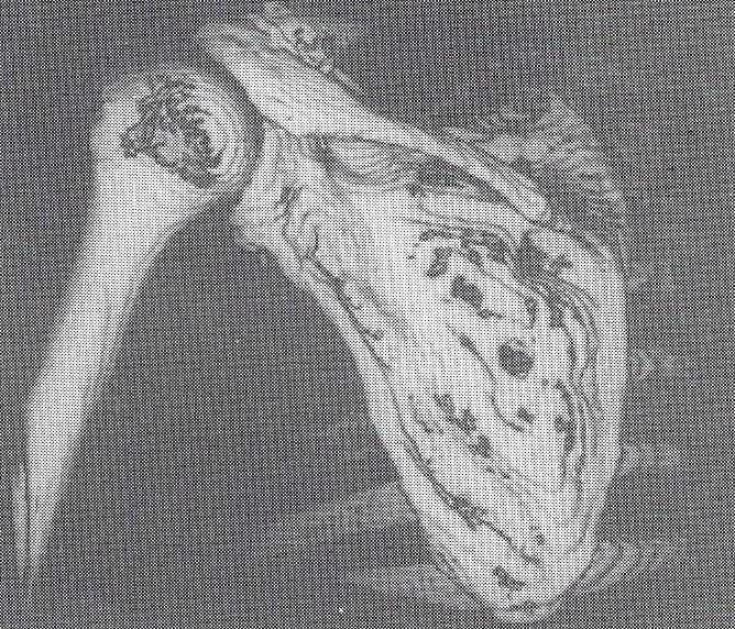 Figure 6. 3-dimensional reconstruction of the series of CT images showing the bone structure of the shoulder. |
Two examples of plastinated slices with the correlating MR, CT, and US images are shown. With the technique described here, accurate positioning for thin slices is possible. We believe this method to be the most advantageous way to obtain exactly matching MRI, CT and anatomic slices for correlative anatomic studies, thus making comparison of structures easier and more accurate.
von Hagens, Gunther, K Tiedemann, W Kriz: The current potential of plastination. Anat Embryol 175:411-421, 1987.
https://doi.org/10.1007/BF00309677