Department of Anatomy, Shanghai Medical University, Shanghai, P. R. China.
Due to the great importance of mummies for archeological research, methods have to be developed to preserve these specimens. Two preserved mummies (died 410 and 380 years ago) were exhumed and plastinated to avoid deterioration from exposure. They were first re-fixed with formalin and dehydrated at room temperature in a graded series of acetone solutions. The corpses were then pre- impregnated, force impregnated with silicone and subsequently cured all at room temperature. Histological studies were performed before and after plastination on pieces of lung, liver, kidney, heart, spleen and skin. Plastination improved the color and flexibility of the mummies and will permanently preserve them.
Su-Yi Chinese Silicone, Archeology, Paleopathology
Zheng Tianzhong, Department of Anatomy, Shanghai Medical University, Shanghai 200032, P. R. China. Tel: 86 21 6213 8681 /Fax: 86 21 6213 8681. Email: xyshen@shmu-email.shmu.edu.cn

![]()


Mummies have an invaluable value for academic re- search of our national culture. Extensive research studies are conducted to develop methods for the preservation of these corpses. There are two types of mummies: dry type and wet type. For the dry type most scientists prefer to keep them in a dry atmosphere, but for the wet type, scientists must dry them before keeping them in dry conditions or just immerse them into bath of preservative solutions. The wet mummies are considered more valuable than the dry ones. Immersing bodies into a bath of formalin is also not considered like the ideal method of long-term preservation. With limited technology in hand, some scientists suggest to keep these mummies underground and to stop exhumating them until an ideal long-term preservation technique has been developed and is available.
Plastination is a technique for permanent preservation of biological specimens (Bickley et al., 1981; Bickley and Townsend, 1984; von Hagens et al., 1987; Dawson et al., 1990). The plastinated specimens retain their original sur- face relief and cellular identity down to the microscopic level (von Hagens et al., 1987; Grondin et al., 1994). Plastination technique is suited for anatomy, zoology as well as for bio- logically oriented museums. To our knowledge, only one case of plastination of an archaeological human specimen has been reported (Wade and Lyons, 1995). In our laboratory, we successfully plastinated two ancient (400 years old) Chinese corpses, through fixation, dehydration, pre-impregnation and forced impregnation (Zheng et al., 1998).
Case 1: ancient corpse discovered near Zhengjaing in July 1997, male age 45, died 410 years ago. The skin of the limbs was dry, but the thorax wall and abdominal wall were still wet and flexible.
Case 2: ancient corpse discovered near Canton in 1992, male age 49, died 380 years ago. The skin of most parts of the body except the back was wet and flexible.
We do not know what methods were used at that time to preserve these bodies (Xu and Hu, 1996).
All internal organs within the thorax and abdominal cavities of these two mummies were removed. The bodies and internal organs were fixed by immersion in 7% formalin for 1 month at room temperature, then kept in a bath of 5% formalin at room temperature (2 months for case 1 and 5 years for case 2).
Dehydration
Dehydration of the bodies was performed at room temperature in a graded series of acetone solutions of increasing concentration of 60%-70%-80%-90%-100% foe more than 8 baths.
After fixation the specimens were rinsed in running tap water for 4 days in order to remove excess formalin. They were then transferred to a 60% solution of acetone. After thorough mixing of the acetone bath with the immersed mummies, an acetonometer was used to monitor the acetone con- centration every day. Once the acetone level was stable, the mummies were moved to the next higher concentrated acetone solution. In this manner, the mummies were gradually brought to the 100% acetone bath. When reading remained at 100% for 5 days we were sure that dehydration was completed and that the mummies were ready for impregnation.
Impregnation
This process was divided into three stages, all done at room temperature.
The mummies were moved from the 100% acetone bath into the Su-Yi Chinese Silicone bath (Su-Yi Plastination factory, Nanjing, China). All parts of the bodies were completely submerged into the silicone solution for 7 days. They were moved around and turned over every other day to allow the acetone and air bubbles to escape and the corpses to equilibrate with the silicone.
After the initial period of equilibration, the mummies were transferred into a vacuum chamber and the intermittent forced impregnated procedure was started. Each working day, the vacuum was re-established and the pressure slowly decreased. At the end of the working day the vacuum was re- leased and the chamber opened to allow the mummies to be moved around to relax them and facilitate further equilibration with the silicone. The pressure was decreased slowly over a period of 2 weeks. The vacuum was monitored by a manometer, and the progress of impregnation checked by observing the release of acetone gas bubbles from the sur- face of the bodies. The acetone gas bubbles rose slowly to the surface of the Silicone. Overtime the pressure was gradu- ally lowered to 10 mbar and the vacuum maintained for 3 more days until no more acetone gas bubbles appeared. This indicated that no more acetone remained in the mummies.
We have observed that at room temperature the silicone retains a much lower viscosity than at -25°C, permitting faster penetration, and easier acetone gas bubble escape.
After the intermittent forced impregnation, the mummies were removed from the vacuum chamber. They were however still kept immersed in the silicone for five additional days to allow them to further equilibrate with silicone.
Curing
Specimens were removed from the silicone and excess silicone was wiped off. All the parts were placed in anatomical position and slow cured. Old silicone solution mixed with different percentage of the hardener (1-5%) was used to smear the different parts of the skin of the ancient corps twice a day for seven days.
Internal thoracic and abdominal organs were plastinated the same way as the entire bodies. Pieces of tissue from the lungs, liver, kidney, heart, spleen and skin were taken out for histological study before and after plastination.

Figure 1. The Chinese mummy (after plastination).
After plastination the mummies retained their original shapes but their weights increased. The colors appear
better than before (figure 1). The soft tissues remain flexible and the specimens present no smell and no toxicity. The surfaces are dry, without oozing of remnant silicone and can be touched by bare hands. These mummies can now be easily kept for a long period of time without special care.
The internal organs within the thoracic and abdominal cavities retained their original shape and the colors are also much better than before the plastination process. The soft tissues are still flexible and it was possible to expand the cavities of the alimentary canal by air-injection (figure 2). The surfaces are dry and can be handled without gloves.
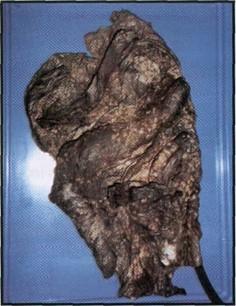 Figure 2. The cavity of the alimentary canal was expanded by air injection (after plastination). |
 Figure 3. Red Cells found in the lung tissue of one mummy (after plastination). |
During the histological study, before and after plastination, we found some red cells in the lung tissue of one of the mummies. The morphology of these 400 years old red cells has been preserved and looks exactly like fresh ones (figure 3).
The preservation of the valuable mummies is a complex problem. Many years of research have been dedicated to this subject. Even after all these years of work by many scientists, there are still no ideal methods or techniques to preserve the exhumed mummies.
We have used the technique of plastination to preserve two exhumed 400 years old mummies and achieved good results. Now it will not be necessary to dry the wet type mummies or immerse them into a bath of preservative solution. Using the plastination technique, the mummies can be preserved easily for a long time without special care. After plastination the weight of the mummies is increased which indicates that the silicone has penetrated down to the tissues of the specimens.
We were very excited to find some red cells in the lung tissue of one of the mummies. The morphology of these 400 years old red cells has been preserved. We do not know how they could have been preserved so well and which methodologies were used for keeping these bodies from decay. It should be another research subject.
Acknowledgment
The authors wish to thank Ms. Dan Hua Lian for help in histological study and Mr. Chen Yu Lian for his help in photography.
Bickley HC, von Hagens G, Townsend FM: An Improved Method for the Preservation of Teaching Specimens. Arch Pathol Lab Med 105: 674-676, 1981.
Bickley HC, Townsend FM: Preserving Biological Material by Plastination. Curator 27 (1): 65-73, 1984.
https://doi.org/10.1111/j.2151-6952.1984.tb01684.x
Dawson TP, James RS, Williams GT: Silicone plastinated pathology specimens and their teaching potential. J Pathol 162: 265-272, 1990.
https://doi.org/10.1002/path.1711620314
Grondin G, Grondin GG, Talbot BG: A Study of Criteria Permitting the Use of Plastinated Specimens for Light and Electron Microscopy. Biotech Histochem 69 (4): 219-234, 1994.
https://doi.org/10.3109/10520299409106291
von Hagens G: Heidelberg Plastination folder: Collection of all Technical Leaflets for Plastination, 2nd Ed, Anatomisches Institut 1, Universitat Heidelberg, Heidelberg, Germany. 1986.
Wade RS, Lyons W: The restoration of anatomical and archaeological specimens using S-10 plastination method: With special reference to preserving the good heart of a good priest. II World Congress on Mummy Studies, Cartagena, Columbia, 1995.
Xu Y, Hu W: The Ancient Corpses in China. Shanghai Scientific and Technological Education Publishing House, pp 229-235, 1996.
Zheng T, Liu J, Zhu K: Plastination at room temperature. Chinese Journal of Anatomy (in press).