Life Science Division, Chaffey Community College, Rancho Cucamonga, CA
An efficient means of storing a plastinated cross-sectioned cadaver was developed utilizing a foam bed. The foam bed fit into a disaster type body bag and is easily transported on a gurney. The storage system allows for easy storage and maneuverability, as well as, allowing students to study a section of the specimen and relate it to the entire cadaver.
cadaver storage; plastination; cross section;
Dan Whitten Life Science Division, Chaffey Community College, Rancho Cucamonga, CA

![]()


The development of Plastination has greatly enhanced teaching methods in many disciplines (Bickley et al., 1981; Baptista et al., 1989; Holladay and Hudson, 1989). The ability to study tissue properties, individual organs and the spatial organization of whole organisms on different planes has proven to be a valuable teaching aid (Lane, 1990; Henry et al., 1991).
Chaffey College offers Cross-sectional anatomy for Radiologic Technologists as a continuing education course. We presently have one entire male cadaver in cross- section which is used in this course. It was plastinated using the standard S10 technique (yon Hagens, 1985).
Due to limited space in our facility, the sections were originally stored in plastic bags, three sections per bag. This system proved inadequate; it was awkward to transport the specimens to the classroom, difficult to tell at a glance what region of the body each specimen belonged to, and some damage to the specimens occurred due to the extra handling required. In an effort to remedy these problems, we devised a system to facilitate storage, transportation and use of the cadaver that was both compact and efficient. The cadaver is now stored in an ester foam "bed".
This method of storing sectioned cadavers will be utilized for specimens which will be plastinated in the future as it offers good protection of the specimen, is easy to move around, is esthetically pleasing, and also allows the students to study the specimen as a whole unit.
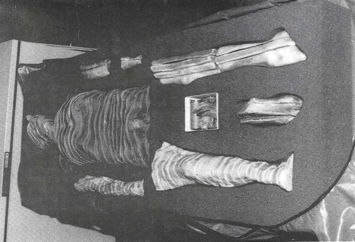
Figure 1. Sectioned cadaver in its "foam bed".
The storage container for the cadaver was constructed from a piece of two pound ester foam, six inches thick. The foam was cut to the dimensions of 25" wide and 75" long. This size allowed the completed container and specimen to fit easily into a standard size disaster type body bag and allowed easy closure of the bag without any tension on the zipper or specimen. The sections were placed as a unit on the foam in their correct anatomical position and the outline of the cadaver traced onto the foam. The sections were removed and the foam cut to allow the sections to be positioned securely into the foam but not too tightly. Approximately one inch of foam was left intact on the bottom of the cut-out to cushion the torso sections and two to three inches of foam was left for the extremities. The entire foam bed project required two hours for completion. As the cadaver is used in different laboratories, the disaster bag, containing the cadaver, is stored on a gurney to facilitate movement. ' Costs for this system were relatively inexpensive: $65.00 for the disaster bag, $50.00 for the foam (Figure 1). The foam was cut easily with razor blades or a long-bladed knife, but a standard electric kitchen knife was found to work the best.
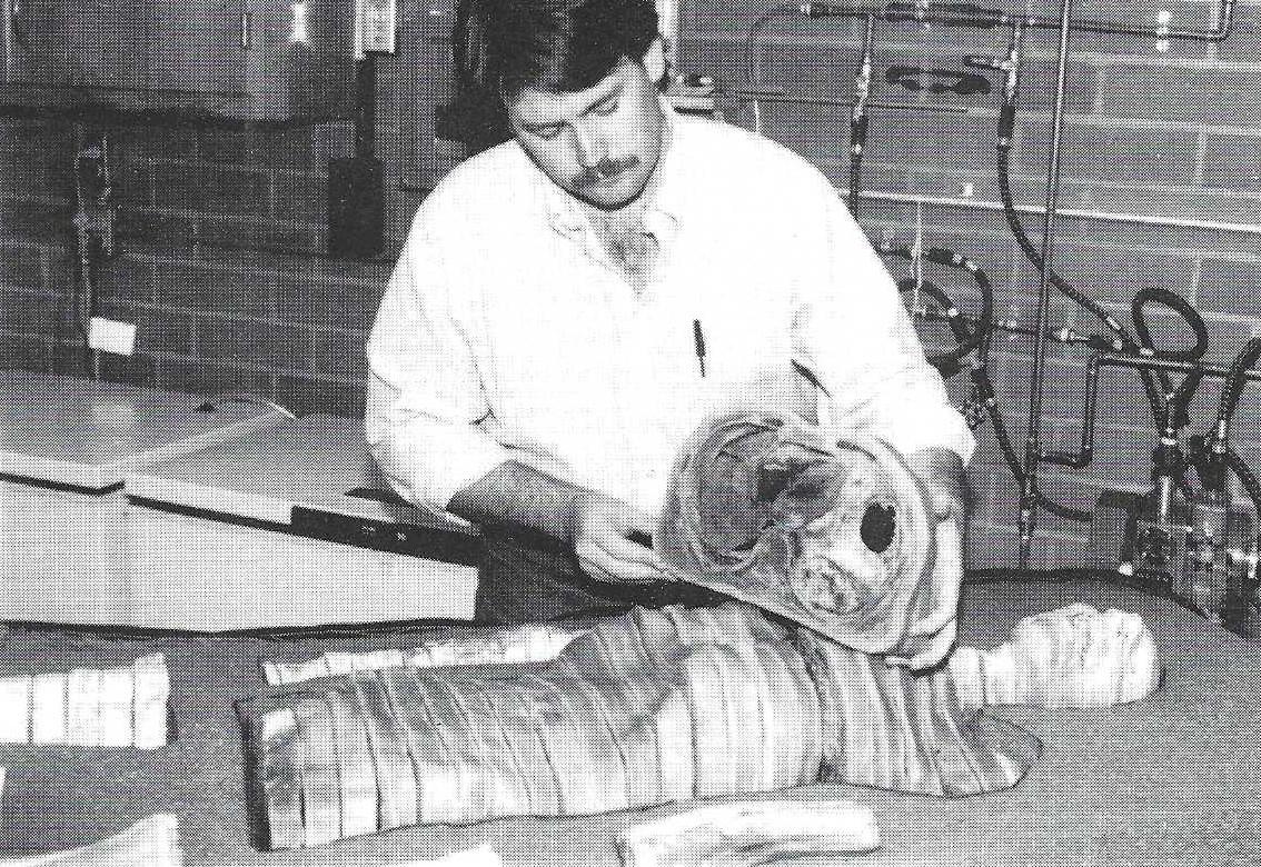
Figure 2. A section of cadaver is easily removed for study.
The finished product holds the sections of the cadaver in anatomic position while still allowing removal of various sections for study. This method of storage allows the various routes of organs and tissues to be traced throughout the body with ease (Figure 2). The foam not only provides a storage system that is convenient but also offers the specimen adequate protection. No chemical reactions have been observed between the plastinated specimen and the foam.
When we began this project, we had several objectives in mind: to provide a convenient method of storage and transportation, while offering the specimen adequate protection. This system does all of the above and has another very important function: it allows the students to see the specimen as a whole. By being able to see exactly where an individual section belongs, the students are better able to visualize the intricate spatial relationships of various systems and thereby, better able to understand the section being studied.
Baptista CAC, M Skie, RA Yeasting, N Ebraheim, WT Jackson: Plastination of the wrist: Potential uses in educational and clinical medicine. J Int Soc Plastination, Vol 3:18-21,1989.
https://doi.org/10.56507/XENF9035
Bickley HC, G von Hagens, FM Townsend: An improved method for preservation of teaching specimens. Arch Pathol Lab Med 105:674-76,1981.
Henry RW, G Daniel, FK AI-Bagdadi, J Butler: Plastinated specimens, an aid in inte rp reting M R I, CT, and sectional anatomy. Anat Histol Embryol 20:1,1991.
Holladay SD, LC Hudson: Use of plastinated brains in teaching neuroanatomy at the North Carolina State University, College of Veterinary Medicine. J Int Soc Plastination 3:15-17,1989.
https://doi.org/10.56507/FEKB4686
Lane Alexander: Sectional anatomy: Standardized methodology. J Int Soc Plastination, 4:16-22,1990.
https://doi.org/10.56507/LYMW2924
Tiedemann K: A silicone-impregnated knee joint as a natural model for arthroscopy. J Int Soc Plastination 2(1): 13-17,1988.
https://doi.org/10.56507/CACT1479
von Hagens G: Heidelberg Plastination Folder: Collection of all technical leaflets for plastination. Anatomisches Institut 1, Universitat Heidelberg, 1985.