Department of Biology, Union University, Jackson, TN 38305, USA
The utilization of plastinated specimens has increased significantly in anatomy instruction, providing self-directed aids for student inquiry. Due to increasing access to plastinated specimens, low-cost identification systems are being developed for the monitoring of their usage, handling, and distribution. Radio frequency identification (RFID) technology has been used in the healthcare field and recent plastination studies for its automated identification and tracking of multiple artifacts. This study demonstrates a streamlined tag system for biological specimens subjected to the S10 cold-temperature plastination process. Four commercially available RFID tag types were selected and embedded in biological specimens, prior to the plastination process. The results indicated that the four types of tags selected are reliable and have the ability to sustain lengthy periods of time in the harsh plastination conditions. Embedding RFID tags in varying tissue types represents a successful small-scale study for seamless tracking of anatomical specimens. Comparison of the four RFID tag types reveals that there was no significant difference in the composition and successful performance following the plastination process. This study further demonstrates that RFID tags are a feasible and low-cost option for the identification and management of plastinated biological specimens.
anatomical specimens; healthcare field; radio frequency identification; RFID tags; student inquiry
Dr. Mark G. Bolyard, Department of Biology, Union University, Jackson, TN 38305, USA. Tel.: +1 731-661-6586;
E-mail: mbolyard@uu.edu

![]()


In response to the heightened demand for health care professionals, the integration of enhanced anatomy curricula has been implemented in North American universities. Experiential learning within the anatomy field has become increasingly difficult, due largely to the inability to find and collect biological specimen resources (Marks, 1996; Older, 2004; Lockwood and Roberts 2007). For decades, anatomists have turned to tissue preservation to produce high quality, long lasting specimens through plastination, which relies on the physical replacement of water and lipids with curable polymer. Although plastination requires chemicals to fix the specimens and dehydrate the tissue, specimens do not require long-term immersion or storage in chemicals.
Cadaveric remains are essential for academic instruction and research, but can be costly and difficult to maintain. Although plastination equipment and materials can also be costly, its utilization allows for permanent preservation of remains, in addition to alleviating the residual costs of fixing additives or repurchasing (von Hagens et. al., 1987). One challenge for plastination is tracking and monitoring specimens during and after the plastination process. Tracking specimens through the plastination process can be difficult because of the limited access during procedural steps, in addition to plastinating indistinguishable specimens (e.g. organs from several donated remains). Likewise, monitoring specimen access following plastination is extremely important due to plastination expenses, in addition to ensuring the integrity and respectful handling of donated remains (Schmitt et al., 2014). Plastinated specimens do not require storage containers and are safe to handle, therefore, they can be misplaced, lost, or stolen, much easier than other fixed specimens. Management of specimens can be improved by utilizing radio frequency identification (RFID) tracking systems, benefitting body donation programs, universities with anatomical studies, and professional health programs (Porzionato et al., 2012). However, a streamlined system for classification and tracking has not yet been established (Noël and Connolly, 2016). A recent study successfully attached over 300 RFID tags onto biological specimens after the plastination process. Analysis of these tags revealed the successful and economically feasible tracking system tracking abilities of RFID tags (Noël and Connolly, 2016).
Tracking and management of plastinated specimens is important not only after specimens have been prepared, but from the time of harvesting. Having the ability to track assets greatly reduces misidentification. Implementation of RFID tagging can be done at the time of organ harvesting or acquisition, however, it was not known how these RFID tags would fare going through the rigorous plastination process. RFID tags are advantageous due to their small, inlaid integration that protects them from harsh environments, and passive tag ability requiring no battery source (Hanna and Pantanowitz, 2015). RFID tags utilize a digitized bar code, which allows multiple tags to be read simultaneously without disruption of signal frequency or directly scanning a visible barcode. A RFID reader sends a high frequency radio wave powering the passive RFID tag, activating its integrated circuit. Each tag stores an electronic product code (EPC) within the integrated circuit on a memory chip. The EPC number is emitted from the RFID tag following activation and read by the network-connected RFID reader. The reader communicates between the tags and a computer database, which inventories the EPCs for storage and tracking purposes. Each RFID tag possesses a unique EPC that is programmed into the memory chip by the manufacturer. The factory EPC typically consists of a unique twenty-four-digit code that is used for specific tag identification. Standard factory EPCs come in a Hex format which can only be coded in numbers zero through nine and the character letters A-F. If a RFID tat possesses “read and write” capability, the Hex format may be altered to ASCII. The ASCII format utilizes the numbers zero through nine as well as letters A-Z. The RFID tags used for this study possessed the “read and write” capability, which can be advantageous when tracking large quantities of specimens.
Plastination requires documentation and close examination of the specimens throughout the entire process, particularly if similar tissues or organs from different donated remains are used (Schmitt et al., 2014). To overcome this difficulty, specimens in this study had a unique RFID tag embedded in the tissue, prior to the plastination process. The objectives of this project were to successfully embed four unique RFID tag types into twenty different specimens prior to the S10 plastination process, implement an RFID classification system for the biological specimens by creating unique, distinguishable EPCs, and assess the durability and functionality of the four RFID tag types following plastination.
The durability of four RFID tag types (Table 1) was assessed following the S10 cold-temperature plastination method outlined by DeJong and Henry (2007). The TSL 1128 Bluetooth UHF RFID Reader and commercially available RFID tags (Omni-ID Fit 400P, Omni-ID Exo 400P, Omni-ID Exo 200, Xerafy Dash-IN XS) were purchased from Atlas RFID Solutions (Birmingham, Alabama, US 35203). Methods for inserting tags are contingent on the specimens used, but for these experiments, tags were inserted following the method demonstrated in Figure 1.
| RFID Tag Type | Manufacturer | Read Range (Fixed Reader) | Read Range (Handheld Reader) | Material Compatibility | Dimensions |
| OMNI-ID FIT 400P | Omni-ID, Rochester, NY, US | 3.5 m (11.5 ft) | 1.75 m (5.7 ft) | Plastic Assets | 17.6 x 7.1 x 4.1 mm |
| XERAFY DASH-IN XS | Xerafy, Dallas, TX, US | 2.0 m (6.6 ft) | 1.5 m (5.0 ft) | Metal Assets | 12.3 x 3 x 2.2 mm |
| OMNI-ID EXO 200 | Omni-ID, Rochester, NY, US | 2.0 m (6.6 ft) | 1.0 m (3.28 ft) | Plastic Assets | 14.5 x 12 x 5.4 mm |
| OMNI-ID EXO 400P | Omni-ID, Rochester, NY, US | 3.5 m (11.5 ft) | 1.75 m (5.7 ft) | Plastic Assets | 23.5 x 13 x 6.9 mm |
Table 1. RFID tag specifications
 Figure 1. Insertion of an Omni-ID Exo 200 RFID tag into a Leopard frog (Rana sphenocephala) and scanning of the inserted tag: (a) making a small incision in the specimen, (b) inserting the RFID tag, (c) suturing the incision, (d) reading the tag with the TSL 1128 Bluetooth UHF RFID Reader. |
The TSL 1128 Bluetooth UHF RFID Reader was selected to read the RFID tags, as it provides a cost-effective way to read and write EPC UHF transponders. The TSL 1128 is capable of communicating with a wide variety of host Bluetooth devices, such as iOS or Android phones/tablets, while being compatible with a variety of database software. The operating frequency range of 902-982 MHz was selected in order to successfully activate and read the RFID tags under study. The maximum read distance of this reader was 4 meters, which was well within the desired range.
Due to the harsh conditions, careful consideration was given to deciding which RFID tags were most capable of surviving the plastination process. The RFID tags were required to operate uniformly in the range of 860 to 960 MHz and contain Alien H3 IC, providing large user memory (96 EPC-bits, extensible to 480 bits) and enhanced IC security. Manufacturer specification, including thermal operating ranges (-20°C to 85°C), thermal and chemical cycling durability, signal output, overall size, and cost effectiveness were also considered. Furthermore, the selected tags had a maximum readable distance of less than 4 meters (short-range RFID tags), which are optimal for the TSL 1128 UHF RFID Reader. Tags were tested individually for performance and the ability to emit readable EPC’s prior to embedding into the specimens to be plastinated.
Each RFID tag arrived with a unique EPC from the manufacturer. The factory specific EPC consisted of a unique twenty-four-digit code that is used for individual tag identification. For logistic purposes, modification was required to enable faster tracking and better categorization of plastinated specimens. The TSL 1128 UHF RFID handheld reader and the RFID Tag Finder iOS application (v 1.0.8.2449) from Technology Solutions UK LTD was used to rewrite the factory EPC’s. Utilizing the read and write capabilities of the TSL 1128, the twenty-four-digit HEX code was converted to a twelve-digit ASCII format. Specimens were given an eight-digit abbreviation for classification. Subsequently, all codex EPCs were used to track and categorize the plastinated specimens.
The cold-temperature BiodurTM S10/S15 plastination technique outlined by DeJong and Henry (2007) was used in this study. A silicone polymer mix (impregnation mixture) was previously prepared consisting of the S10 silicone polymer, S3 catalyst and chain extender. Mixing prior to impregnation allows the silicone molecule elongation to start, resulting in longer silicone chain length and a more viscous impregnation mixture. The impregnation mixture was stored in a freezer at -15° C to retard chain elongation until the impregnation phase. Embalmed (formalin- fixed) specimens were placed in 50 % ethanol for seven days, removing excess embalming fluids prior to the plastination process (DeJong and Henry, 2007).
After tag assessment and S10 cold-temperature plastination preparations, 20 tags (5 of each type) were embedded into 20 formalin-fixed specimens (Table 2). Because the tags differ slightly in size, tags were selected to match each specimen based on the size of the specimen compared to the tag. After insertion of the tags, several specimens required suturing to seal openings. The specimens were then placed in a flushing water bath for 7 days to remove any residual formalin (DeJong and Henry, 2007).
| Specimen | Binomial Classification | RFID Placement Site | Abbreviation for Codex |
| Pigfish | Orthopriatis chrysoptera | Abdominal cavity | PigFish1 |
| Fetal Pig | Sus scrofa domesticus | Superior femoral region (Right) | FetalPg1, FetalPg2 |
| Axolotl | Ambystoma mexicanum | Abdominal cavity | Axolotl1 |
| Southern Flying Squirrel | Glaucomys volans | Superior femoral region (Right) | SFSquir1 |
| Bichir | Polypetrus retropinnis | Abdominal cavity | Bichir01 |
| Southern Brook Lamprey | Ichthyomyzon gagei | Abdominal cavity | SBLampe1 |
| Mole Salamander | Ambystoma talpoideum | Abdominal cavity | MSalama1 |
| Shrew | Cryoptotis parva | Superior femoral region (Right) | Shrew001 |
| Atlantic NeedleFish | Strongyura marina | Abdominal cavity | ANeedle1 |
| Long-tailed Weasel | Mustela frenata | Superior femoral region (Right) | LtWease1 |
| Grey Squirrel | Sciursus carolinensis | Superior femoral region (Right) | GSquir01 |
| Leopard Frog | Rana sphenocephala | Abdominal cavity | LeoFrog1 |
| White Footed Mouse | Peromyscus leucopus | Superior femoral region (Right) | Wfmouse1 |
| Mink | Mustela vison | Superior femoral region (Right) | Mink0001 |
| Eastern Box Turtle | Terrapene carolina | Superior to Right leg, Ventral to Carapace | EBTurtl1, EBTurtl2 |
| Alligator | Alligator mississippiensis | Superior femoral region (Right) | Alligat1, Alligat2, Alligat3 |
Table 2. Specimens used for plastination, RFID tag placement sites, and edited Codex for each specimen. Codex abbreviations must be 8 alphanumeric characters.
The specimens were then placed in a chemical-resistant receptacle and submerged in a 90% acetone bath. The receptacle was placed in a freezer (-15° C) for 8 days. An acetonometer was used to determine the acetone concentrations for the next 45 days of specimen submersion as the concentration was gradually increased to 99% (DeJong and Henry, 2007). The receptacle containing the specimens was removed from the freezer and placed at room temperature for 6 days to undergo the defatting process. The gradual increase in solvent temperature initiates defatting, removing excess fat/lipids, resulting in increased tissue permeability, allowing for better polymer penetration and distribution, and producing a more life-like and durable specimen. Monitoring was conducted by observing the color of the acetone solvent that the specimens were submerged in. The transition in the color of the acetone from clear to yellowish/brown color signified the defatting process is complete (DeJong and Henry, 2007).
After the defatting process, specimens were immediately placed in a freezer containing a vacuum chamber designed to withstand one atmosphere decrease in pressure. This chamber contained the S10 silicone polymer, S3 catalyst and chain extender. Specimens were left for one day to stabilize at -14° C. The vacuum pump was turned on and the pressure was reduced to 22 cm Hg. The pressure was slowly lowered over the next 14 days to 4 cm Hg, resulting in the removal of acetone vapor from the specimens, allowing for the forced impregnation of the S10 polymer mix. Pressure was further reduced to 1 cm Hg (0.013 atm), resulting in further gas removal, for the next 20 days. On day 36, gas release ceased, signifying the completion of forced impregnation. The pressure was slowly returned to normal atmospheric pressure and left to equalize for 2 days until the specimens were removed (DeJong and Henry, 2007).
The specimens were then placed out at room temperature for 19 days, allowing excess impregnation mix to drain, and silicone molecules to undergo chain extension, linking their S3 extender portions together during the pre-curing process. The pre-curing phase was extended to 30 days to yield more pliable, flexible specimens. Finally, specimens were placed in a chemical-resistant curing chamber, containing a desiccant and the S6 cross-linker during the gas curing phase, resulting in the connection of adjacent silicone molecule chains (DeJong and Henry, 2007).
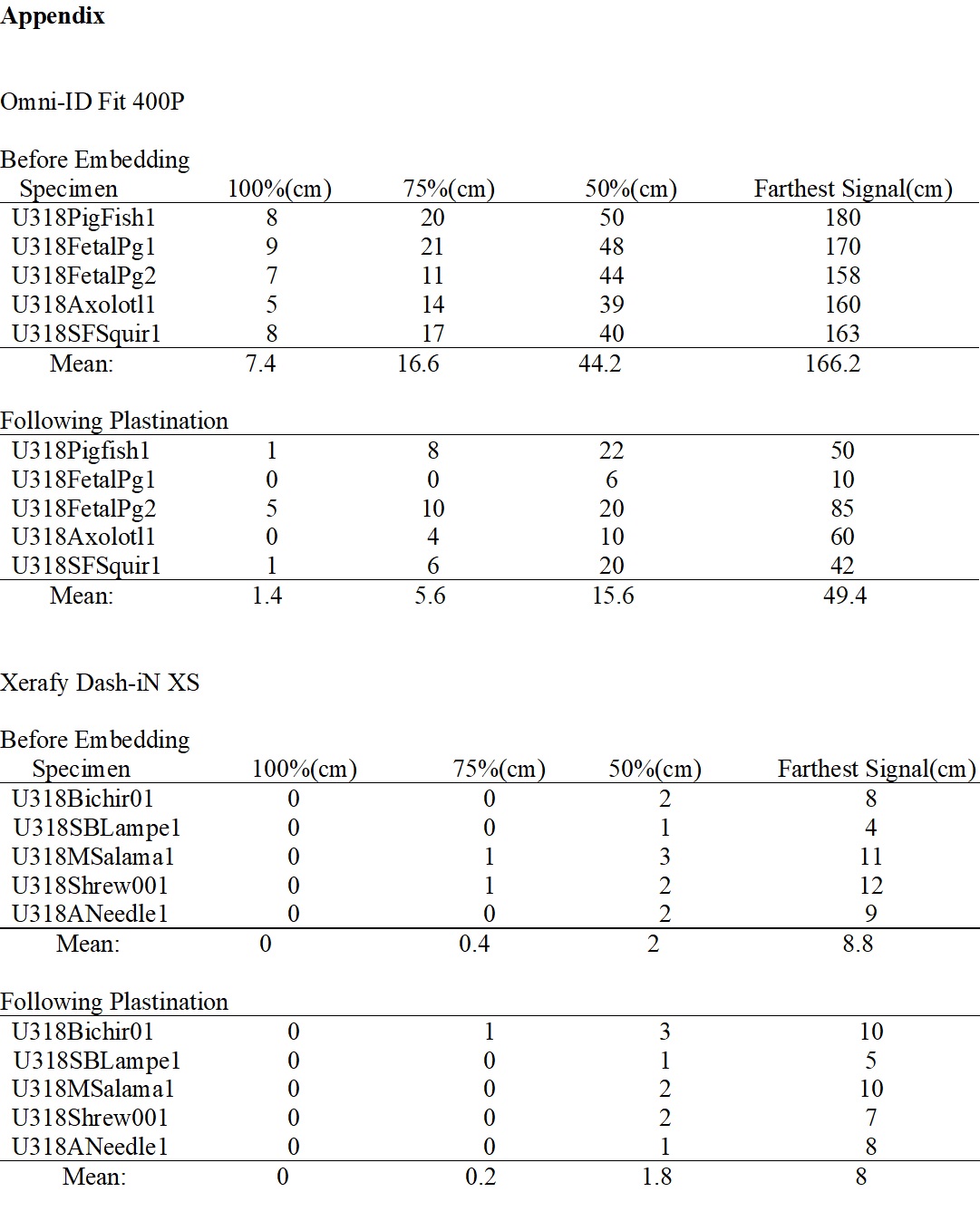
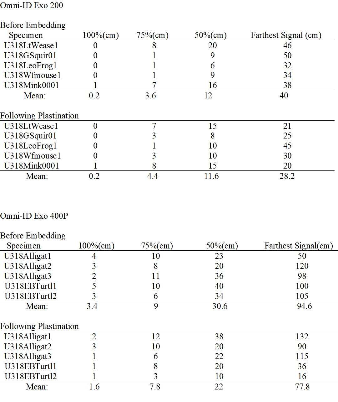
During the five-month plastination process, the RFID tags were assessed for readability after the following plastination steps: flushing, dehydration, impregnation, and curing. Each specimen containing an RFID tag was scanned to ensure each tag would emit a readable EPC to the TSL 1128 Bluetooth UHF RFID Reader (Fig. 1d). RFID tag read distances were measured at 100%, 75%, and 50% signal strength, as well as the farthest distance to a readable signal, both before and after plastination. Statistical analyses were performed on the mean differences of the distances before and after plastination for each RFID tag used, regardless of the sample tested.
Normally distributed data were analyzed using a paired t-test, otherwise a Wilcoxon signed ranks test was used. All analyses were performed at an a-level of 0.05 using R statistical software.
Prior to insertion and plastination, all tags provided readable EPCs at distances up to 160 mm, although the Omni-ID tags (Fit 400P, Exo 400P, Exo 200) provided readable EPCs at greater distances than the Xerafy Dash-IN tags (see data in the Appendix). All twenty RFID tags emitted an accurate and readable EPC following flushing. Four of the Xerafy Dash-IN tags, and one Omni-ID Fit 400P tag did not emit an accurate and readable EPC following dehydration. However, following impregnation and curing, all twenty RFID tags once again emitted an accurate and readable EPC. After read range capabilities were collected at 100%, 75%, and 50% signal strength, as well as the farthest distance to a readable signal, average distances and standard deviations were calculated (Table 3).
| Tag Type | Signal Strength | X Difference(cm) | S difference(cm) | t- Value | V- value | df | P-Value |
| Omni Fit 400 | 100% | -6.0 | 2.65 | 5.071 | 4 | 0.0071* | |
| 75% | -11.0 | 7.11 | 3.461 | 4 | 0.0258* | ||
| 50% | -28.6 | 8.29 | 7.710 | 4 | 0.0015* | ||
| Farthest | 116.8 | 32.63 | 8.004 | 4 | 0.0013* | ||
| Omni Exo 200 | 100% | 0.0 | 0.00 | 0.000 | 4 | NA | |
| 75% | 0.8 | 1.30 | 1.500 | 4 | 0.2652 | ||
| 50% | -0.4 | 3.29 | 0.272 | 4 | 0.799 | ||
| Farthest | -11.8 | 16.30 | 1.619 | 4 | 0.1808 | ||
| Omni Exo 400 | 100% | -1.8 | 1.48 | 2.714 | 4 | 0.0533* | |
| 75% | -1.2 | 3.11 | 0.862 | 4 | 0.4375 | ||
| 50% | -8.6 | 16.02 | 1.2000 | 4 | 0.2963 | ||
| Farthest | -16.8 | 68.04 | 0.552 | 4 | 0.6103 | ||
| Xerfy | 100% | 0.0 | 0.00 | 0.000 | 4 | NA | |
| 75% | -0.2 | 0.84 | 4.000 | 4 | 0.7728 | ||
| 50% | -0.2 | 0.84 | 0.535 | 4 | 0.6213 | ||
| Farthest | -0.8 | 2.68 | 0.667 | 4 | 0.5415 |
Table 3. Statistical analyses of signal strength differences before and after plastination, including means ( difference), standard deviations (S), and sample statistics (paired samples t-test [t] or Wilcoxon signed-ranks test [v]) with associated P-values are presented, with 4 degrees of freedom. The * denotes an level of less than 0.05.
F.2 Omni-ID Fit 400P Tag Analysis
Differences in means before and after plastination were significantly different among all read strengths, and for the greatest distance to a readable signal (p <0.05; Table 3), indicating that, in this study, plastination had a significant impact on the function of these RFID tags. However, even after plastination these tags had the second highest mean distance to a readable signal (Appendix), indicating that they were still very useful for this application.
F.3 Xerafy-IN XS RFID Tag Analysis
No significant difference was detected in mean differences across read strengths, indicating that the plastination process did not have a significant impact on the function of these tags (Table 3), although these tags had the lowest mean distance to a readable signal of the tags tested following plastination (Appendix).
F.4 Omni-ID Exo 200 RFID Tag Analysis
No significant difference was detected in mean differences across read strengths, indicating that the plastination process did not have a significant impact on the function of these tags (Table 3), although these tags had the second lowest mean distance to a readable signal of the tags tested following plastination (Appendix).
F.5 Omni-ID Exo 400P RFID Tag Analysis
Differences in distances at 100% signal strength approached a statistically significant difference before and after plastination (p = 0.0533; Table 3), and additional testing may indicate significance, but no significant difference was detected among any of the other signal strengths. These tags had the highest mean distance to a readable signal of the tags tested following plastination (Appendix).
This study demonstrates a cost-effective approach to tagging specimens prior to the plastination process. Tagging these specimens with RFID sensors prior to plastination provides an efficient tracking system that maintains correct identification and categorization through the entire preservation process, and throughout the life of the specimen. It is also more straightforward to insert the tags prior to plastination rather than after the process is complete. The twenty RFID tags emitted readable EPCs after flushing. However, four of the Xerafy Dash-IN XS and one of the Omni-ID Fit 400P RFID tags did not produce EPCs following the dehydration step. This was likely due to diminished read range capabilities while being submerged in preservation fluid, which was also observed by Noël and Connolly, (2016). The four tags were unable to be scanned at close enough distances for detection due to the solvent’s corrosive effects on the scanner. In order to prevent damage, and maintain the integrity of the handheld scanner, plastinated samples were scanned at a distance of 30 cm (1 ft). Although five tags were unable to be read after defatting, all twenty RFID tags emitted accurate and readable EPCs following impregnation and curing. The 4 commercially-available RFID tag types tested provide a standard for large-scale tagging of biological specimens undergoing the plastination process. The twenty RFID tags used in this study will continue to be monitored for any long-term disruption or malfunction after being exposed to the harsh conditions of plastination.
With regard to the function of the four RFID tags relative to each other, there are several factors to consider. First, there is the loss of signal at certain points during the plastination process when using the Xerafy Dash-IN XS and the Omni-ID Fit 400P. Second, the Omni-ID Fit 400P showed statistically significant differences in signals before and after plastination. Third, when considering the differences in the mean of the farthest signal detected after plastination, the order of distances (from the Appendix) is Omni Exo 400 (77.8 cm), Omni Fit 400 (49.4 cm), Omni Exo 200 (28.2 cm), and Xerafy (8 cm). Therefore, selection of the appropriate RFID tag will be based on particular applications of the researchers.
Future investigation could explore optimal depths for implantation of RFID tags, using multiple samples of the same tissue, and integrating all four RFID types into the same sample. These could allow for differences to be calculated among specimens of varying tissues, providing useful data for optimal tag choice regarding individual tissues. Also, multiple RFID tag types embedded in a single specimen could provide a much closer comparative analysis between RFID tag types. Due to RFID capabilities and technology, close proximity of multiple tags does not have an effect on surrounding RFID tag signals. Additional RFID tags, such as the Fit 220 HT (Atlas RFID), should also be evaluated. It is also our hope to compare the effectiveness of this wider range of RFID tags in cold- vs room-temperature plastination systems, as each temperature system is beneficial for different types of specimens.
As universities continue to enhance their anatomy curricula to aid in the development of aspiring healthcare providers (Marks, 1996; Older, 2004; Lockwood and Roberts 2007) methodologies for specimen management continue to be important. Demonstrating the distinct characteristics of each anatomical specimen continues to be pedagogically important. Plastination provides a means to turn these valuable, perishable specimens into non-perishing, reusable teaching tools. These tissues are also accurate in terms of structure and approximate in terms of color, are chemical free, odor free, maintenance free, safe for handling, and are able to retain integrity/clarity throughout the specimens handling. Protecting donated remains and costly anatomical specimens is essential (Schmitt et al., 2014). Implementation of RFID tagging promotes proper handling and care, due to its monitoring and tracking capabilities, potentially saving plastinated resources as well as funding for medical universities or programs. With RFID technology, monitoring and tracking difficulties become alleviated, allowing maximum confidence that specimens will sustain the quality of preservation (Wakefield, 2007). It may also be possible to use RFID tags with greater information storage capacity to provide educational information in addition to identifiers. The data provided in this study demonstrates the possibility of embedding RFID tags within various biological tissues, categorizing plastinated specimens, tracking and monitoring samples during the plastination process, and managing the usage of valuable plastinates following plastination. Furthermore, this study promotes a stronger relationship with body donation and medical programs. Plastination continues to serve as a valuable tool for the future for anatomical research and education, and can be enhanced through the implementation of RFID technology.
 Page 1 |
 Page 2 |
Acknowledgment
The authors wish to acknowledge Tyler Lockard and the Atlas RFID team for dedicating their time, resources, and support throughout this project. We also appreciate the work of Drs. Marc Lockett and Michael Schiebout in editing Mr. Vandezande’s original Masters manuscript, Dr. Micah Fern for assistance with photography, and Ms. Anna Laura Livingston for assistance with manuscript and table preparation.
DeJong K, Henry RW. 2007: Silicone plastination of biological tissue: Cold-temperature technique BiodurTM S10/S15 technique and products. J Int Soc Plastination 22:2-14.
https://doi.org/10.56507/ZLMJ7068
Hanna MG, Pantanowitz L. 2015: Bar coding and tracking in pathology. Surg Pathol Clin 8:123-135.
https://doi.org/10.1016/j.path.2015.02.017
Lockwood AM, Roberts AM. 2007: The anatomy demonstrator of the future: An examination of the role of the medically-qualified anatomy demonstrator in the context of tomorrow's doctors and modernizing medical careers. Clin Anat 20:455-459.
https://doi.org/10.1002/ca.20427
Marks SC Jr. 1996: Information technology, medical education, and anatomy for the twenty- first century. Clin Anat 9:343-348.
https://doi.org/10.1002/(SICI)1098-2353(1996)9:5<343::AID-CA8>3.0.CO;2-D
Noël G, PJC, Connolly CC. 2016: Monitoring the use of anatomical teaching material using low-cost radio frequency identification system: A comprehensive assessment. Anat Sci Educ 9:197-202.
https://doi.org/10.1002/ase.1575
Older J. 2004: Anatomy: A must for teaching the next generation. Surgeon 2:79-90.
https://doi.org/10.1016/S1479-666X(04)80050-7
Porzionato A, Macchi V, Stecco C, Mazzi A, Rambaldo A, Sarasin G, Parenti A, Scipioni A, De Caro R. 2012: Quality management of body donation program at the University of Padova. Anat Sci Educ 5:264-272.
https://doi.org/10.1002/ase.1285
Schmitt B, Wacker C, Ikemoto L, Meyers FJ, Pomeroy C. 2014: A transparent oversight policy for human anatomical specimen management: The University of California, Davis experience. Acad Med 89:410-414.
https://doi.org/10.1097/ACM.0000000000000135
Von Hagens G, Tiedemann K, Kriz W. 1987: The current potential of plastination. Anat Embryol (Berl) 175:411-421.
https://doi.org/10.1007/BF00309677
Wakefield D. 2007: The future of medical museums: Threatened but not extinct. Med J Aust 187:380-381.
https://doi.org/10.5694/j.1326-5377.2007.tb01304.x