Departement de chimie-biologie, Universite du Quebec a Trois-Rivieres, Trois-Rivieres, Quebec, Canada
The western anatomical descriptions of the human cerebral meninges still available date back to the mid-fourteenth century. Since that time, numerous anatomists made more or less accurate descriptions of the human meninges and tried to understand their functions. Unfortunately, the belief in animal spirits led to esoteric interpretations which hindered the understanding of these structures up to the early eighteenth century. This paper summarizes the contributions of Antonio Pacchioni and Giovanni Fantoni to the anatomy and functions of the meninges, respectively.
Meninges - Arachnoid granulations - Antonio Pacchioni - Giovanni Fantoni.
Dr. R. Olry, Departement de chimie-biologie, Universite du Quebec a Trois-Rivieres, C. P. 500, Trois-Rivieres, Quebec, CanadaG9A5H7. Telephone: 819 376 5053 /Fax: 819 376 5084. Email: Regis_Olry@uqtr.uquebec.ca

![]()


Though Galen probably outlined the anatomy of the meninges and its sinuses in the early first millennium (Dumont, 1894), the description of this part of neuroanatomy remained for a long time marked with esotericism (see Clarke and Dewurst, 1984 and Corsi, 1990 for review). One of the very first anatomical illustrations depicting the human meninges is to be found in Guido da Vigevano's manuscript "Liber notabilium" (1345): four plates show the chest of a trephined body and both dura and pia mater are outlined (Olry, 1997). Subsequently, many
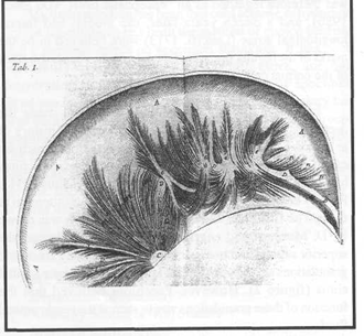
Figure 1. Plate 1 of Pacchioni's 1701 treatise. The radiate fibres of the falx cerebri were believed to be muscular bundles.
anatomists tried to describe the structure of the meninges and to understand their functions. Johannes Dryander (1536, PI. 4) described two meningeal layers (probably the dura and pia mater). Some years later, Andreas Vesalius (1543) depicted the dura mater with its vessels (sinuses and middle meningeal artery) and the pia mater. In the mid-seventeenth century, Jean Riolan denied the existence of both layers of the cerebral dura mater (meningeal and endosteal, respectively), but their existence was confirmed by Isbrand van Diemerbroeck (1695) and Philippe Verheyen (1708).
Antonio Pacchioni (1665-1726), a friend and pupil of Marcello Malpighi, was particularly concerned with the anatomy and function of the dura mater (Kemper, 1905;
Norman, 1983; Eimas, 1990). In his 1701 treatise, he described in detail the tentorial incisure (Pacchionian foramen) and the structure of the falx cerebri, including its radiate fibres (figure 1). Unfortunately, he mistook them for muscle and tendon bundles which were supposed to contract the falx and therefore compress the cerebral cortex. According to Pacchioni, this compression was intended to make the cerebral glands secrete the animal spirits. The dura mater was therefore regarded as an "encephalic heart" (De Smet, 1986), and a dozen years later, the medial and lateral longitudinal striae (Lancisi, 1713) were believed to be the marks of the regular impacts of the falx on the superior surface of the corpus callosum.
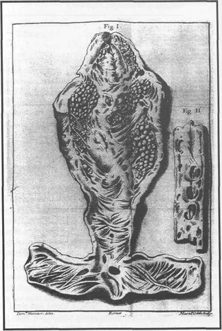
Figure 2. Plate 1 of Pacchioni's 1705 dissertation. The superior sagittal and transverse sinuses are open, and many arachnoid granulations are depicted.
In a later dissertation (1705), Pacchioni made the decription which made him find his place in the history of anatomy: he described the arachnoid granulations which are called "Pacchionian glands" since that time. These structures had been previously depicted by Andreas Vesalius (1543, Plates 7 and 66), but the author did not pay much attention to them. The first plate of Pacchioni's dissertation was drawn by D. Moratori and engraved by N. Oddi. It depicts the superior sagittal and transverse sinuses, and many arachnoid granulations are to be seen in the lumen of the superior sagittal sinus (figure 2). However, Pacchioni believed that the function of these granulations was to secrete the cerebrospinal fluid.
Antonio Pacchioni made therefore accurate anatomical descriptions, but misunderstood the functions of the dura mater and arachnoid granulations.
Some decades later, Giovanni Fantoni (1675-1758) corrected both Pacchioni's mistakes: on the one hand, he showed that the dura mater does not contain any muscular bundles, and on the other hand he asserted that the arachnoid granulations are in charge of the reabsorption, and not secretion, of the cerebrospinal fluid: "The humoral flow is sent to the superior sagittal sinus rather than to the hemisphere convexity. This is more in line with the laws of nature, and the sinus itself, and not the meninges, is irrigated by this liquid, and the blood will therefore be diluted" (1738).
Though he misunderstood the nature of the fibrous tracts in the falx cerebri and the real function of the arachnoid granulations, Antonio Pacchioni has to be regarded as a pivotal figure in the history of the anatomy of human meninges. He described the tentorial incisure and the arachnoid granulations which had only been mentioned by his predecessors, and his name rapidly appeared in the studies of his contemporaries (Heister, 1719). That is why Antonio Pacchioni became eponymous in the medical profession.
Clarke E, Dewurst K: Histoire illustree de la fonction cerebrale. Paris: R. Dacosta, 1984. Corsi P: La decouverte du cerveau. De l'art de la memoire aux neurosciences. Milan: Electa, 1990.
De Smet Y: La neuropsychologic "pre-corticale". Histoire de la localisation et de la constitution des "fonctions mentales superieures" de Thales de Milet a Franz Josef Gall. Bruxelles: Ciaco editeur, 1986.
Diemerbroeck I van: L'anatomie du corps humain. Lyon: Anisson & Posuel, 2 volumes (French translation by J. Prost), 1695.
Dryander J: Anatomia capitis humani. Marburg: E. Cervicornus, 1536. Dumont J: Les sinus posterieurs de la dure-mere et le pressoir d'Herophile chez l'homme. Nancy: A. Voirin et L. Kreis, 1894.
Eimas R: Heirs of Hippocrates. The Development of Medicine in a Catalogue of Historic Books in the Hardin Library for the Health Sciences, the University of Iowa, 3rd Ed. Iowa City: University of Iowa Press, 1991.
Fantoni G: Diss. de structura & motu durae membranae cerebri, de glandulis ejus, & vasis lymphaticis piae meningis. In: Opusculamedicaetphysiologica. Geneva: Pellissari, 1738.
Heister L: Compendium anatomicum. Altdorf & Nuremberg: Kohlesiano et Adolphiano, 1719.
Kemper GWH: The World's Anatomists. Philadelphia: P. Blakiston's Son & Co., 1905.
Lancisi GM: Diss. II. quarum prior est de physiognomia, altera de sede cogitantis animae. Venice, 1713.
Norman J: Medicine and the life sciences. Catalogue thirteen. San Francisco: J. Norman & Co., 1983.
Olry R: Medieval Neuroanatomy: the Text of Mondino dei Luzzi and the Plates of Guido da Vigevano. J Hist Neurosci 6 (2): 113-123, 1997. https://doi.org/10.1080/09647049709525696
Pacchioni A: De durae meningis fabrica & usu disquisitio anatomica. Rome: typis D. A. Herculis, 1701.
Pacchioni A: Dissertatio epistolaris ad Lucam Schroeckium de glandulis conglobatis durae meningis humanae, indeque ortis lymphaticis ad piam meningem productis. Rome: Giovanni Francisco Buagni, 1705.
Verheyen P: Anatomie oder Zerlegung des menschlichen Leibes. Leipzig: Thomas Fritsche, 1708.
Vesalius A: De humani corporis fabrica libri septem. Basle: Oporinus, 1543.
Vigevano G da: Liber notabilium illustrissimi principis Phiippi septimi, Francorum regis, a libris Galieni per me Guidonem de Papia, medicum suprascripti regis atque consortis ejus indite Johanne regine, extractus, anno Domini millesimo CCC° XLV°, papa vivente Sexto Clemente. Latin manuscript No. 569, Muse'e Conde de Chantilly, 1345.