Department of Biology,
Union University,
Jackson, TN 38305
USA
Plastinated tissues have gained popularity due to their structural integrity and the ease in handling and storing. Specimens can lose natural (or added) color throughout the plastination process, and so one objective of coloration is to restore natural color. A second objective of this study was to measure color change, both objectively and by subjective visualization of coloration. Coloration was investigated using 3 pigments added at 4 defined points during the 5-stage plastination process, using squirrel leg muscle samples. Each of the 12 experimental groups, and an untreated sample, were analyzed by measuring the Euclidean distance and CIE2000 DE values. Statistical analysis indicated that none of the coloration treatments restored natural coloration. However, the coloration did show subjective improvement in the contrast among tissues. The results showed that pigment addition prior to acetone dehydration, or prior to silicone impregnation, resulted in the best coloration, and that the use of Rit dye was most effective. Use of this approach was subsequently validated using human arm tissue, which consists of more tissue variety than squirrel legs.
Brea Reese ink; color; dyes; human arm; pigments; pigments for silicone; Rit All-purpose liquid dye; squirrel leg
Dr. Mark G. Bolyard, Department of Biology, Union University, Jackson, TN 38305, USA. Tel.: +1 731-661-6586;
E-mail: mbolyard@uu.edu

![]()


Plastinated tissues are effective study models for students, physicians, and scientists, and are great anatomical study resources today. Usefulness of plastinates stems from their durability, lack of moisture in finished specimens, and absence of harmful chemicals (von Hagens et al., 1987). Loss of color during the fixation and dehydration steps of plastination can detract from the usefulness and realism of these tissues. A common method used in previous research into the coloration of plastinated tissues has been the direct addition of pigments to the surface of specimens after the completed plastination process (Steinke and Spanel-Borowski 2005; McCreary et al., 2013; Raoof et al., 2013; Steinke et al., 2017).
Previous coloration studies have focused on the addition of pigments at 1 or 2 points within the 5-step process of plastination, and only one previous study quantitatively evaluated the change in color. McCreary et al. (2013) focused on addition of pigments post-plastination, while Steinke and Spanel-Borowski (2005) added pigments prior to the first step of plastination. Several other studies focused on additions either prior to curing, or immediately before or during silicone impregnation (Sakamoto et al., 2006; Mendez et al., 2008; Raoof et al., 2013). The materials used across each of these studies varied widely, including Biodur pigments, acrylics, dyes, traditional histological stains, and organic compounds.
McCreary et al. (2013) studied the addition of pigments post-plastination by evaluating 7 test solutions with varying amounts of Biodur pigments mixed with silicone and methyl ethyl ketone. They found that the mixtures worked best on muscle tissue when compared to vasculature, due to the greater surface area. Raoof et al. (2013) tested the addition of 6 pigment mixtures of differing colors, bases, and catalysts to the specimens prior to curing. The success of these mixtures was determined through physical wear tests, and it was found that addition of the paints prior to curing allowed for greater durability of the color. Steinke and Spanel-Borowski (2005) added pigments onto vasculature in their study prior to dehydration, and most endured dehydration without severe discoloration. Interaction among the different pigment mixtures on the surface of the plastinates resulted in color loss.
Steinke and coworkers (2017) used a periodic acid-Schiff (PAS) reaction on pelvic, thigh, and foot samples post-dehydration, with the purpose of defining fasciae across the specimens. A Sudan III stain was added prior to the PAS process, in order to stain fatty areas of the tissue. They found that the PAS stain produced staining that helped to easily distinguish between muscle tissue and the surrounding tissues, and the addition of the Sudan III stain did not negatively impact the PAS stain, whatsoever. However, the cost of these reagents may make it challenging for routine use.
Sakamoto et al. (2006) used the Romanhanyi technique of reactivation of hemoglobin in tissues to produce color in the plastinated specimens, in order to give the specimens a natural red color. The imidazole-ethanol mixture was added to the polymer mixture and allowed to enter during the impregnation process. The added mixture resulted in authentically red specimens with visible areas of hemorrhage and erythema. Exposure to the atmosphere over the years post-study resulted in some loss of surface color.
Mendez et al. (2008) used a similar blood color reactivation method to Sakamoto et al. (2006). In their study, half of the samples received the imidazole mixture prior to impregnation. The added imidazole mixture resulted in obvious red coloration of kidney specimens, but not lung tissue, and, ultimately, there was no significant difference between the control and experimental groups. However, their study highlights that this red color was unnatural, and exposure to the atmosphere resulted in discoloration.
Mendez et al. (2008) also obtained quantitative data from the color of the samples. They compared saturation and hue changes of the colored plastinated tissue to the same tissue prior to coloration using ImageJ software. They measured colors within defined color spaces that bring together color attributes such as hue, saturation, and brightness or intensity of specific colors. Two color spaces, RGB (red, green, blue) and L*C*h (lightness, chroma axis, hue), utilize these characteristics to define specific colors. Represented in 3D form as a cube, the RGB color space consists of the three primary colors, red, green, and blue as well as a grayscale (Ibraheem et al., 2012). The L*C*h color space is a spherical representation of color, based on luminance, chroma, and hue (Okada et al., 2007). Color positions within these color spaces can then be compared to other color positions giving a distance between colors. Distance between colors within the RGB color space is typically defined through the Euclidean distance (Koschan and Abidi, 2008). Euclidean distance offers a simple approach to color distance, but it does not relate to human color perception. Euclidean distance in the RGB space is expressed as values between 0-255. A value of 0 indicates that the two colors are exactly the same. Values above 0 signify that there is a difference between the two colors. Distance within the L*C*h color space is also measured through a variation of the Euclidean distance formula, including the values of luminance, chroma, and hue angle (Koschan and Abidi, 2008). The International Commission on Illumination developed a specific formula using the L*C*h color space and Euclidean distance in 1994. The most updated version of this formula, CIEDE2000, fixes luminance issues by weighting them correctly within the formula (Schuessler, 2014). The CIEDE2000 outputs color distance as the distance metric DE which is based on visual human color perception (Schuessler, 2014). Values of DE fall between 0-100, with a distance value less than 1 indicating no perceptible difference between two colors.
Each of these studies approaches coloration of plastinated specimens in very different ways. Most of these coloration studies, however, focus on pigment addition at only 1 or 2 points in the plastination process. The study reported here examined the addition of three types of color, at four points in the plastination process. We have evaluated the coloration process of squirrel legs using the CIEDE2000 process, as well as a visual comparison of samples, which led to a subsequent comparison of human arm tissue, using the method that was most effective for squirrel tissue.
Dissection and Fixation
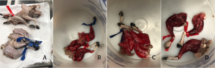 Figure 1. A: squirrel leg samples immediately prior to the dehydration process; B: painted with Rit Dye; C: painted with Silc Pig; D: painted with Brea Reese |
|
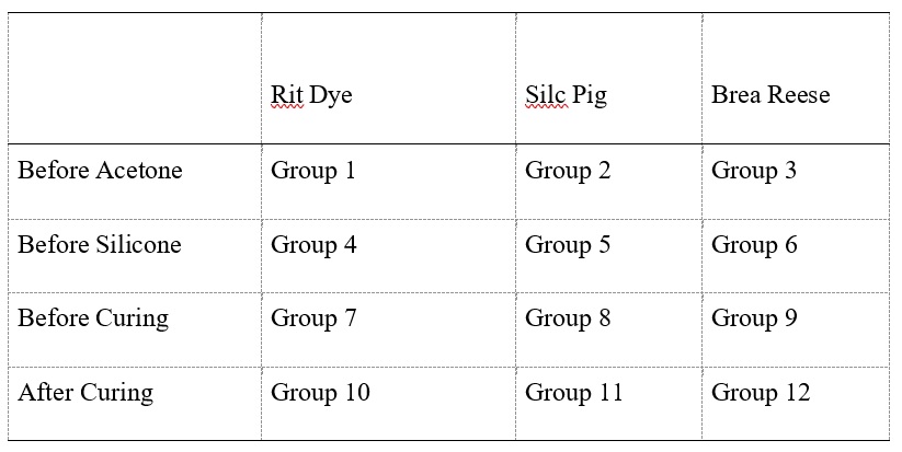 Table 1. Squirrel Leg Experimental Group Number. Breakdown by addition point and color added |
Twenty squirrels that had been previously collected, euthanized, and frozen were donated to the University. Later, specimens were thawed, skin removed, and the pelvic limbs were removed from the body (Fig.1). Each leg was opened on its cranial aspect to expose deep muscle. Some fascia was left on each leg. After fixation in 10% formalin for 7 days, the limbs were flushed with water for 11 days, and divided into 13 groups of 3 leg specimens (Table 1). Embalmed human transverse sections were prepared using the same methods to compare with the squirrel plastinates. Based on the results from the squirrel tissue, only processes using Rit Dye and Brea Reese pigments were completed.
Plastination procedures were followed according to de Jong and Henry (2007). Prior to pigment addition, each specimen was air dried to allow a uniform coat of pigment to be applied with a brush. Coloration at each step involved adding a coat of the pigment to the entire surface of the samples. Each pigment was gently mixed prior to addition at each step to ensure homogeneity. After flushing, the 3 legs (muscle, fascia, and other exposed structures) in the first three groups (Groups 1-3) were painted with the appropriate undiluted pigment, either Rit All-Purpose Liquid Dye in the shade Scarlet; NC brand Red silicone pigment (Silc Pig), or Brea Reese ink in the shade Crimson (Table 1).
Dehydration
After coloring, the 3 colored groups of specimens were placed in -12° C acetone in separate plastic buckets to avoid pigment mixing (Fig. 1). The remainder of the samples were placed into a large tub of -12° C acetone to dehydrate in a freezer. For the first bath, 96% acetone was used. The acetone-to-water ratio was checked with an acetonometer, and was changed when the acetone percentage equilibrated. The acetone was changed 5 times over a period of 47 days. The samples were removed from the acetone tanks once the acetone equilibrated at 97%.
Defatting (degreasing)
After dehydration, the samples in acetone were moved to room temperature for 2-5 days to defat the samples. The change from -12° C to room temperature allows for the removal of excess fats from the specimens.
Forced Impregnation
Pigments were added to three additional groups (Groups 4-6) of samples prior to silicone impregnation (Table 1). All samples were submerged in liquid S10 silicone mixed with S3 (the catalyst with chain extender) in a forced impregnation tank. The pressure of the vacuum chamber was decreased to 686 mmHg (27 in Hg) and allowed to stabilize for 1 day at -12° C. The pressure of the tank was then continuously but slowly decreased to 762 mmHg (30 in Hg). When impregnation was finished, pressure was returned to atmosphere and the specimens removed from the polymer-mix.
Manicuring
The surface of each leg sample was cleaned twice a day for 5 days, to remove excess silicone, until minimal amounts of silicone were seen on the surface of the specimens. At this time, the pigment was applied to three additional groups of samples (Groups 7-9), prior to curing (Table 1).
Curing/Hardening/Cross-linking
For curing, S6 cross-linker was vaporized by bubbling air through an aliquot of S6 for 7 days in a closed plastic tank, to harden the specimens. After curing, pigments were added to the final three sets of samples (Groups 10-13) (Table 1).
Analysis
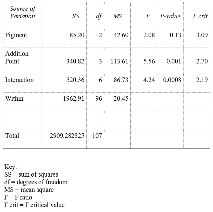
Analysis of the squirrel results was based on the distance between colors defined in two ways: the Euclidean distance and DE. In order to carry out analysis of these two values, the images of each specimen were uploaded into the ImageJ software system. An area of muscle was defined with the polygon tool, and the pixel coordinates within that area were exported to Microsoft Excel. Three pixel locations for each specimen were randomized from the Excel file. The ImageJ color picker tool was then used to find the hexadecimal code for the colors in each of the 3 pixels. A single hexadecimal value was determined for the fresh squirrel muscle tissue by averaging 6 randomized pixel hexadecimal codes within a defined area of muscle on 2 separate legs. A JavaScript library containing embedded functions for working with colors was used for calculating the Euclidean distance (Aisch, 2013). The JavaScript library generated an RGB value between 0-255, giving the distance between the natural squirrel muscle tissue and the experimental color. The DE value was calculated through an online color difference calculator (Lindbloom, 2012). The original CIE formula utilized the L*a*b color space; therefore, the CIEDE2000 formula used converts L*a*b values to the appropriate L*c*h values before determining the DE (Ibraheem et al., 2012). The hexadecimal codes for each specimen, including the fresh squirrel muscle tissue, were converted to L*a*b values through the ColorHexa website (Color Hexa, 2012). The CIEDE2000 formula generated a DE value between 0-100, giving the distance between the fresh squirrel muscle tissue and experimental tissue color. A two-way ANOVA test was used to test significance of the data using a p-value <0.05. This test was part of the Microsoft Excel data analysis tool pack.
Application of squirrel results to human tissue
Based on the results of the squirrel data, it was decided to prepare three transverse sections of an embalmed human arm to evaluate and compare with squirrel data, following the same methods as were used for the squirrel legs above. One slice was left untreated, one was painted with Rit Dye prior to the acetone step, and one was painted with Brea Reese prior to the acetone step. Color was reapplied after each acetone change (usually 2-3 applications). Each sample was kept in a separate tank so that the stain would not cross-react with the other specimens. The plastination process for these samples was completed as described above for the squirrel tissue. For this trial with human tissue, the process using Silc Pig was not completed because it did not absorb into the tissue at all, but would be present only superficially on the tissue and easily wipe off without staining the tissue. The results were not indicative of the desired natural presentation of plastinated specimens (data not shown).
The impact of the coloration was evaluated quantitatively, and by visual/physical inspection. Euclidean distance and CIE2000 DE values were averaged within the same treatment (Table 2). Analysis of the Euclidean distance and CIE2000 DE by addition point indicated that the addition of Silc Pig and Brea Reese dyes at later points in the process produced results that were quantitatively further from fresh tissue. Conversely, Rit Dye demonstrated essentially the same results at each point of addition. Although there was a large visual difference between the specimen groups, two-way ANOVA analysis revealed that there was no significant difference (p >0.05) between pigment groups, but there was a significant difference (p <0.05) between addition points for both Euclidean distance (Appendix A) and CIE2000 DE (Appendix B).
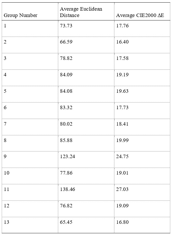 Table 2. Average Euclidean Distance and CIE2000 E Values for Each Experimental Squirrel Muscle Group (group 13 is untreated tissue). |
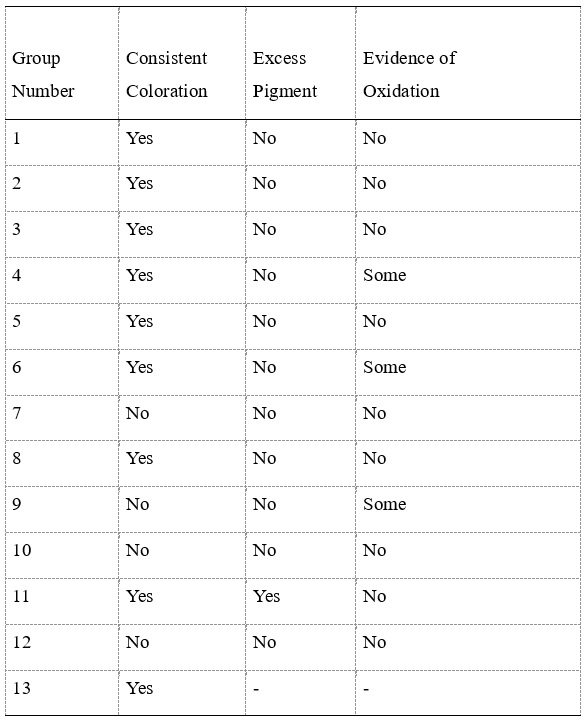 Table 3. Visual Results for Each Experimental Squirrel Leg Group Based on Consistent Coloration, Excess Pigment, and visual observation of Oxidation. |
Visual/physical results showed that most specimen groups maintained consistent coloration (Table 3). However, Rit Dye and Brea Reese “before curing” and “after curing” specimen groups showed evidence of beading up of the pigment on the surface upon visual inspection post-plastination. Excess pigment was seen only in the Silc Pig specimen group at the “after curing” addition point (Table 3). Visual inspection of the Rit Dye and Brea Reese “before silicone impregnation” groups (Table 3) showed possible oxidation of these pigments on the surface of the squirrel legs, as evidenced by darkening of the sample color. Further visual inspection indicated that the Rit Dye and the Brea Reese gave a natural, fresh tissue appearance to the musculature of the squirrel legs.
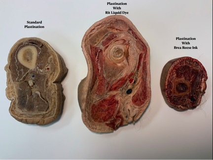 Figure 2. Human arm sections following the completion of the process, including curing. Untreated (left); treated with Rit Dye (middle); treated with Brea Reese (right) |
Because squirrel leg tissue consists almost entirely of muscle and bone, human arm tissue was used to evaluate the performance of the Rit Dye and Brea Reese dye in a specimen with a greater variety of tissue types. When compared to untreated arm tissue by visual inspection, Rit Dye remained in muscle tissue but was washed from fat and connective tissue during the acetone phase, giving strong contrast between the tissues, and a realistic appearance. However, the Brea Reese dye remained in all of the tissues, and did not provide the tissue contrast noted with Rit Dye (Fig. 2).
Plastination has been demonstrated to be a valuable process due to the long-lasting, durable specimens it produces. However, the preservatives and fixatives used during the process can alter the color of the specimens. This discoloration tends to result in a bleaching effect on the skin and a severe lightening of the muscle tissue.
Our work supports previous efforts (Steinke and Spanel-Borowski, 2005; Sakamoto et al., 2006; Mendez et al., 2008; McCreary et al., 2013; Raoof et al., 2013; Steinke et al., 2017) that indicate promising possibilities for coloration of plastinated specimens.
 Appendix A. |
 Appendix B. |
Aisch, G. (2013, May). Chroma.js. https://gka.github.io/chroma.js/.
Color Hex. (2012). Retrieved July 25, 2020, from https://www.colorhexa.com/.
de Jong, K. and Henry RW. 2007: Silicone plastination of biological tissue: cold temperature technique BiodurTM S10/S15 technique and products. J Int Soc Plastination 22:2-14.
https://doi.org/10.56507/ZLMJ7068
Ibraheem NA, Hasan MM, Khan RZ, Mishra PK. 2012: Understanding color models: a review ARPN J Eng Appl Sci 2: 265-275.
Koschan A and Abidi M. 2008. Digital Color Image Processing. John Wiley & Sons.
https://doi.org/10.1002/9780470230367
Lindbloom, B. J. (2012, January 13). Color Difference Calculator. http://www.brucelindbloom.com/index.html?Eqn_DeltaE_CIE2000.html.
McCreary J, Iliff, S, Hermey D, McCreary K, Henry RW. 2013: Silicone-base coloration technique developed to highlight plastinated specimens J Plastination 25: 13-20.
https://doi.org/10.56507/XLBR3803
Mendez BA, Romero RL, Trigo FJ, Henry RW, Candanosa AE. 2008: Evaluation of imidazole for color reactivation of pathological specimens of domestic animals. J Int Soc Plastination 23:17-24.
https://doi.org/10.56507/HZTR8339
Okada K, Ueda Y, Oyabu J, Ogasawara N, Hirayama A, Kodama K. 2007: plaque color analysis by the conventional yellow-color grading system and quantitative measurement using LCH color space. J Interven Cardiol 20: 324-334.
https://doi.org/10.1111/j.1540-8183.2007.00276.x
Raoof A, Marchese C, Marchese LA, Falk KC, Mirafzali N. 2013: Painting plastinated neurovascular pathways: evaluation of coloring techniques. J Plastination 25: 21-26.
https://doi.org/10.56507/LJZQ6496
Sakamoto Y, Myake Y, Kanahara K, Kajita H, Ueki H. 2006: Chemically reactivated plastination with shin-etsu silicone KE-108. J Int Soc Plastination 21:11-16.
https://doi.org/10.56507/BSRA2644
Schuessler Z. 2014: Delta E 101. http://zschuessler.github.io/DeltaE/learn/#toc-introduction.
Steinke H, Spanel-Borowski K. 2005: Coloured plastinates. Ann Anat 188:177-182.
https://doi.org/10.1016/j.aanat.2005.10.001
Steinke H, Wiersbicki D, Speckert M, Merkwitz C, Wolfskampf T, Wolf B. 2017: Periodic acid-Schiff (PAS) reaction and plastination in whole body slices. A novel technique to identify fascial tissue structures. Ann Anat 216:29-35.
https://doi.org/10.1016/j.aanat.2017.10.001
von Hagens G, Tiedemann K, Kriz W. 1987: The current potential of plastination. Anat Embryol 175: 411-421.
https://doi.org/10.1007/BF00309677