1 Departement de chimie-biologie, Universite du Quebec a Trois-Rivieres, Trots-Rivieres, Quebec, Canada
2 Medical Museum, Medical Department, University of Tokyo, Hongo, Bunkyoward, Tokyo, Japan.
The collaboration of artists and anatomists marked the evolution of anatomical illustration, and is a liaison which dates back as far as the sixteenth century. The most striking anatomical masterpieces were drawn and engraved as illustrations in noteworthy books, but could unfortunatelly not be preserved due to the lack of effective, long-term preservation methods. Plastination therefore finds in museography, potential new developments, and is capable of putting new life into the history of artistic anatomy. This process can exhibit three-dimensional plastinated specimens which have been inspired by some of the most impressive plates of previous centuries.
Exhibition - Baroque anatomy - History of anatomical illustrations - Plastination.
R. Olry, Departement de chimie-biologie, Universite du Quebec a Trois-Rivieres, CP 500, Trois-Rivieres, Quebec, Canada G9A 5H7. Telephone: 819 376 5053 /Fax: 819 376 5084. Email: Regis_Olry@uqtr.uquebec.ca

![]()


Plastination is the process accepted to be the most promising preservation method for educational use in anatomy and related fields. Numerous publications and conferences emphasized its exceptional potential for teaching anatomy (for review, see Grondin and Olry 1996). However, a three- dimensional plastinated specimen is first, a specimen which must be dissected. The pedagogic features rely more upon skillful dissection than on the plastination procedure itself. The evolution of the promise of plastination, therefore, re- minds us of the history of anatomical illustrations. The talent of drawers and engravers was often the basis for the renown of the anatomists by whom they were employed. We, therefore, feel that the baroque anatomical masterpieces of previous centuries, should effectively serve as models for future plastinated specimens.
Pietro Betrettini da Cortona
Pietro Berrettini (1596-1669), usually known as Pietro da Cortona after his birthplace, was perhaps the most influential painter of the Italian Baroque movement (Norman 1986).
His anatomical plates, most of them probably engraved by Luca Ciamberlano in 1618, were published over 100 years after their completion by the Italian surgeon Caetano Petrioli (Berrettini 1741). A second and final edition was published in 1788 by the Italian professor of medicine and philosophy, Francesco Petraglia (Berrettini 1788).
The twenty-seven plates dealt mainly with the muscular and peripheral nervous systems, but plates XXI-XXVI appear to have been added later and drawn by another artist (Duhme 1981). On plate XII of the 1741 edition (figure 1), a kneeling eviscerated body holds a medallion in his right hand depicting the anterior cervical region which displays the nerves of the tongue and vicinity. In his left hand, is held a bone which replaced a piece of wood, seen in the original drawing (figure 2). The ribs were resected along the midaxillary line, so that the intercostal nerves became evident. Brachial and lumbosacral plexi are depicted, and some muscles of the upper and lower limbs were removed to demonstrate the course of median, ulnar and femoral nerves. Though the accuracy of some muscles could be questioned, numerous nervous rami could be traced to their respective muscular masses. The intercostobrachial nerve (Hyrtl 1873), can be seen on the left side of the specimen, but its origin cannot be accurately determined as the thorax interestingly possesses thirteen ribs.
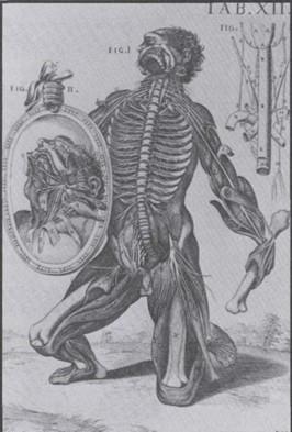 Figure 1. Berrettini's Plate XII, probably engraved by Luca Ciamberlano towards 1620 (taken from Norman 1986). |
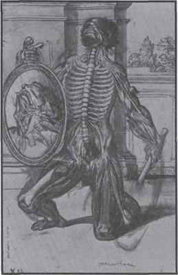 Figure 2. Original drawing for Berrettini's Plate XII, University of Glasgow Library, Hunterian Collection (taken from Norman 1986). |
Claude Nicolas Le Cat
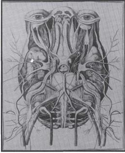
Figure 3. The brain and related structures in Le Cat's essay, engraved by Herisset (taken from Rubin and Nor- man 1987).
Claude Nicolas Le Cat (1700-1768) could be regarded as one of the most extravagant ophthalmologists of the seventeenth century. Having once invited his fellow member "Chevalier" John Taylor (1703-1772) to dinner, he "served him a splendid lunch capped off with a covered dessert that turned out to be, when uncovered, a dissection of the nerves to the extraocular muscles which blatantly proved that they could not have been excised by Chevalier's method" (Rubin and Norman 1987). Le Cat's treatise (1740), part of a work in progress on physiology which never was completed, contains a very interesting view of the brain and nearby vascular,
peripheral and autonomic nervous structures (figure 3). This plate, engraved by Herisset, exhibits the base of the brain, but the superior third of the face was preserved in- tact. The floor of the orbital cavities was removed so that the extraocular muscles and ciliary ganglia were revealed. Most of the cranial nerves are depicted, especially the trigeminal nerve, including the ophthalmic, maxillary and mandibular branches, and its voluminous ganglion. Surprisingly, the discovery of this ganglion is usually attributed to Gasser's pupil Anton Raymund Balthasar Hirsch in his study of the fifth cranial nerve published only twenty-five years later (Olry 1995). The superior cervical ganglia and many of their rami (including the internal carotid nerve) roughly compare to the description made by the celebrated Jacques Benigne Winslow (Olry 1996). The main arterial trunks (internal carotid, external carotid, vertebral and basilar arteries) are present, and part of the course of the intracranial segment of the left internal carotid artery was severed so that both oculomotor and abducent nerves could be dis- played.
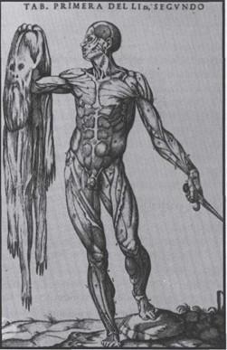
Figure 4. Valverde's «flayed man», engraved by Nicolas Beatrizet after drawings by Caspar Beccera (taken from Roberts and Tomlinson 1992).
Juan Valverde de Amusco
The Spanish anatomist Juan Valverde de Amusco (c.l525-c.l587) illustrated a book (1556) whose contents were derived from the Vesalian woodcuts (Choulant 1852; Roberts and Tomlinson 1992). However, his reputation as a plagiarist could be questionned as he corrected or improved many anatomical details in these plates (Guerra 1967). A plate, engraved by Nicolas Beatrizet after drawings by Caspar Beccera, is probably one of the most striking anatomical illustrations of the sixteenth century (figure 4). A flayed man holds in his right hand, his own skin, and in his left hand a dagger which appears to have been used as the skinning knife. This plate resembles Vesale's plate XXVI, but the abdominal musculature is better depicted here. The other plates of Valverde's book also have a quite "surrealist" appearance: dissected torso clothed in armour, dissected man dissecting another man (in turn plagiarized by Jan Wouters in 1569), standing man, holding between his teeth, his own elevated abdominal wall so that the small intestine and greater omentum can be seen (Roberts and Tomlinson 1992).
Thomas Bartholin
Thomas Bartholin (1616-1680) descended from a renowned family of Danish anatomists (Bouchet and Picault 1956): his father Caspar Bartholin (1585-1629) was a professor of anatomy and rector of the Copenhagen University, and his uncle Ole Worm (1588-1654) is known as the dis- coverer of the so-called "wormian" or sutural bones (Olry 1994). For the 1655 revision of his father's classic anatomy text, Thomas Bartholin used a very surprising frontispiece, engraved by Jacob van Meurs (figure 5). The entire skin was removed from a body, except the head, hands and feet, which apparently still maintain their osseous skeleton, and the specimen (an "inside out" ecorche) was nailed to what appears to be a wooden frame. On the anterior aspect of the trunk was engraved the title of the book.
This strange frontispiece was introduced seventy years later in the Danzig edition of Johann Adam Kulm's famous handbook (Figure 6). It is noteworthy that the 1734 Dutch version of this book was one of the first Western medical texts translated into Japanese: Gempaku Sugita (1733-1817) and Ryotaku Maeno (1723-1803) edited the Japanese translation in 1774, and Sugita's pupil Gentaku Otsuki (1757- 1827) published a revised and corrected edition in 1826 (Rubin 1991).
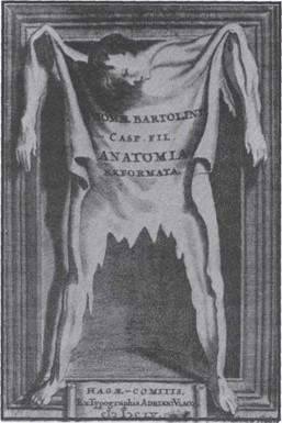 Figure 5. Frontispiece of Thomas Bartholin's treatise of 1655, engraved by Jacob van Meurs (taken from Nor- man 1982). |
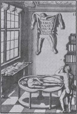 Figure 6. The same drawing introduced in Kulm's frontispiece of 1725 (taken from Wolf-Heidegger and Cetto 1967). |
Described in this article, are some of the most striking masterpieces of anatomical illustrations of previous centuries. Though some celebrated anatomists never wanted to acknowledge the necessity of including illustrations in their books (Jean Riolan, Xavier Bichat, etc...), most authors, as far back as the sixteenth century (for review, see Wolf- Heidegger and Cetto 1967), understood the importance of iconography. In order to increase the quality and the accuracy of their dissections, anatomists often employed the most renowned artists of their time whom they sometimes personally paid. Bernard Siegfried Albinus (1697-1770) ex- pended twenty-four thousand florins for his illustrations (Corner 1964), and Jacques Gamelin (1739-1803) expended his own personal fortune for the publication of an atlas, which bankrupted him.
However, the successful collaboration of Andre Vesale, an anatomist, and Joannes Stephan of Calcar, an illustrator (Titian's pupil), paved the way for a new trend in the his- tory of anatomical illustration. Covert Bidloo (1649-1713) had his specimens drawn by the famous Gerard de Lairesse, artist of Prince of Orange Guillaume III; the frontispiece of Realdo Colombo's treatise was the work of the celebrated Venetian painter Paul Veronese; and the title page of Fran9ois Michel Disdier's masterpiece was drawn by Fran9ois Boucher, artist to the King of France, Louis XV.
The striking specimens described in this paper had been dissected by renowned anatomists and illustrated by no less renowned artists and engravers. Unfortunately, the lack of any effective preservation methods doomed these masterpieces to a very short time span. Plastination, therefore, could prove to be the most recent development in the field of museography and breathe new life into the history of artistic anatomy, by preserving three-dimensional specimens inspired by the most celebrated anatomical plates of previous centuries.
Acknowledgments
The authors express their sincere appreciation to Mr Peter Kogon who criticized this manuscript, carefully scrutinized the English and offered numerous valuable suggestions.
Bartholin T: Anatomia, ex Caspari Bartholini... reformata... Accessit... appendix de lacteis thoracicis & vasis lymphaticis. Hagae-Comitis: Ex Typographia Adriani Vlacq, 1655.
Berrettini da Cortona P: Tabulae Anatomicae a celeberrimo pictore Petro Berrettino Cortonensi delineatae, & egregie aeri incisae nunc primum prodeunt, et a Cajetano Petrioli Romano doctore, Regis Sardiniae chirurgo, publico anatomico, & inter arcades Erasistrato Coo notis illustratae. Impeasis Fausti Amidei Bibliopolae in Via Cursus. Romae: Ex Typographia Antonii de Rubei apud Pantheon in via Seminarii Romani, 1741.
Berrettini da Cortona P: Tabulae Anatomicae Ex Archetypis Egregii Pictoris Petri Berrettini Cortonensis Expressae et In Aes Incisae Opus Chirurgis et Pictoribus Apprime Necessarium Alteram Hanc Editionem Recensuit Nothas Iconas Expunxit Perpetuas Explicationes Adjecit Franciscus Petraglia, Philosophiae Et Medicinae Pro- fessor. Romae: Impensis venantii Monaldi Bibliopolae Praesidum Facultae, 1788.
Bouchet A, Picault E: Une famille d'anatomistes danois, les Bartholin, et 1'histoire de la glande vulvovaginale. Lyon: Imprimeries R6unies, 1956.
Choulant L: Geschichte und Bibliographic der anatomischen Abbildung nach ihrer Beziehung auf anatomische Wissenschaft und bildende Kunst. Leipzig: Rudolph Weigel, 1852.
Corner GW: Anatomy. Clio Medica. New York: Hafner Publishing Company, 1964.
Duhme L: Die Tabulae anatomicae des Pietro Berrettini da Cortona. Koln: Institut fur Geschichte der Medizin, 1981.
Grondin G, Olry R: Current Plastination Index. Publica- tion of the International Society for Plastination, Trois- Rivieres, Canada, 1996.
Guerra F: Juan Valverde de Amusco. Clio Med 2: 339-362, 1967.
Hyrtl J: Lehrbuch der Anatomie des Menschen, mit Rucksicht auf physiologische Begründung und praktische Anwendung. Wien: Wilhelm Braumiiller, 12th edition, 1873.
Kulm JA: Anatomische Tabellen. Danzig, 1725.
Le Cat CN: Traite des sens. Rouen: n. p. (English transla- tion: A physical essay on the senses. London, 1750, R. Griffiths), 1740.
Norman J: Medicine and the Life Sciences. San Francisco: Jeremy Norman & Co., Inc., 1982.
Norman J: The Anatomical Plates of Pietro da Cortona. 27 Baroque Masterpieces. New York: Dover Publication, Inc., 1986.
Olry R: Os des Incas, os e'pactal, os wormien: histoire d'une trilogie mal comprise. Acta Belg Hist Med 7 (3): 137- 141, 1994.
Olry R: Dictionary of anatomical Eponyms. Jena, Stuttgart, New York: Gustav Fischer Verlag, 1995.
Olry R: Winslow's Contributions to our Understanding of the Cervical Portion of the Sympathetic Nervous Sys- tem. J HistNeurosci 5: 190-196,1996.
https://doi.org/10.1080/09647049609525666
Otsuki G: Jutei Kaitai Shinsho. Edo: Touto Shoritsu.
Senshobo, 1826.
Roberts KB, Tomlinson JDW: The Fabric of the Body. Eu- ropean Traditions of Anatomical Illustration. Oxford: Clarendon Press, 1992.
Rubin D: Medicine and the Life Sciences. Catalogue Twenty-One. San Francisco: Jeremy Norman & Com- pany, Inc., 1991.
Rubin D, Norman J: Medicine and the Life Sciences. Cata- logue Eighteen. San Francisco: Jeremy Norman & Com- pany, Inc., 1987.
Sugita G, Maeno R: Kaitai Shinsho. Edo: Tobu Shorin: I. Suharaya, 1774.
Valverde de Amusco J: Historia de la composici6n del cuerpo humano. Rome: Antonio de Salamanca and Antonio Lafreri, 1556.
Wolf-Heidegger G, Cetto AM: Die anatomische Sektion in bildlicher Darstellung. Basel, New York: S. Karger, 1967.
Wouters J: Dat Epitome ofte Cort Begriip der Anatomien, Andr. Vesalii Wt Het. Te Brugghe: Pieter de Clerck, 1569.