1Department of Animal Morphology & Physiology, São Paulo State University (Unesp), School of Agricultural and Veterinarian Sciences, Jaboticabal, São Paulo, Brazil
2Department of Biomechanics, Medicine and Rehabilitation of Locomotor System, College of Medicine, University of São Paulo (Usp), Ribeirão Preto, Brazil
3Department of Exact Sciences, São Paulo State University (Unesp), School of Agricultural and Veterinarian Sciences, Jaboticabal, São Paulo, Brazil
Formaldehyde is a fixative and preservative widely used in anatomy laboratories, but it is harmful to health, and poses an environmental risk. Ethyl alcohol (EtOH) has also been used for effective fixation of bird muscles, and sodium chloride has been successfully tested for the preservation of anatomical parts for more than five years. The objective of this present study was to evaluate a new anatomical technique for teaching surgical techniques using dog cadavers fixed in EtOH, and preserved in a 30% aqueous solution of sodium chloride (ASSC). In addition, we aimed to determine the ideal time to stop the fixation, so that the skin and jejunum present biomechanical characteristics as close as possible to the control group of fresh animals. Five groups were used: a control group (fresh animals without fixation or conservation), and the other 4 groups which differed in the time of fixation in EtOH (30, 60, 90 and 120 days). Except for the controls, all groups were conserved in 30% ASSC for 120 days. Statistical analysis of variance (ANOVA) revealed no difference between treatments and times (P > 0.05) relative to the skin, and showed at least one time significantly different from the others (P < 0.01) in relation to the jejunum. The non-linear modeling test showed differences in the group fixed in EtOH for 30 days, suggesting that this was the best time period for fixing dog cadavers for use in surgical training.
preservation, biomechanics, jejunum, dog
Fabrício S Oliveira, Department of Animal Morphology and Physiology, São Paulo State University (UNESP), Via de Acesso Paulo Donato Castelane, Jaboticabal, SP, 14884-900, Brazil. Telephone: 551632097333, e-mail: singaretti@fcav.unesp.br

![]()


Good conservation of anatomical specimens prevents deterioration, and also prevents the proliferation of pathogens that can spread diseases to laboratory personnel (Corrêa, 2003). Formaldehyde is widely used as an anatomical fixative and preservative, because it is inexpensive and rapidly penetrates the tissues (Rodrigues, 2010). However, it is hazardous to health, and can contaminate the environment through improper handling and through disposal of carcasses and effluent (WHO, 1991). In 2006, the International Agency for Research on Cancer (IARC) classified formaldehyde as carcinogenic and teratogenic (IARC, 2006). Glycerin is a preservative with dehydrating and antiseptic properties, preventing fungi and bacterial growth (Alvarenga, 1992). It does not have harmful fumes such as those released by formaldehyde (Cury et al., 2013), but it is up to ten times more expensive than formaldehyde (Krug et al., 2011).
There are a number of other examples of fixative solutions that are capable of preserving cadavers for surgical training, among them Thiel solution (composed of boric acid, ethylene glycol, potassium nitrate, (chloromethyl)phenol, sodium sulfate, and formalin) (Groscurth et al., 2001), Klotz's solution (sodium chloride, sodium bicarbonate, chloral hydrate, formalin and water) and Jores’ solution (distilled water, formaldehyde, sodium sulfate, potassium sulfate, sodium chloride, sodium bicarbonate, glycerin, and sodium or potassium acetate) (Rodrigues, 2010), all of which contain formalin. Modified Larssen solution contains 100 ml of 10% formaldehyde, 400 ml of glycerol, 200 g of chloral hydrate, 200 g of sodium sulfate, 200 g of sodium bicarbonate, 180 g of sodium chloride, and 2 liters of distilled water (Silva et al. al., 2004). The original Larssen solution did not contain liquid glycerin (Menezes, 2012). Laskowski solution contains 800 ml of glycerine, 200 ml of ethanol, 50 g of phenolic acid and 50 g of boric acid, and requires the preservation of cadavers at 0° C until they are used in surgery classes. Between classes, they must remain frozen, and have to be thawed in water for 24 hours prior to each class. With this solution, the tissues become excessively dark (Silva et al., 2007). Of these, Larssen solution is considered the best solution for maintaining the original consistency, color and characteristics of the biological material, and can also remove blood clots (Sampaio, 1989).
Ethyl alcohol (96%) has been found to be efficient for fixing and preserving the pectoral muscles of hens, although they became almost five times more rigid during the first six months, and three times more rigid after one year of immersion in the preservative agent (Nunes et al., 2011). The use of a 30% aqueous solution of sodium chloride (ASSC) in the preservation of anatomical specimens previously fixed by formaldehyde was successfully evaluated for 5 years, with no visual contamination, presence of putrefaction odors, or alteration of tissue color and softness (Oliveira, 2014).
There is a need to find a preservation technique that preserves the body realistically for a long time, serving not only anatomy classes, but also surgical and clinical studies, testing of new radiographic equipment, minimally invasive surgery, and encouraging more scientific research to develop protocols that will assist anatomists in the preparation of preserved cadavers, in order to meet these demands for high quality parts (Balta et al., 2015).
The objective of this study was to determine the viability of a new anatomical technique using EtOH for fixation, and 30% ASSC for conservation; and to establish the best time to stop fixation in EtOH for the optimal tissue resistance in comparison to fresh cadavers without fixatives/preservatives.
The animals
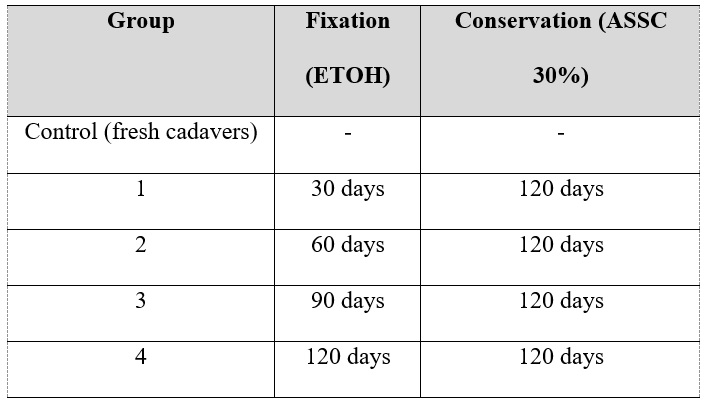 Table 1: Division of dog cadaver groups in relation to fixation time in ethyl alcohol (ETOH) and storage in a 30% sodium chloride solution (ASSC 30%). Control group consisting of cadavers of dogs not subjected to fixation or conservation (fresh). |
 Figure 1. Plastic box of 310 liters filled with 180 litres of EtOH (A). Dogs cadavers kept during ethylic fixation (B). |
Forty (40) frozen, adult, dog cadavers, 14 male and 26 female were used. The dogs all died due to causes that did not involve evident morphological alterations, such as large tumors, extensive lacerating traumas or bone fractures. All were from the Zoonoses Control Center of Ribeirão Preto, São Paulo, Brazil, after approval from the Municipal Law Department (process 02.2014/ 000027-1). The ages of the animals were determined from their death certificates.
The selected cadavers had a mean body weight of 7.6±2.7 kg, mean age 5.6±4.1 years, and body score of 4 to 5, which is considered ideal on a scale of 1 to 9 (Laflamme, 1997). They were thawed in a horizontal refrigerator at 8° C, weighed, and then randomly distributed into groups (Table 1).
For fixation, 96% EtOH containing 5% glycerin was infused with a 60 ml plastic syringe, via the common carotid artery, at a rate of 120 ml/kg. Each group consisted of eight animals that were fixed for different times (except for the control group).
After the fixative was injected, running water was used to flush out surplus liquid accumulated in the cavities via two incisions, one in the thorax (between the fourth and fifth right intercostal space), and a median abdominal incision. The cadavers were then placed in plastic tanks (1 tank per group) with a threaded cap (total capacity 310 liters – Fig. 1), containing 180 litres of EtOH. The abdomen and thorax were washed with water for 5 consecutive days. The plastic boxes were kept in an area and with abundant ventilation, away from sources of ignition, to avoid any risk of accident. Storage in 30% ASSC for 120 days followed the fixation period. Sodium chloride solution was placed in plastic tubs with a capacity of 310 liters, and the same volume (180 liters) was used for each fixation group.
Collection of Material
A 1 x 7cm stainless steel template was made for the collection of the skin and jejunum samples (chosen due to the higher tissue sample availability and frequent use in teaching of surgical techniques) during both the fixation in EtOH and 30% ASSC conservation solution. A total of 1,152 samples were taken.
The cadaver was initially placed in right lateral decubitus (left antimere, facing upwards). Using a scalpel (blade number 23) and the template, three sequential, equally-sized samples were collected transverse to the dog's skin tension line (Kirpensteijn and Haar, 2013), on the lateral side of the thorax, parallel to, and 5 cm from, the median plane (Figure 2). Shaving had been performed throughout the thorax. Samples from the control group were also collected in the left antimere and immediately submitted to biomechanical testing. During the fixation phase in EtOH, samples were collected from the 4 groups in the left antimere and, during the conservation phase in 30% ASSC, from the right antimere.
For the collection of the jejunum samples, the animals were positioned in the right lateral decubitus, exteriorizing the jejunum by manual traction. After identification of the duodenojejunal flexure, the steel template was positioned to delimit the specimen, which was then sectioned longitudinally with Metzenbaum scissors. Subsequently, section of the mesenteric edge was performed, exposing the lumen, under which the incision template was positioned with a scalpel blade (Figure 3).
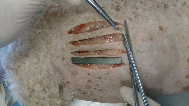 Figure 2. Skin samples collected transversally to the dog's skin tension line, on the lateral side of the thorax and using a steel mold, parallel to and 5 cm from the median plane after trichotomy. |
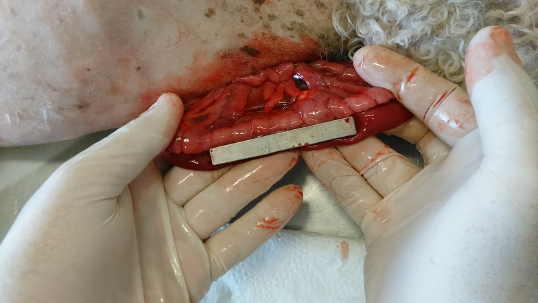 Figure 3. Jejunum sample collected with the cadaver positioned in the right lateral decubitus, after manual traction and using a steel mold. |
 Figure 4. Universal testing machine (A) for biomechanical analysis. A dog´s skin sample during a traction test (B). |
Tissue Strength Analysis
To evaluate tissue resistance, a Universal Testing Machine (Instron®- EMIC® - DL2000) was used, with a 500 N load cell and electromechanical drive support, with a speed of 100 mm/min. Traction claws were also used by manual compression, in the Laboratory of Surgical Anatomy of the Department of Morphology and Animal Physiology of FCAV - UNESP, Jaboticabal Campus, SP, Brazil (Figure 4). Tensile testing was conducted up to the point of rupture of the skin and jejunum samples, obtaining the values of the maximum force applied, in N.
Statistical Analysis
The data for the maximum force for the rupture of skin and jejunal tissues at different times and treatments were analyzed by analysis of variance (ANOVA) with significance set at 0.05.
For a more detailed analysis of the maximum jejunum breaking point as a function of time, nonlinear models were fitted in exponential form by the ordinary least squares methodology, according to the following equations:
 |
where Y is the maximum force (N), Yo is the mean in the stabilization of the maximum force (N) at the final times of the treatment, A1 is the weighted amplitude of the values of Y, Xo is the time of greatest decay rate of the maximum force (time), t1 is the mean decay rate (N time-1), and X is the measured time. MAPE is the mean percentage error, Yesti is the estimated value of maximum force at time i, Yobsi is the observed value of maximum force at time i and N is the number of data.
An ANOVA test was also performed to verify the differences between the adjusted parameters in the different treatments.
The preservation technique employed proved to be efficient for the fixation and conservation of the animals throughout the experiment. During the storage period in the plastic boxes in ASSC 30%, there was a gradual release of fat from cadavers in groups 3 and 4 (fixed 90 and 120 days in EtOH respectively). Because they remained longer in the alcohol fixative, they were more greasy and viscous at the end of storage in 30% ASSC, making it difficult to manipulate them during sample collection. During fixation in EtOH, stiffening of the skin and jejunum was observed when handling the samples, and later, there was an increase of the tissue malleability during the period of conservation in 30% ASSC. At the end of the period of alcohol fixation, the alcohol concentration ranged from 80 to 83%, which is an excellent amount of alcohol in the solution, and an appropriate degree of fixation of the cadaver for placement in the 30% ASSC for 120 days.
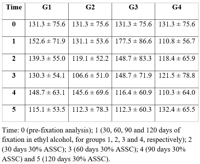 Table 2 - Absolute mean (± SD) of the maximum strength of rupture (N), of skin samples of groups 1, 2, 3 and 4, submitted to different fixation times in ethyl alcohol and preservation in an aqueous solution of sodium chloride. |
From the general data of the maximum rupture forces of the skin and jejunum samples submitted to the biomechanical tensile test, the absolute mean and respective standard deviation of groups 1, 2, 3 and 4 were obtained at different times of fixation in EtOH, and conservation in 30% ASSC (Tables 2 and 3). The maximum force required to rupture the skin and jejunum samples (at all times) ranged from 106.7 N to 177.5 N (mean 142.1 N) and from 12.9 N to 27.6 N (mean 20.2 N), respectively.
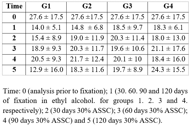 Table 3 - Absolute mean (± SD) of the maximum force of rupture (N) of jejunum samples of groups 1, 2, 3, and 4 submitted to different fixation times in ethyl alcohol and preservation in an aqueous solution of sodium chloride at 30% (30% ASSC). |
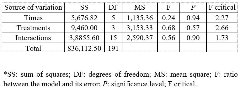 Table 4 - Analysis of variance (ANOVA) between treatments and time for skin samples from dog cadavers fixed at different times in ethyl alcohol and preserved in a 30% aqueous solution of sodium chloride for 120 days. There was no difference between treatments (P > 0.05). |
 Table 5 - Analysis of variance (ANOVA) between treatments and times for jejunum samples from dog cadavers fixed for different times in ethyl alcohol and preserved in a 30% aqueous solution of sodium chloride for 120 days. There was at least one different time in the treatments (P < 0.01). |
For the skin samples, ANOVA showed that there were no differences between treatments and times (Table 4), because the P value was always higher than 0.05. The ANOVA test also indicated that there were no interactions between the treatments and times. For the jejunum samples, ANOVA showed that at least one time was significantly different from the others (p <0.01) and that there were no differences between treatments (G1 to G4) (Table 5).
As a different time was identified in the statistical analysis of the jejunum, nonlinear modeling was performed, aiming to determine the time. In the modeling, the exponential decay model (equation 1) explained the variation of the maximum force for rupture of jejunum samples as a function of the different times in the treatments (Figure 5), since general MAPE was 9.6%. All values of Yo between the models were statistically similar, indicating that the mean maximum strength at the final times of the treatments were equal, thus confirming the efficiency of the 30% sodium chloride solution as a preservative. All other adjusted parameters showed at least one difference between the treatments, indicating that the response of the treatments varied in relation to the mean amplitude (A1), the time of the highest decay rate (Xo), and the decay rate (T1), to the maximum force values.
In the jejunum samples, a gradual decay of the values was observed for the maximum rupture force until the final fixation time in ethyl alcohol and a later stabilization of the maximum force for up to 120 days, thus confirming the preservative effect of the 30% ASSC for this type of tissue. Thus, in relation to jejunum, G1 presented A1 and Xo differently from the other groups, as this is the time of significance (Table 6).
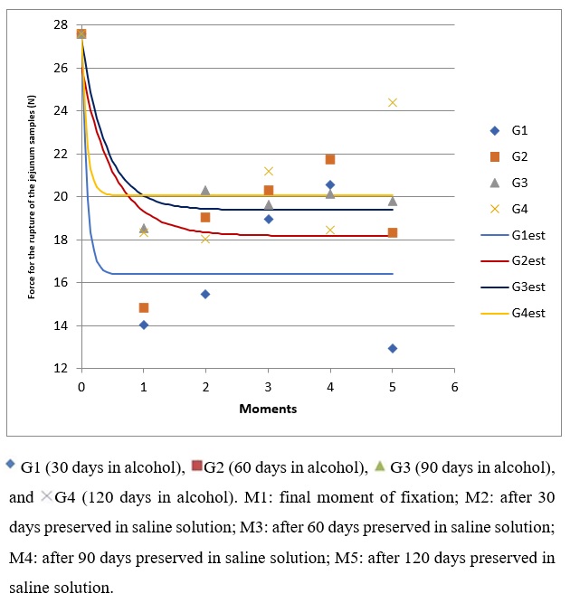 Figure 5. Nonlinear models of exponential decay relating to the maximum force as a function of different treatment times and different fixation times in ethyl alcohol and preservation for 120 days in a solution of 30% sodium chloride. Note the stabilization of the values during storage in saline solution. |
 Table 6 - Adjusted values of the parameters of the exponential non-linear decay model. The general model indicates the fit for all treatments together. The letters in superscript indicate the ANOVA test verifies at least one difference of the parameters between treatments of different fixation times in ethyl alcohol and preservation for 120 days in a 30% aqueous solution of sodium chloride. G1 (30 days in alcohol), G2 (60 days in alcohol), G3 (90 days in alcohol) and G4 (120 days in alcohol). |
Ethyl alcohol was effective as a fixative of dog cadavers, providing good conservation, and avoiding deterioration of the material, per Rodrigues (2010). The method described here is similar to the study involving alcohols used to fix human cadavers for 6 months to 1 year, which left the tissue quality like that of fresh tissue (Goyri-O’Neill et al., 2013).
The 30% ASSC solution was extremely effective in the preservation of the fixed tissues, as described by Oliveira (2014). There was no apparent contamination similar to that described in canine pericardium preserved for a minimum period of 90 days in hyper-saturated sodium chloride solution (Brun et al., 2002), and that described for the canine phrenic center preserved in glycerin (Brun et al., 2004). The 30% ASSC may be successful because of the difficulty of survival for microorganisms in a medium that requires an enormous osmoregulation capacity, similarly to the oceans (Munro et al., 1989), and the Dead Sea (Nissenbaum, 1975). Several concentrations of fixatives and preservatives have been evaluated for preservation of anatomical specimens, but the use of a sodium chloride solution below 20% has failed to preserve parts for use in tissue dissection (Friker et al., 2007). Statistical modeling analysis evidenced the stability of the data in this study, using 30% ASSC as a tissue preservative for skin and jejunal samples for up to 120 days.
There was no generation of contaminated effluent, commonly observed when toxic preservatives are used (WHO, 1991), nor any health-damaging fumes, such as those released by formaldehyde (Cury et al., 2013). In addition, the appropriate waste management of formaldehyde is costly in both financial and environmental terms, requiring the search for low-cost and non-risky alternatives (Janczyk et al., 2011).
Cadavers prepared for anatomical dissection were described as having better quality when a mid-line abdominal incision was performed (to enable the preservative to enter the abdominal cavity, to improve perfusion of the abdominal tissues) as was done in this study, instead of not opening the abdominal cavity (Janczyk et al., 2011).
Tissues become stiffer with formaldehyde, which, in biomechanical shear tests, caused a considerable stiffening in chicken breasts fixed for up to one year (4.4 to 5 times higher) (Guastalli et al., 2012). When EtOH was used as fixative, tissue stiffness increased, making them almost five times more lilely to rupture during the first six months, and three times more likely after one year of immersion (Nunes et al., 2011). However, in this study, the maximum tensile strength in the skin traction test (mean of 142.1 N) and jejunum (mean of 20.2 N) did not show a great variation in relation to the maximum strength of skin (131.3 ± 75.6 N) and jejunum (27.6 ± 17.5 N) of the unfixed cadaver control group.
Conventional procedures for fixing cadavers using formaldehyde are of limited use for surgical practise, due to the profound alteration in the staining, resistance, and fragility of organs and tissues. Artificial anatomical models are an alternative, and can be used mutiple times (Groscurth et al., 2001). However, with the method described here, each cadaver, prepared without the use of formaldehyde, can be used for training in surgical techniques for an entire semester, with life-like softness and tissue malleability.
ANOVA showed that there were no differences between treatments and times (P > 0.05 in all cases). Thus, among the evaluated time periods (30, 60, 90 or 120 days), there is no specific time that is better than others. Therefore, we suggest the shortest time (30 days - G1) for the preparation of the dog cadavers, because, due to the rapidity of preparation and the lower occupational risk that it confers, it is the best for cutaneous surgery.
Acknowledgement: FAPESP, process 2015/08259-9.
Alvarenga J. 1992: Possibilidades e limitações da utilização de membranas biológicas preservadas em cirurgia. In: Daleck CR (editor). Tópicos em cirurgia de cães e gatos. Jaboticabal: Fundação de Estudos e Pesquisas em Agronomia da Universidade Estadual Paulista. p 33-39.
Balta JY, Cronin M, Cryan JF, O'Mahony SM. 2015: Human preservation techniques in anatomy: a 21st century medical education perspective. Clin Anat 28: 725-734.
https://doi.org/10.1002/ca.22585
Brun MV, Pippi NL, Dreimeier D, Contesini EA, Beck CAC, Cunha O, Pinto Filho SL, Roehsig C, Stedile R. 2002: Solução hipersaturada de sal como conservante de pericárdio canino utilizado na reparação do músculo reto abdominal de ratos Wistar. Ciên Rur 32: 1019-1025.
https://doi.org/10.1590/S0103-84782002000600016
Brun MV, Pippi NL, Driemeier D, Contesini EA, Beck CAC, Cunha O, Pinto Filho SL, Roehsig C, Stedile R, Silva TF. 2004: Solução hipersaturada de sal ou de glicerina a 98% como conservantes de centros frênicos caninos utilizados na reparação de defeitos musculares em ratos Wistar. Ciên Rur 34: 147-153.
https://doi.org/10.1590/S0103-84782004000100022
Corrêa WR. 2003: Isolation and identification of filamentous fungi found in anatomical pieces preserved in 10% formalin solution. Dissertation (Biological Sciences). Institute of Research and Development, University of the Valley of Paraíba. 59p.
Cury FS, Censoni JB, Ambrósio CE. 2013: Técnicas anatômicas no ensino da prática de anatomia animal. Pesq Vet Bras 5: 688-696.
https://doi.org/10.1590/S0100-736X2013000500022
Friker J, Zeiler E, McDaniel BJ. 2007: From formalin to salt. Development and introduction on a salt-based preserving solution for macroscopic anatomic specimens. Tierärztl. Praxis 35: 243-248.
https://doi.org/10.1055/s-0038-1622624
Guastalli BHL, Nunes TC, Gamón THM, Carmo LG, Del Quiqui EM, Oliveira FS. 2012: Análise da textura de músculos submetidos à fixação em formaldeído e conservação em benzoato de sódio 0,5% e ácido acético 0,5%. Acta SciVet 40: 1041.
Goyri-O'Neill J, Pais D, Freire De Andrade F, Ribeiro P, Belo A, O'Neill A, Ramos S, Neves Marques C. 2013: Improvement of the embalming perfusion method: The innovation and the results by light and scanning electron microscopy. Acta Med Port 26: 188-194.
https://doi.org/10.20344/amp.4234
Groscurth P, Eggli P, Kapfhammer J, Rager GJ, Hornung P, Fasel JDH. 2001: Gross anatomy in the surgical curriculum in Switzerland: improved cadaver preservation, anatomical models, and course development. Anat Rec 265: 254-256.
https://doi.org/10.1002/ar.10030
IARC (International Agency for Research on Cancer). 2006: Formaldehyde, 2-butoxyethanol and 1-tertbutoxypropan-2-ol. IARC Monogr Eval Carcinog Risks Hum 88: 1-478.
Janczyk P, Weignera P, Luebke-Beckerb A, Kaessmeyera S, Plendla J. 2011: Nitrite pickling salt as an alternative to formaldehyde for embalming in veterinary anatomy: a study based on histo- and microbiological analyses. Annals Anat 193: 71-75.
https://doi.org/10.1016/j.aanat.2010.08.003
Kirpensteijn J, Haar G. 2013: Reconstructive Surgery and Wound Management of the Dog and Cat. Manson Publishing Ltd. London UK. p.12.
https://doi.org/10.1201/b15201
Krug L, Pappen F, Zimmermann F, Dezen D, Rauber L, Semmelmann C, Roman LI, Barreta MH. 2011: Conservação de Peças Anatômicas com Glicerina Loira. Concórdia: Instituto Federal Catarinense p 1-6.
Laflamme DP. 1997: Development and validation of a body condition score system for dogs. Can Pract 22: 10-15.
Menezes CLM. 2012: Preservation of rabbit cadaver with modified Larssen's solution for training in videolaparoscopic surgery. Federal University of Rio Grande do Sul: Brazil. Dissertation of Veterinary Sciences. 82p.
Munro PM, Gauthier MJ, Breittmayer VA. 1989: Influence of osmoregulation processes on starvation survival of Escherichia coli in seawater. Appl Environ Microbiol 55: 2017-2024.
https://doi.org/10.1128/aem.55.8.2017-2024.1989
Nunes TC, Oliveira FS, Gamon THM, Guastalli BHL, Carmo LG, Del Quiqui EM. 2011: Análise da textura de músculos peitorais submetidos á fixação e conservação em álcool. Braz J Vet Res An Sci 48: 464-467.
https://doi.org/10.11606/S1413-95962011000600004
Nissenbaum A. 1975: The microbiology and biogeochemistry of the Dead Sea. Microbiol Ecology 2: 139-161.
https://doi.org/10.1007/BF02010435
Oliveira FS. 2014: Assessing the effectiveness of 30% sodium chloride aqueous solution for the preservation of fixed anatomical specimens: a 5-year follow-up study. J Anat 225: 118-121.
https://doi.org/10.1111/joa.12185
Rodrigues H. 2010: Técnicas Anatômicas. Vitória-ES, Brazil: GM Gráfica & Editora.
Sampaio FJB. 1989: Study of the human kidney growth during fetal life. Thesis (Doctorate of Morphology). São Paulo State Medicine College, Brazil.
Silva RMG, Matera JM, Ribeiro AACM. 2004: Preservation of cadavers for surgical technique training. Vet Surg 33: 606-608.
https://doi.org/10.1111/j.1532-950x.2004.04083.x
Silva RMG, Matera JM, Ribeiro AACM. 2007: New alternative methods to teach surgical techniques for veterinary medicine students despite the absence of living animals. Is that an academic paradox? Anat Histol Embryol 36: 220-224.
https://doi.org/10.1111/j.1439-0264.2007.00759.x
WHO 1991: World Health Organization - IPCS International Programme on Chemical Safety - Formaldehyde - Health and Safety Guide. n. 57. Available at http://www.inchem.org.