The Journal of Plastination

Published in J. Int. Soc. Plast. 10 (1): 6-7 (1996)

Brain Preparation to Show Fibre Tracts and Nuclei

AUTHORS:
M. R. Haffajee
affiliations:

Department of Anatomy University of Durban Westville, South Africa

ABSTRACT:

The use of brain sections for the teaching of neuroanatomy is well known. Using brain sections along with stained sections to illustrate the differences between white and grey matter is common. In addition many texts contain illustrations outlining the various tracts and nuclei. However, alone or in combi- nation, all of these aids tend to complicate rather than simplify the understanding of neuroanatomy for the undergraduate.

The purpose of this paper is to outline a process of producing 3D models of the brain's fibre tracts and nuclei using Klinger's and von Hagens' procedures.

It should be noted that after Klinger's preparation the brains appear more porous under magnification possibly due to the presence of ice-crystals formed when thawing. This also assisted in making the tracts more definable for dissection.

The procedure described above may be supplemented by using a number of other useful techniques such as: opacification of the arterial tree or filling of the ventricles with a transparent resin (Thompsett, 1970).

KEY WORDS:

Fiber Tracts; Brain; Silicone: S10; Biodur; Plastination

*CORRESPONDENCE TO:

M. R. Haffajee Department of Anatomy University of Durban Westville, South Africa

Print Friendly, PDF & Email

Article Statistics

Volume: 10
Issue: 1
Allocation-id: 0000

Submitted Date:May 30, 1996
Accepted Date: June 27, 1996
Published Date: July 31, 1996

DOI Information:       https://doi.org/10.56507/LDTQ2301

Loading


Copyright 2022 International Society for Plastination

Copyright

This work is licensed under a Creative Common Attribution-NonCommercial-ShareAlike 4.0 International License.

Article Citation

The Journal of Plastination (May 5, 2024) Brain Preparation to Show Fibre Tracts and Nuclei. Retrieved from https://journal.plastination.org/articles/brain-preparation-to-show-fibre-tracts-and-nuclei/.
"Brain Preparation to Show Fibre Tracts and Nuclei." The Journal of Plastination - May 5, 2024, https://journal.plastination.org/articles/brain-preparation-to-show-fibre-tracts-and-nuclei/
The Journal of Plastination - Brain Preparation to Show Fibre Tracts and Nuclei. [Internet]. [Accessed May 5, 2024]. Available from: https://journal.plastination.org/articles/brain-preparation-to-show-fibre-tracts-and-nuclei/
"Brain Preparation to Show Fibre Tracts and Nuclei." The Journal of Plastination [Online]. Available: https://journal.plastination.org/articles/brain-preparation-to-show-fibre-tracts-and-nuclei/. [Accessed: May 5, 2024]

INTRODUCTION

The use of brain sections for the teaching of neuroanatomy is well known. Using brain sections along with stained sections to illustrate the differences between white and grey matter is common. In addition many texts contain illustrations outlining the various tracts and nuclei. However, alone or in combi- nation, all of these aids tend to complicate rather than simplify the understanding of neuroanatomy for the undergraduate.

The purpose of this paper is to outline a process of producing 3D models of the brain's fibre tracts and nuclei using Klinger's and von Hagens' procedures.

MATERIALS AND METHODS

This process can be divided into three stages:

STAGE 1: Klinger's method of brain preparation
STAGE 2: Fibre tract and nuclei dissection
STAGE 3: Plastination of brains

Stage 1: Klinger's method of brain preparation

  • Select 3 brains per dissection required
  • Immerse and suspend the brains in 10% formalin overnight
  • Add fresh 10% formalin after 24 hours
  • Pass a wide bore needle into both ventricles of each brain to assist in the passage of fixative to deeper brain tissues
  • Place brains in 10% formalin and refrigerate at -20°C for 8 days
  • Remove frozen blocks of formalin containing brains from freezer and let thaw under running tap water for 24 hours
  • When thawed immerse brains in 5% formalin to await dissection

Stage 2 : Fibre tract and nuclei dissection

The above process renders the fibre tracts and nuclei easily distinguishable by blunt dissection. The fibre tracts can be peeled off in strands and the grey matter takes on a granular "brown sugar" like texture. It is delicate and easily removed if meticulously dissected.

  • Decide on a tract to It is best to pick a large tract such as the pyramidal tract (A number of neuroanatomy texts have photographs illustrating this tract)
  • Have a clear knowledge of the path of the tract to be dissected and an illustration of it for reference Roughly define the tract by excising all brain tissue around it
  • It is useful to begin where the tract is easily definable (i.e. the pyramidal tract in the pyramids of the medulla) If dissecting the pyramidal tract dissect inferiorly to superiorly along the tract towards the internal capsule Define the basal nuclei of the grey matter bordering the internal capsule
  • Follow the tract through the corona radiata to the precentral gyrus of the cerebral cortex, excising all non- pyramidal fibres as you dissect Instruments required are blunt nosed forceps, to peel off brain tissue and sharp dissecting scissors, to trim the tract fibres neatly
  • It is helpful to practice on one or two other brains before finally selecting a brain for dissection of the finished product
  • A magnifying glass, for dissection of small nuclei and fibre tracts, is required

Stage 3 : Plastination of the brain

The brains were plastinated using the standard S-10 technique (von Hagens, 1987).

RESULTS

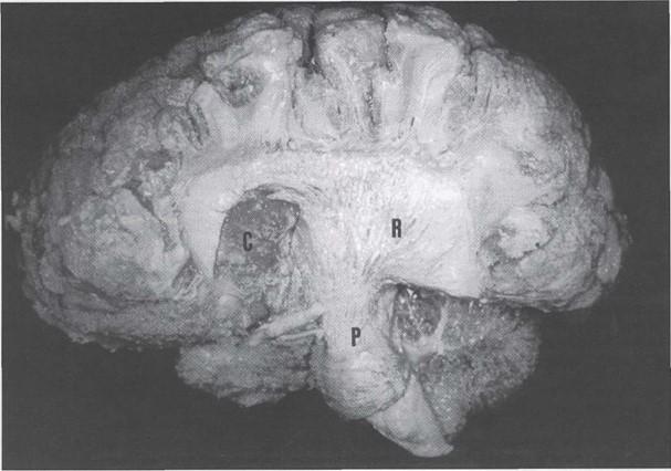
Fig. 1: Left cerebral hemisphere showing pyramidal tract (P), head of caudate nucleus (C) and corona radiata (R).

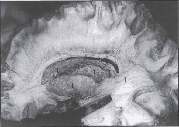
Fig. 2: Left cerebral hemisphere showing lentiform nucleus (L), thalamus (T), caudate nucleus (C) and inferior longitudinal fasciculus (I). Arrow indicates location of fibres of the internal capsule.

REFERENCES

Thompsett, D.H., 1970. Anatomical Techniques, 2nd Edition

von Hagens, G. 1985. Heidelberg Plastination Folder

Online ISSN: 2311-777X
Contact Us
Copyright 2022
bookmarkcrosslist