1 Medical Museum, Kawasaki Medical School, 577 Matsushima, Kurashiki, Okayama, 701-0192, Japan.
2 Department of Life Science, College of Life Science, Kake Educational Institution, Kurashiki University of Science and the Arts, 2640 Nishinoura Tsurajima, Kurashiki, Okay ama, 701-8505, Japan.
3 Department of Universal Design, Faculty of Health and Welfare Services Administration, Kawasaki University of Medical Welfare, 288 Matsushima, Kurashiki, Okayama, 701-0193, Japan.
Since von Hagens developed the plastination technique in 1978, plastination has gained world wide popularity. During this time, new techniques have been developed and an array of experiences has been accumulated. Plastinated specimens are invaluable for teaching and learning gross pathology and anatomy. However, for gross pathology, not only their natural shape but also the authentic color of normal and diseased tissues is desirable. Unfortunately information is scarce and not very helpful on how to prepare such plastinate specimens. In this report, a new technique for chemically reactivated blood color in plastinated specimens using Shin-Etsu Silicone KE-108 and imidazole is presented.
plastination; silicone; KE-108; imidazole; color
Telephone: 86-462-5600-33206 (dial in) /86-462-1111 ext. 33206; e-mail: elena@med.kawasaki-m.ac.jp

![]()


Since Kaiserling (1896) reported the development of a technique to reactivate wet mount specimens to enhance color, other techniques have been developed and reported. Rornhanyi 's technique has proven to be one of the best techniques to preserve specimen color (Rornhanyi, 1956; Sandhyamani et al., 2005). This technique, based on the hemochromogen reaction, uses pyridine and nicotine as bases and sodium dithionite as a reducing agent to restore a somewhat natural red color to formalin-fixed gross specimens. We modified this technique by adding sodium dithionite and irnidazole , which are a reducing agent and a base (Sakamoto et al., 1993), to the mounting medium fluid (water, formalin, glycerin and sodium acetate). Specimens which were recolorized with this modified and improved Rornhanyi technique have remained stable and continue to display a natural red color for over 20 years. Using this basic reaction as the key to color restoration, this wet technique was modified for plastination of specimens using only irnidazole of the wet mount ingredients.
Shin-Etsu silicone polymer KE-108 combined with CAT-108 (catalyst plus cross-linker) and room temperature vulcanizing (RTV) thinner has been used for over a decade for plastination (Miyake et al., 1990; Sakamoto et al., 1995). KE-108 polymer is a transparent, colorless, two-component RTV silicone. Industrially, KE-108 polymer is used for potting (sealing) electrical/electronic equipment and optical equipment. Its curing mechanism is via condensation polymerization similar to other popular plastination silicones.
Fixation: Pathological specimens used for this project were fresh tissues fixed in 10 to 20% buffered formalin for one week at room temperature. As well, longer term fixed specimens in varying formalin solutions were plastinated. In preparation for dehydration, specimens were washed in running tap water for 24 hours and then photocopied to record specimen size. The perimeter of each of the resulting images was traced and the area of each was calculated using the image measuring system (IBAS). After photocopying the specimens, they were precooled to +5°C.
Dehydration: Dehydration of specimens was carried out using the classic freeze substitution method in 100% cold (-25°C) acetone (Tiedemann and Ivic-Matijas, 1988). Specimens were immersed in acetone bathes and at least three weekly changes were performed. After dehydration was complete, more lipid was removed from lipid-rich specimens by placing the specimens in a fresh acetone bath at room temperature for at least one week.
Forced impregnation : A polymer reaction-mixture was prepared by mixing 100 parts Shin-Etsu silicone polymer (KE-108), 2 parts catalyst (CAT-108) and 10 parts RTV thinner, and 2 parts of an imidazole-ethanol mixture (polymer reaction-mixture ratio 100:2:10:2). An appropriate quantity of imidazole-ethanol mixture was prepared in advance using a ratio of 1:3 by weight. Ethanol was decanted and stirred with a stick into the imidazole.
This polymer/catalyst/thinner/imidazole reaction mixture was poured into a disposable acetone-resistant bag. The dehydrated specimens were submerged in this polymer reaction-mixture inside the bag. The bag was loosely closed and placed in the vacuum chamber in a -25°C freezer. A small weight was placed on top of the bag to keep specimens submerged in the polymer and the chamber was closed.
Two defined stages of impregnation were used. Stage one was carried out at -25°C. The vacuum pump was turned on and pumped at full vacuum (no throttling via an intake valve) and pressure was lowered to approximately Imm Hg by the end of day one. At the end of the day, the pump was turned off and pressure was returned to ambience. Two acetone traps placed in line between the vacuum chamber and the vacuum pump condensed and collected the vaporized acetone. Traps were emptied as needed. Stage one of impregnation varied from one week for thin specimens to one month for large specimens and was judged complete when no acetone was collected in the traps. In stage two, the vacuum chamber and its contents were brought out to room temperature during the day. As in stage one, the pump was turned on in the morning and the pressure lowered to approximately lmm Hg. At the end of the day, the pump was switched off and the chamber and its contents were placed back in the freezer over night. This routine was repeated for usually 4 days or until no acetone was collected in the traps. Stage two time varied from 1 day for thin specimens to 4 days for large thick specimens (liver and brain).
Curing: After forced impregnation, the specimens were removed from the vacuum chamber and polymer reaction-mixture and allowed to drain at room temperature. Daily, specimens were gently wiped of excess polymer and adjusted to a desired form. Curing took two to three days at room temperature. If any areas of the specimen remained sticky, CAT-108 was applied to the area to finish curing. The remaining polymer reaction-mixture usually was not reused but allowed to cure inside the bag at room temperature and discarded.
After the specimens had been cured, they were photocopied to record size. The perimeter of each of the resulting images was traced and the area of each was calculated using the image measuring system (IBAS). After photocopying, the specimens were used many different ways : Exhibition in room atmosphere or in display cases in the museum, used for demonstration during lecture in the lecture hall, or placed in plastic bags for storage.
After plastination, the specimens retained their original shape and some flexibility. Their grayish formalin color appeared authentically red. Areas of hemorrhage and erythema were visible (Figs. 1, 2, 3, 4). Hemorrhagic edema of the lungs was demonstrable (Figs. 5, 6). Red colored erythrocytes were visible in vessels (Fig. 7). Comparison and analysis of images of post-fixed specimens with images of specimens after curing showed shrinkage of most specimens ranged from 2 to 5%. Whole brain shrinkage was less than 10%. After many years of exposure to the atmosphere, some specimens lost surface color (Fig. 8). However, the reactivated color was maintained inside the specimen.
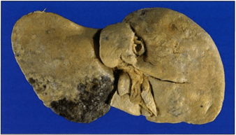 Figure 1. Visceral surface of formalin fixed/ stored liver with multiple tumors. |
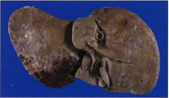 Visceral surface of liver shown in figure 1 after reactive plastination . Note the more natural appearance of the organ and the hemorrhagic area of the left lobe. |
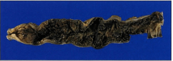 Opened lumen of formalin fixed/stored colon with psuedomembranous colitis. |
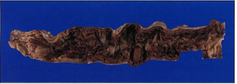 Figure 4. Opened lumen of colon with psuedomembranous colitis shown in figure 3 after reactive plastination. The hemorrhagic colitis is demonstrated. |
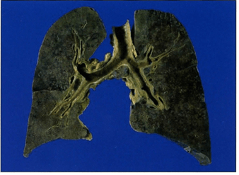 Figure 5. Anterior view of formalin fixed/stored lungs with hemorrhagic edema. |
 Figure 6. Anterior view of formalin fixed/stored lungs with hemorrhagic edema shown in figure 5 after reactive plastination. Observe the nearly uniform hemorrhagic edematous nature of the lungs. |
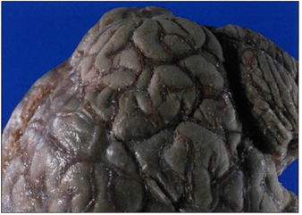 Figure 7. Left lateral view of brain after reactive plastination demonstrating red blood in the vessels. |
 Figure 8. Pictoral history of a reactivated specimen: Chemically reactivated-plastinated, 1996 (A); demonstrating color loss after continual room environment display, 1998 (B); near complete color loss with continued display, 2006 (C). |
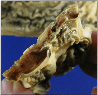 Figure 9. Section of specimen shown m figure 8c demonstrating sub-surface color retention. |
Plastinated specimens have become a great asset in teaching pathology and anatomy (von Hagens et al., 1987; Bickley et al., 1981; Nicaise et al., 1990; Hirokawa et al., 1994; Latorre et al., 2001; Alpar et al., 2005). Modifications of the original plastination equipment and process have been varied (von Hagens, 1979; Miyake et al., 1990; O'Sullivan and Mitchell, 1995; Smodlaka et al., 2005). However, loss of color is a distraction from the effectiveness and aesthetics of plastinated specimens (Alpar et al., 2005). Surface stains have been used with minimal success (Henry et al., 1997). This process demonstrated a method for restoration of specimen color during the plastination process.
During forced impregnation, a hemochromogen reaction occurs from the presence of imidazole within the specimens. It reacted with the hemoglobin to change the dark color due to oxidation to a reactivated more normal red. This color is similar to the color obtained in restored wet specimens. Thus, specimen color appeared authentic. Unfortunately, this active red color on the surface, over time, will revert back to the dark color if plastinated specimens are kept in the atmosphere. On the surface of the plastinated organ, the ferrohemochrome/hemochromogen (bright red color) may be easily oxidized and revert to ferrihemochrome/parahematin (burnt umber) when exposed to air. However internally, the reactivated color remains. This reverting back to the fixed color can be slowed dramatically by exhibiting the plastinated specimens in display cases or storage in bags, when not in use. Using these precautions, has kept the renewed red color in plastinated specimens more than three years. Research on this subject should be continued to find a method to preserve and/or restore the surface activated blood color longer term. If a more suitable reducing agent can be identified , the resulting color could likely be improved to be even more life-like.
Other silicones have not been used for this process. However, we believe other silicones could be used. It is possible that specimens preserved by specialized preservation solutions other than formalin alone might interfere with this reactivation process. Plastination with chemical reactivation offers advantages over both routine plastinated and wet mount specimens. There is no odor or oozing and the specimens may be handled without gloves. In addition, the specimen is preserved in a more natural color tone. This methodology results in excellent specimens for teaching and exhibition.
Plastination of color reactivated wet mounted specimens does not work for reactivation of the color. When the specimen is removed from a display container and reintroduced to air to flush away chemicals and to dehydrate, the reactivated color is lost. Once the reactivated color is lost (oxidized), it is difficult to reactivate the color again using current techniques. Also the presence of glycerin, sodium dithionite and sodium acetate in the wet mounted specimens would likely leave residue on the surface of the plastinated specimens as previously reported, unless they undergo ethanol bathes and flushing (von Hagens, 1985).
Importing the Biodur silicone polymer series, to determine if plastination of our museum specimens would be feasible, was expensive and needed a lot of time and effort to obtain such. Therefore, we researched locally available Japanese silicones to see if one might meet the conditions necessary for plastination: Room temperature vulcanizing (RTV) type silicone, Colorless and transparent, Specific gravity similar to water, Room temperature cure, Flexibility after curing, and No known curing inhibitor. KE-108 met these criteria and was chosen to use for plastination of the museum specimens. CAT-108 (catalyst) was the standard curing agent for this polymer. Its condensation type curing of RTV silicone has no known curing inhibitors, such as water, organic solvents, organic metal salts, sulfur, phosphorus , nitrogen compounds, etc.
The recommended mixture of catalyst (catalyst and hardener) with the KE-108 polymer is 5:100. To ensure adequate impregnation time, the rate of cure needed to be prolonged. Therefore, only 2 parts of catalyst were used. To decrease the polymer reaction-mixture viscosity, 10 parts of RTV thinner was added to the 100 parts of the KE108 polymer. This decreases polymer viscosity from 700 centipoise to around 600 cps. RTV thinner is nonvolatile and was used to dilute the KE-108 polymer. Polymer dilution enhances the influx of polymer into the specimen and may extend the workable time of the polymer reaction-mixture. The workable time for this reaction-mixture, which contains both catalyst and cross-linker, is over three months when kept at -25°C. However at room temperature, pot-life is decreased to a few days. Impregnating specimens in a bag within the vacuum chamber served several purposes: Kept specimens submerged, Reduced the amount of reaction-mixture needed for each load, Served as liner for the vacuum chamber, Prevented aspiration of exploding silicone bubbles into the vacuum line and pump which can clog them when it hardens, Ease of removing specimens and polymer reaction-mixture from the vacuum chamber, and Ease of disposal of thick silicone.
It was necessary to add imidazole to the polymer-mixture to reactivate the natural color of the blood. Imidazole is not soluble in silicone. Therefore, an ethanol-saturated mixture was prepared and used in the polymer reaction-mixture. Imidazole remained suspended in the reaction-mixture. Shin-Etsu silicone polymer KE-108 mixed with CAT-108 is a useful plastination mixture to impregnate specimens. Measured shrinkage of 2 - 10% was less than the previously reported 8 - 29% by Brown and colleagues (2002). Part of this discrepency is likely due to the difference of units used: Brown's group used volume as opposed to area in this project. Imidazole is the key to preparing dry specimens with natural coloring. The exact minimal amount of imidazole to specimen volume has not been determined. However, at the determined level (0.5 pts imidazole/ 100 pts KE-108 polymer) it is likely to be an adequate amount for impregnation of more than one load of specimens. We chose not to reuse the mixture because of the increased viscosity of the polymer reaction-mixture after one load of specimens had been impregnated. Much higher concentrations of imidazole will likely yield a much darker unnatural red and is much more expensive.
Alpar A, Glasz T, Kalman M. 2005: Plastination of pathological specimens - A continuing challenge. J Int Soc Plastination 20:8-12.
https://doi.org/10.56507/TFCD4856
Bickley HC, von Hagens G, Townsend FM. 1981: An improved method for the preservation of teaching specimens. Arch Pathol Lab Med 105(12):674-676.
https://doi.org/10.1093/labmed/12.11.676
Brown MA, Reed RB, Henry RW. 2002: Effects of dehydration mediums and temperature on total dehydration time and tissue shrinkage. J Int Soc Plastination 17:28-33.
https://doi.org/10.56507/XNQM4606
Henry RW, Janick L, Henry C. 1997: Specimen preparation for silicone plastination. J Int Soc Plastination 12(1):13-17.
https://doi.org/10.56507/HVSK9838
Hirokawa M, Yamashita K, Miyake Y, Kanahara K, Sakamoto Y. 1994: Pathologic gross specimens in medical education (Japanese). Kawasaki Igakkai Shi 20(Supp1):41-48.
Kaiserling C. 1896: Ueber die konservirung von sammlungspraparaten mit erhaltung der naturlichen Fraben. Berliner Klinsche Wochenschrift 23:775-777.
Latorre R, Vazquez JM, Gil F, Ramirez G, L6pez-Albors 0, Orenes M, Martinez-Gomariz F, Arenciba A. 2001: Teaching anatomy of the distal equine thoracic limb with plastinated slices. J Int Soc Plastination 16:23-30.
https://doi.org/10.56507/ACRF7155
Miyake Y, Sakamoto Y, Hirokawa M, Yamashita K. 1990: Modified von Hagens' method using silicone KE108 (Japanese). Pathology and Clinical Medicine 8(11): 1439-1441.
Nicaise M, Simoens P, Lauwers H. 1990: Plastination of organs: A unique technique for the preparation of illustrative demonstration Vlaams Diergeneeskd Tijdschr (Flemish Veterinary Journal) 59(4):141-146.
https://doi.org/10.1159/000147145
O'Sullivan E, Mitchell BS. 1995: Plastintion of gross anatomy teaching using low cost equipment. PMID 17(3):277-281.
https://doi.org/10.1007/BF01795063
Romhanyi G. 1956: Einfaches verfahren zur konservierung in naturlichen Fraben. Virchows Archiv, Bd. 328:573-575 .
https://doi.org/10.1007/BF00955072
Sakamoto Y, Miyake Y, Hirokawa M, Yamashita K. 1993: Gross specimens in Medical Museum of Kawasaki Medical School (Japanese). Japanese Society of Pathological Technology 48:10-12.
Sakamoto Y, Miyake Y, Hirokawa M, Yamashita K. 1995: Gross pathological specimens - A new method for plastination (Japanese). Japanese Society of Pathological Technology 51:15-16.
Sandhyamani S, Sindhu JK, Sriramachari S. 2005: Recolorization of museum specimens: A modification of Romahanyi' s technique based on pyridine/nicotine hemochoromogen reactions. Virchows Arch. 447(1):94-98.
https://doi.org/10.1007/s00428-005-1273-8
Smodlaka H, Latorre R, Reed RB, Gil F, Ramirez G, Vazquez JM, L6pez-Albors 0, Ayala MD, Orenes M, Cuellar R, Henry RW. 2005: Surface detail comparison of specimens impregnated using six current plastination regimens. J Int Soc Plastination 20:20-30.
https://doi.org/10.56507/FIHG2408
Tiedemann, K, D lvic-Matijas. 1988: Dehydration of macroscopic specimens by freeze substitution in acetone. J Int Soc Plastination 2(2):2-12.
https://doi.org/10.56507/SCLL2742
von Hagens G. 1985: Heidelberg Plastination Folder: Collection of all technical leaflets for plastination. G. von Hagens, editor, Anatomisches Institut I, Universitat, D-6900 Heidelberg, Germany.
von Hagens G. 1979: Impregnation of soft biological specimens with thermosetting resins and elastomers. Anat Rec 194(2):247-255.
https://doi.org/10.1002/ar.1091940206
von Hagens, G, Tiedemann K, Kriz W. 1987: The current potential of plastination. Anat Embryol 175(4):411-421.
https://doi.org/10.1007/BF00309677