1 Department of Anatomy, Lincoln Memorial University-DeBusk College of Osteopathic Medicine, Harrogate, TN 37752 USA.
2College of Veterinary Medicine, Lincoln Memorial University, Harrogate, TN USA
Plastinated specimens, which have been painted or stained to highlight anatomical structures, can be helpful materials for medical students as well as others interested in studying anatomy. However, one problem with these specimens is that the paint often chips off due to being handled by many students and teachers. It may also wear off naturally if the specimen is kept for a long time. In this experiment, specimens were colored prior to the curing stage in an attempt to improve durability and better adherence of the color to the specimen. The results were then compared to techniques used by McCreary (2013). Colored specimens were challenged using methods which might be similar to routine handling during study. It was expected that the new coloring technique of applying the solution prior to the cross-linking stage instead of after the cross-linking stage would provide a stronger adhesion to the specimen. Although the color on the specimen applied prior to the cross-linking step demonstrated a more polished appearance compared to the color applied after the cross-linking stage, various durability tests confirmed no better efficacy or durability in adhesiveness of the silicone color to the specimen. Rather, some of the color came off when rubbed with fingers or latex-gloved hands.
S10 plastination; color; muscle, vessels
Stanley Iliff, telephone: (423) 869-6337; Fax: (423) 869-6006; Email: stanley.iliff@lmunet.edu

![]()


Coloring of plastinates can serve as a great learning tool for students who have difficulties distinguishing certain vascular structures and tissues. Various coloring techniques to highlight structures of the plastinated specimen have been developed by researchers, each with its own advantages (enhanced clarity of structures) and disadvantages (no method is student proof) (e.g. Marchese et al., 2008; McCreary et al., 2013; Raoof et al., 2013). However, recent staining techniques used in our lab show long lasting durability (Concha et al., 2014).
The goal of this research was to introduce the coloring technique at a different stage of the plastination process developed by von Hagens (1979a; 1979b, 1985) and implement various test methods to compare the durability of the coloration on a freshly-prepared lower limb specimen, and an upper limb prepared and described previously by Mc Creary et al. (2013) as well as the advantages and disadvantages of the new coloring procedure described here. In the upper limb specimen, color was applied after the cross-linking/curing stage of the plastination process (McCreary et al., 2013). In this modified protocol, however, color was applied to the muscles of the pelvis and lower limb (including the thigh, knee, and foot of the specimen) prior to cross-linking of the impregnation-mix (polymer-catalyst). In addition, the blue pigment, BiodurTM AC52 was used to color the veins of the leg specimen instead of the BiodurTM AC40 which was used in the previous research (McCreary et al., 2013) to color the upper extremity veins. Our expectation was that application of the color before curing/hardening the silicone polymer-mix in the specimen would result in a stronger adhesion of the paint to the specimen, and therefore result in better durability.
Specimen Preparation
The specimen for plastination was procured through the Lincoln Memorial University-DeBusk College of Osteopathic Medicine/Anatomical Donation Program. The left lower extremity of a human male was used. The fresh-frozen lower limb was harvested and immediately fixed by injecting 5% formalin solution into the tissue and submersion into a 5% formalin bath. The pelvis, femoral triangle, knee and foot were dissected. Dissection required about 100 hours. Due to the large size and thickness of the tissue, the specimen was stored in a 5% formalin bath for an extended period and maintained in this solution throughout the extended dissection in order to inhibit decay of the tissue during dissection. After dissection was completed, the specimen was rinsed with tap water for 7 days to rid the specimen of formalin and all embalming chemicals. For plastination, the cold BiodurTM S10 plastination technique was used (deJong and Henry, 2007). In order to minimize tissue shrinkage, the specimen was dehydrated by submersion into cold -20° C acetone. Acetone baths were changed 4 times at weekly intervals. After 5 weeks of cold dehydration, the specimen and acetone bath were brought to room temperature for 3 weeks for lipid removal. Next, forced impregnation was carried out by placing the specimen into the silicone polymer/catalyst (S10/S3) mixture in the vacuum chamber at -15°C and gradually reducing the pressure. Impregnation time was five weeks.
Application of color to the specimen
Following forced impregnation, the coloring solution was applied to the surface of the muscles using a paint brush, to create a life-like, aesthetic appearance. Arteries and veins were colored to make the structures more distinguishable. The method of coloration described by McCreary (2013) was utilized for this experiment except that BiodurTM AC52 was used instead of BiodurTM AC40 to color the veins. The color solution for the muscles was made by mixing: 0.3 g of silicone (Akzo Nobel Clear All-Purpose Silicone Sealant, general residential caulking), 5 ml of MEK (methyl ethyl ketone), and 40 μL of BiodurTM AC51 (brown) dye paste. The solution mixture of 0.3 g of silicone, 5 ml of MEK, and 80 μL of BiodurTM AC50 (red) was mixed and applied to the arteries. The veins were colored with a solution containing 0.3 g of silicone, 5 ml of MEK, and 120 μL of BiodurTM AC52 (blue) dye paste.
Because of variation in muscle surface texture, color penetration into the surface of the muscle varied and repeated coats were required to achieve consistent coloration. In cross-sectioned muscles, the color solution was more easily absorbed, and fewer coats were required. In longitudinal muscles, however, several coats were necessary to gain color appreciation depending on size, thickness, and texture of the muscles. Similarly, the arteries and veins both required several coats to achieve the desired color.
Curing/Cross-linking
In the final step, the specimen was cured by using gas cure S6 at room temperature. Throughout the curing process, the specimen was manicured for polymer residue removal. Once the curing procedure was complete, the specimen was ready for results capture.
Durability and Testing
Color durability was investigated, taking into account the specimen’s expected handling for teaching and learning purposes. There are no known scales for rating the levels of abuse or handling of painted specimens. The tests were based on anticipated student and faculty handling mimicking a normal review period. Testing time was based on a normal specimen handling time by a student or professor during a class period.
Durability tests were carried out using the following four methods. The first involved using a latex-gloved hand and rubbing the surfaces of the colored structures of both proximal and distal portions of the specimens. In the second test, colored structures were rubbed with fingers.
For the third test and fourth tests, anatomy dissection instruments, specifically the blunt probe (third) and serrated forceps (fourth), were used to scratch both proximal and distal colored areas of the specimen. These methods engage or test the durability of the paint in real-life hands-on teaching scenarios.
Application of color to the specimen
Different numbers of colored paint coats were necessary to achieve optimal color on the structures of each specimen. Two coats of color were applied to the cross-sectioned muscles of the specimen and 2 to 5 coats were applied to intact muscles. For the arteries, 5 coats of color solution were required while 4 to 6 coats were applied to the veins (Figs. 1-3).
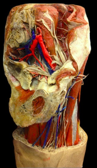 Figure 1. Male, left lower limb, medial view, after coloring but before application of S6 (cross-linker) |
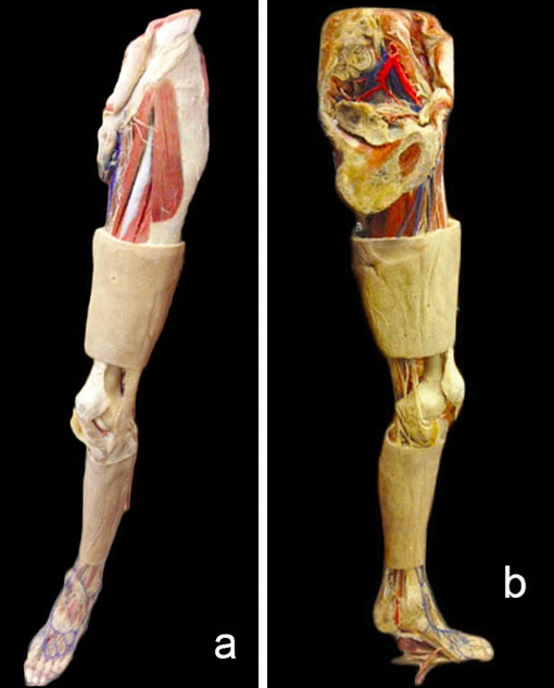 Figure 2. Male, left lower limb, anterolateral (a) and medial (b) views, before application of S6 (cross-linker) |
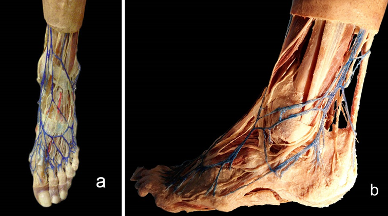 Figure 3. Male, left foot, dorsal (a) and lateral (b) views, after coloring but before application of S6 (cross-linker) |
Durability Testing
The first test (using a latex-gloved hand to rub the surfaces of the colored structures), resulted in a small amount of the brown paint peeling off when the proximal leg muscle was rubbed multiple times. The rest of the colored muscles, arteries and veins showed no peeling of the color, nor were stains observed on the gloved hands.
In the second test (rubbing the colored structures with fingers), as in the first test, a small amount of the brown color on the same muscle came off. Also, the blue color of the small veins on the foot started to peel off as well. The rest of the color on the muscle and vessels did not peel.
For the third and fourth tests (scraping with anatomy dissection instruments), the blunt probe (third) and serrated forceps (fourth), resulted in no peeling of the color on these regions.
Comparison with upper limb specimen
When tests 1 to 4 (as described above) were applied to the upper limb prepared by McCreary et al. (2013), no peeling of the paint resulted.
This coloring technique demonstrated beautiful color appearance on the specimen (Fig. 4), however it did not meet our expectations for durability.
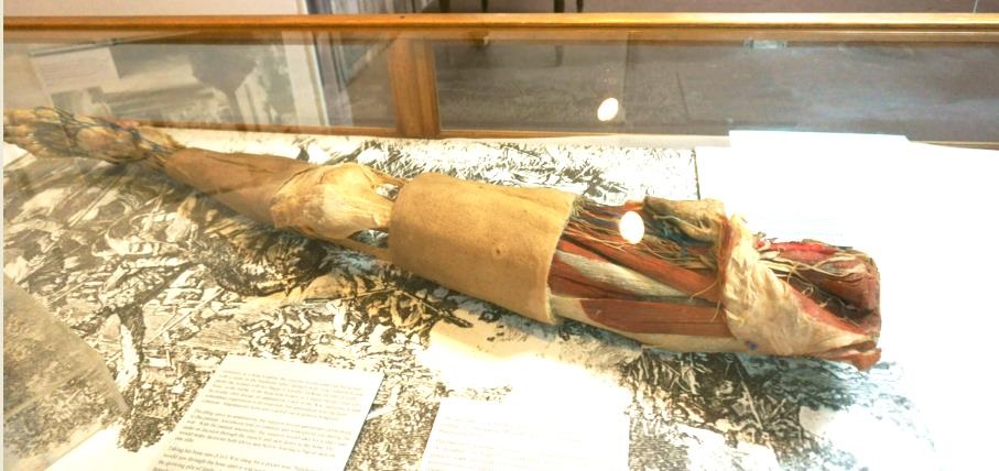
Figure 4. Male, left lower extremity anterior view, after coloring and application of S6 (cross-linker) as displayed at Abraham Lincoln Library, LMU, Harrogate TN.
The goal of this research was to develop better permeation and stability of the color to the specimen for long-term use in education. However, it was discovered that applying the color to the specimen prior to the cross-linking/curing stage resulted in no better durability when compared to the prior painting technique used by McCreary et al. (2013). For example, when the painted muscles and vessels of McCreary et al.’s upper limb specimen (to which the original technique was applied) were rubbed with gloved hands, fingers, blunt probe and serrated forceps, no peeling of the paint resulted. Comparably, in this study, a small amount of the paint peeled off on the lower limb specimen.
Perhaps the different number of applied paint coats could have played a minor role in this unexpected result. For instance, while 2 to 5 coats to the muscles and 4-6 coats to the vessels of the leg specimen were applied in order to gain color appreciation, 2 coats of color on the muscles and 6 coats of the color solution were applied to vessels of the upper limb specimen (McCreary et al., 2013). The color tone was the same for both the lower and upper limb specimen except that the lower limb specimen appeared slightly more polished.
In the past, researchers from the University of Michigan Medical School have developed methods of coloration and plastination of specimens in order to produce stronger adhesion of the paint to the specimen (Marchese et al., 2008; Raoof et al., 2013). Their procedures involved applying the acrylic paint to the specimen prior to the curing process of plastination. As a result, these methods have been shown to improve the durability of the paint on specimens.
Although application of the paint to specimens prior to the curing stage demonstrated a greater durability of the paint in Marchese et al.’s (2008) experiment (Marchese et al., 2008; Raoof et al., 2013), it can be concluded from the research described here that the result could be influenced by a variety of factors. These factors include: use of different specimens containing different tissues with various textures, size, thickness, different number of applied color coats and different coloration material.
Concha I, Iliff S, Henry RW. 2014: New multidimensional stain for plastination. Abstract presented at The 17th International Conference on Plastination Saint Petersburg, Russia July 14-18, 2014. J Plastination 26(1):24.
de Jong K, Henry RW. 2007: Silicone plastination of biological tissue: cold-temperature technique BiodurTM S10/S15 technique and products. J Int Soc Plastination 22:2-14.
https://doi.org/10.56507/ZLMJ7068
Marchese A, Marchese L, Wischmeyer A, Falk K. 2008: Enhancing the understanding of anatomy through the coloration and plastination of anatomical specimens. U of Mich Undergraduate Research Forum Issue 5, Winter.
McCreary J, Henry RW, Iliff S, Hermey D. 2013: Silicone-based coloration technique developed to highlight plastinated specimens. J Plastination 25 (2):13-20.
https://doi.org/10.56507/XLBR3803
Raoof A, Marchese C, Marchese LA, Falk KC, Mirafzali N. 2013: Painting plastinated neurovascular pathways: evaluation of coloring techniques. J Plastination 25(2):21-26.
https://doi.org/10.56507/LJZQ6496
von Hagens G. 1979a: Impregnation of soft biological specimens with thermosetting resins and elastomers. Anat Rec 194(2):247-255.
https://doi.org/10.1002/ar.1091940206
von Hagens G. 1979b: Emulsifying resins for plastination. Der Praparator 25(2):43-50.
von Hagens G. 1985: Heidelberg Plastination Folder: Collection of all technical leaflets for plastination. Anatomisches Institut I, Universitat, D-6900 Heidelberg, Germany.