Department of Anatomy (Dir. Prof. G. Marinozzi) State University of Rome "LA SAPIENZA", Via Alfonso Borelli 50, Italy
The interpretation of CAT and NMR images is some- times very difficult and requires the support of normal morphology to reach the formulation of certain unequivocal diagnosis. Following a careful radiological indication we performed a study of the conservation of cadaver's serial sections of some human body regions. The cadaver, frozen in a refrigerator at -25YC was cut in serial cross sections, by a circular belt disk saw, after indicating points on the anatomic specimen. The sections ob- tained, with a thickness of 0,5-1 cm., after cleaning were mounted on a metal grid. Next, they were subjected to the standard plastination (BIODUR S-10) The careful study of obtained sections and the correspondent NMR-CAT images, provided the direction of the cross-sectional images and the anatomical cuts coincide, and the anatomical support can be clearly useful for the diagnosis.
MRI; CT; CAT scan; Biodur-S10, Silicone
Ripani, M Department of Anatomy (Dir. Prof. G. Marinozzi) State University of Rome "LA SAPIENZA", Via Alfonso Borelli 50, Italy

![]()


The complexity of the results from CAT and NMR examinations very often require the aid of normal morphology support to get to certain and undoubted diagnosis. For this purpose, the study and preservation of "slices" from the head of a corpse, after a precise radiography diagram was made.
In this study, fixed corpses were used. The corpse was frozen at -25°C in order to allow head resection along a transverse-cervical-thoracic plane. After carefully studying the radiography the following points were found: repere, glabella and opisthocranium, necessary to carry out the first transverse section. Other sections, parallel to the first one were then cut. Each "slice", to either cranial or caudal direction, and was 1 cm thick. After cutting the frozen specimens by means of a circular band saw, the sections were treated to remove any residual organic substance on the cut surface. The sections were then set on blotting-paper to absorb defrosted liquid. A soaked pad with hydrogen peroxide (H202) was used to liquify the coagulant on the structures. A solution of hydrogen peroxide was also injected into the blood vessels to remove further obstructions. After this treatment, perfectly cleaned sections, from an anatomical (no residual organic substance) and chromatic viewpoint, were obtained, and the surfaces were photographed with a professional camera (LINHOF). At the end of this phase, each section was set on a suitable metallic support to maintain its morphology.
A cylindrical lattice, 50 cm. high was built. Some trans- verse planes, made up of metal grids, were attached to a steel wire. The planes were 2 cm. apart from each other and were covered by blotting-paper. The sections were at a distance of 4- 5 cm. from one another in order to be thoroughly immersed into the substances used in the next phases. Dehydration was by means of acetone at -25°C. The sections were immersed in that solution for 24 hours. The control of the percentage of acetone in the solution follows. If the value of acetone is less than 98%, it will be replaced with new absolute acetone (99%).
The controls were done every 24 hours until acetone remains unchanged (i.e.,not absolute acetone) to avoid section shrinking. After this dehydration, forced impregnation with S- 10 resin and S-3 hardener start. The lattice was immersed in the resin in the vacuum room and the pump was brought into action. After 5-10 minutes, the resin gradually begins to replace acetone. As this happens, bubbles moving toward the surface of the substance can be observed through the glass sealing the room.
The pump operator should ensure that a constant amount of bubbles be present without pressure changes. In fact, a large quantity of bubbles means that forced impregnation of the sections is too quick. This causes a quantitative change between the amount of acetone going out and the resin replacing it, which causes a shrinking of the organs. Impregnation at room temperature lasts seven days and finishes whenever pressure at 25 mbar doesn't produce any more bubbles. The sections displayed on the lattice grids are taken out of the vacuum to be drained of resin excess present on the surface. Then, the last phase, the cure, begins. For this purpose a slow cure was used. The sections were arranged in two layers in a vessel at a distance of 10 cm. from each other. A small vessel, filled with KOH (Potassium hydroxide), was set on each layer. The potassium hydroxide absorbs humidity therefore catalyzing the hardening reaction of resin. Each day the sections dried because of continuous secretion of small amounts of resin. After one week, when the sections were dry, this phase was complete.
 |
 |
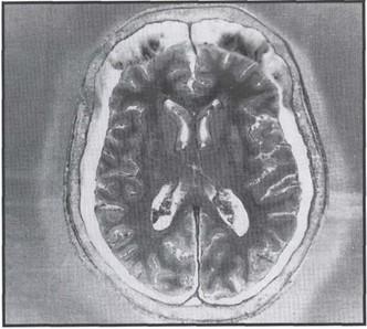
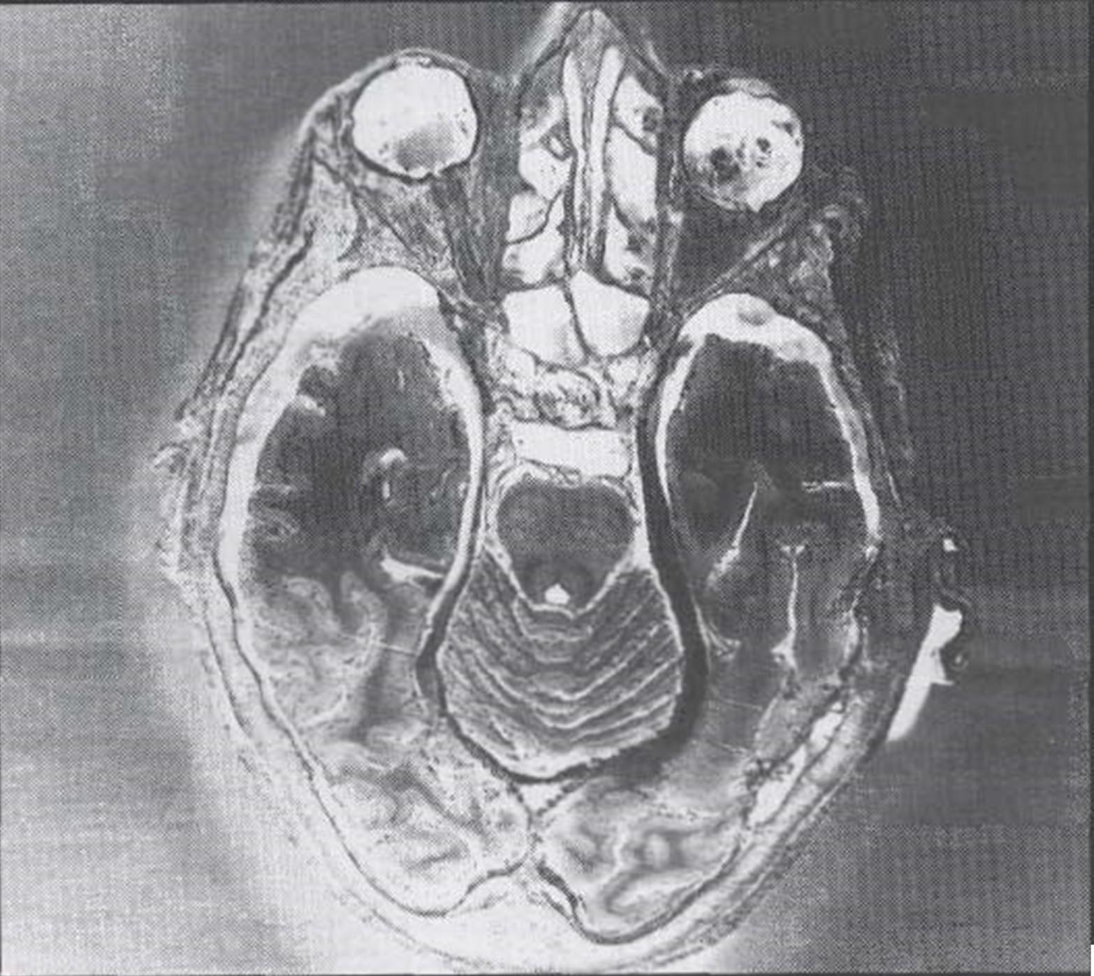
| Fig. 1 - Meningioma of chorioid plexus ventriculi lateralis | |
 |
 |
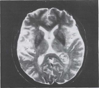
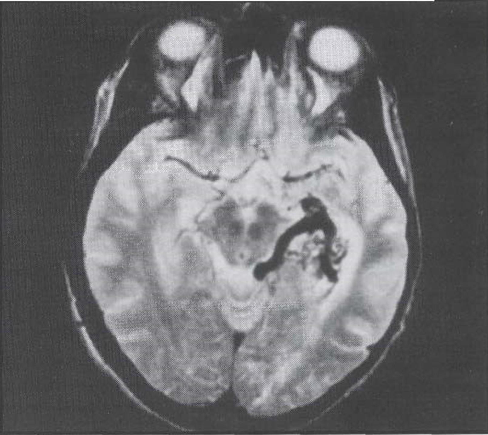
| Fig. 2 - AVM (arteriovenosus malformation) on the mesial surface of temporal lobe | |
 |
 |
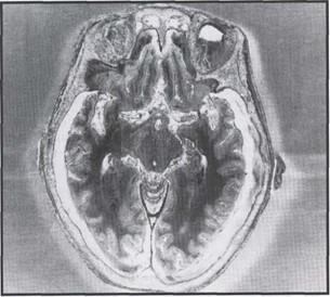
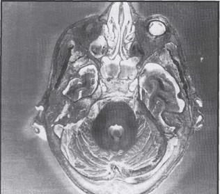
| Fig. 3 - Sphenotentorial meningioma: compression of temporal lobe and cerebral peduncle | |
 |
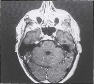 |
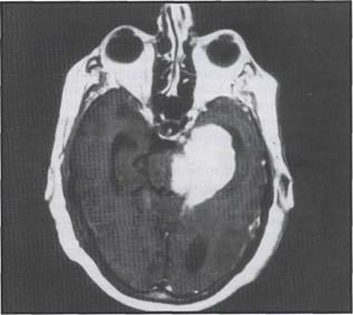
| Fig. 4 - Schwannoma in relation to the fifth cranial nerve | |
 |

|
| Fig. 5 - Schwannoma | |
Terrible Weil Marin,V.; Corrain.C.; Pratiche imbalsamatorie in Europa Rivista: Pathologica Volume 78:107-121, 1986.
Cimmino, F., Vita quotidiana degli Egizi Milario-Ott. 1973
Gelmetti Girolamo Segato, P., La pietrification des parties anatomiques et d' animaux Da Internazionale di storia delia medicina Budapest 1974
Bouchet, A., "L'embaumentent et la conservasion des cadavers humains au corus des siecles" Rivista: Lyon Medical Anno: 1972
Baptista, C.A., Skie, M., Yeasting, R.A., Ebraheim, N.A., & Jackson, W.T., Plastination of the wrist: potential uses in education and clinical medicine. J. Int. Soc. Plastination 3:18-21,1989
https://doi.org/10.56507/XENF9035
von Hagens, G., Plastination Technique Da: Heidelberg Plastination Folder 1989
Tiedmann, K., von Hagens, G., The current potential of plastination AnatEmbryol 175:411-421
https://doi.org/10.1007/BF00309677
Tiedmann, K. The technique of heart plastination Da Anat. Rec. 204 1982
https://doi.org/10.1002/ar.1092040315
Wolfe Plastic embedded hearts DA: Arch. Pathol. 61 1956
https://doi.org/10.1016/S0021-9258(18)65228-7