1 Department of Anatomy Ben Carson [Snr.] School of Medicine, Babcock University, Nigeria
2The National Postgraduate Medical College of Nigeria, Lagos, Nigeria
3 Internal Medicine, Yale University, USA
Plastination is a modern method of preservation of biological specimens, including human cadavers. This study elucidated how temperature might affect plastination, noting that there is sparse scientific literature on this technique, especially from Africa. It is also relevant to the feasibility of adapting and adopting the technique as a feasible and useful laboratory technique in developing countries, where technological advancement, finance, and socio-cultural factors are suspected to be strong determinants to this effect. The S10 plastination technique is usually done at cold temperature (-25° C), but this study investigated and compared the effects of plastinating at room temperature (~25° C). The four main stages of plastination were carried for the control group while the ‘diffusion’ principle was employed for Group B. The forced impregnation process is typically carried out under vacuum at cold temperature (-25° C) with the use of an additional, relatively costly, refrigerated impregation chamber. Ten adult (n=10) human brains were randomly assigned to two groups (A and B), comprising 5 brains each. Forced impregnation of the Group A brains was performed at -25° C (cold temperature), and the ‘diffusion’ impregnation procedure was carried out for the Group B brains at 25° C (room temperature). The Group B brains required less time for draining compared to Group A. Both methods yielded brain plastinates with the basic features of plastination outcomes. The weights of the brains (g) were recorded at each stage of the process using the digital Sartorius ENTRIS 4202-1S balance. The volumes were also measured at each stage using Archimedes’ principles of fluid displacement in a calibrated glass jar (cm³). The room temperature specimens yielded better specimens in terms of relative weight loss, relative colour preservation, physical properties, and texture and preservation of surface features and brain surface topographies.
anatomy; brain; plastination; organ preservation; temperature; S10 technique
Joshua O Owolabi: Department of Anatomy Ben Carson [Snr.] School of Medicine, Babcock University, Nigeria

![]()


The word ‘embalming’ is often associated with the preservation and conservation of the human body or remains; however, conservation and preservation include other methods than the conventional process of embalming (Manzoli et al., 2011). Plastination is a modern method of whole body, or body part, preservation, whereby water and lipids in biological tissues are replaced by curable polymers (silicone, epoxy, or polyester), which are subsequently hardened, resulting in dry, odorless and durable specimen (Klaus et al., 2018). Plastination, simply put, is a process used in the study of the structure of bodies, to preserve parts or the whole body, yielding specimens that are durable, odorless, and lifelike. Polymers used in this process, and the class of polymer used determines the optical qualities (transparent or opaque), and mechanical properties (flexible or firm), of the impregnated specimen. The advantage of plastination over other preservation methods lies in the ease with which it is possible for the resins to move between the macroscopic and microscopic levels of the tissues (Sora and Genser-Strobl, 2005). Also, plastinated specimen are very important in cultural knowledge, education, research and other applied medical areas (Jones and Whitaker, 2009).
Plastination is therefore an unusual method of permanently preserving tissue in a life-like state, in which biological specimens are preserved by replacing the fluids of the body (fat and water) with synthetic materials. This method produces ‘plastic-like’ bodies or organs, which remain very lifelike, non-toxic, odorless, dry and durable and may be handled easily for examination (Singh et al., 2013). There are different types of plastination techniques which include the S10, E12, P35, and the P40 techniques (Anant and Madhavi, 2015). There are four basic stages involved in plastination: fixation, dehydration/defatting, forced impregnation, and curing (hardening) (von Hagens, 1979). The S10 plastination technique is not commonly used for the preservation of whole brains, however, it has been much used for whole-body, and body-part specimens, and it has shown positive properties, such as durability, longevity, non-toxicity, and life-like appearance.
It is important to emphasize the importance of plastination to medical education and how it can be used to mitigate the negative effects of resource constraints, especially in developing countries. This is because plastinates can last for many years and can be used by many generations of students. Additionally, they are life-like specimens that offer close-to-real life features, unlike models and drawings. Unfortunately, in Africa for instance, plastination has not really been established as a routine procedure in preservation, learning, and for producing teaching aids (Azu et al., 2013). The poor appreciation and understanding of the techniques, limited financial resources and facilities, and the difficulties in adapting the conditions required for the procedures might have limited its acceptance and applications in Africa. This is a major reason why this investigation is crucial to advancing this technique, and to appreciate how it might be adapted to various conditions and settings. It also enriches the available literature on the technique, thus providing more scientific and empirical literature on the technique. This is absolutely the best means of optimising its use.
The standard temperature for S10 plastination is at cold temperature (-25° C). This research, however, compared the effects of temperature variation on the results of the S10 plastination technique of the brain at cold temperature (-25° C), and a room temperature (25° C) ‘plastination’ technique.
The brain specimens: a total of 10 adult human brains [n=10] were used for this research work. The brains were procured from the Learning Resource Department (museum unit) of the National Postgraduate Medical College of Nigeria and were randomly grouped into two groups of five brains each, designated Group A and Group B.
Chemicals used: 10% formalin, tap water, distilled water, cold acetone, S10 resin (Biodur), S3 (Biodur), S6 (Biodur) and calcium chloride (CaCl2).
Equipment used: acetonometer, deep freezers, stainless steel drum, stainless steel basket, digital thermometer, conveyor pumps, Heidelberg plastination kettle, replacement glass plate, replacement lid sealer, separator for oil and solvent, Bennert manometer, digital manometer, vacuum lifting pad, mixing rod, vacuum adjustment valve, vacuum adjustment unit, vacuum pump, vacuum pump oil, vacuum tubing, gas curing chamber, membrane pump, stainless steel collecting tray, fan, air tubing, glass jar and power cord.
Experimental room housing facility: the laboratory procedures were carried out at the Plastination Laboratory, museum unit of the National Postgraduate Medical College of Nigeria, under standard conditions.
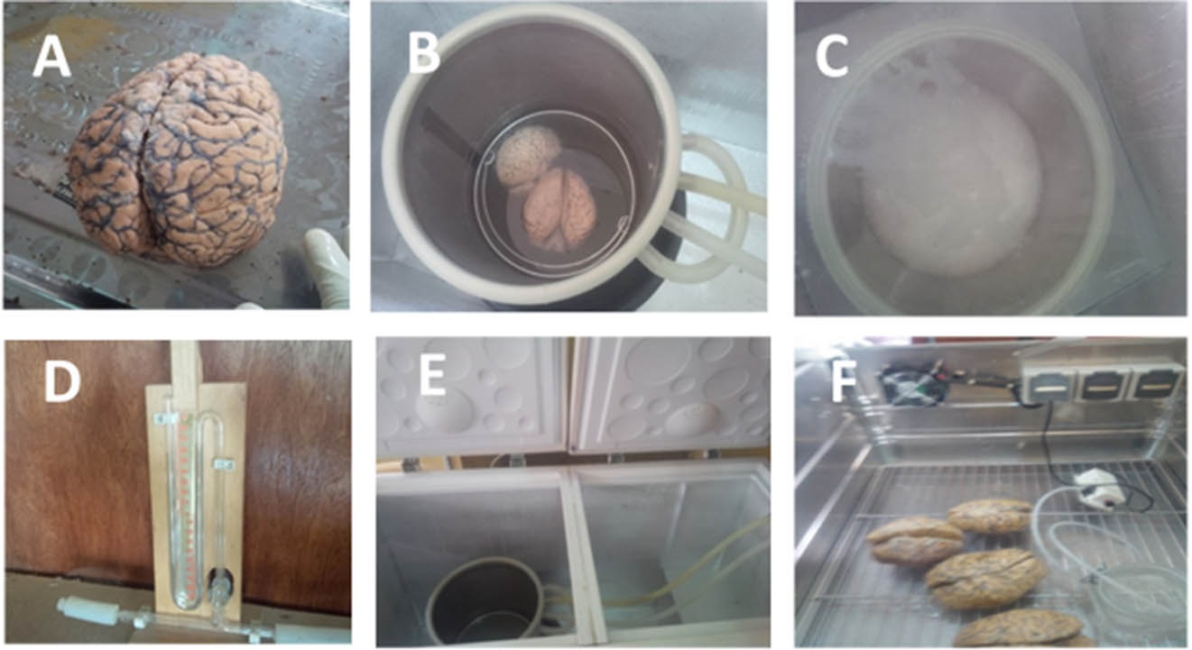 Figure 1. Equipment, materials and brains being plastinated. A brain specimen at the start of plastination [A]; brains in plastination kettle [B]; bubble formation during plastination process [C];The Bennert manometer showing pressure measurement [D]; the forced impregnation chamber [E]; brain samples undergoing gas curing [F]. |
Plastination processes: the four basic processes involved in the plastination technique as prescribed by von Hagens (1979) were observed for Group A brains (Fig. 1).
Fixation: freshly excised brain were fixed at the time of harvesting, by perfusion and immersion in 10% formalin, for four weeks. To ensure proper fixation before the plastination process commenced, the brain were re-fixed (perfused and immersed) in 5% formalin again for 5 days to prevent autolysis, putrefaction, and to harden the brain tissue.
Dehydration: Absolute (100%) pure acetone was used throughout. Three changes of absolute (100%) acetone were used. All brain specimens in both groups were dehydrated by immersion in 100% acetone at -20° C. The ratio of brain specimen weight to volume of dehydration bath was 1:10 or more. All brain specimens were initially immersed in pure acetone for one week, and then transferred to two consecutive pure acetone baths for another two weeks. The total duration of dehydration was thus three weeks. The degree of brain tissue dehydration was monitored with an acetonometer on a daily basis. The dehydration was considered to be completed when the water content was below 1%.
Degreasing: Degreasing of brain specimens of both groups was carried out at room temperature in a bath of absolute (100%) acetone for two days.
Forced impregnation: Forced impregnation is the central and most important step in plastination.
Forced impregnation at cold temperature (-25°C) (group A): the S10 standard technique for forced impregnation was employed for group A, at -25° C. The group A brains were removed from the degreasing tank after two days, and the specimens, (designatedA1, A2, A3, A4, A5) were placed in the plastination kettle in a deep freeze at -25° C, containing silicone impregnation mixture of S10 and S3 (99:1 by volume) at -25° C. The brain specimens were impregnated under vacuum for three weeks until no further acetone bubbles could be observed, and the absolute pressure reached 5 mmHg.
Diffusion of S10/S3 mixture at room temperature (25°C) (group B): the group B brains (B1, B2 ,B3 ,B4 ,B5) were removed from the degreasing tank, and immersed in polymer mixture of S10+S3 (99:1 by volume) at room temperature. No vacuum or external force was employed. The plastination kettle was covered. Impregnation of the S10 + S3 mixture was by diffusion, which took 4 weeks. The process was completed when physical observation of the brains and the chamber were like the standard observations in the forced impregnation procedure - especially, disappearance of bubbles. A digital thermometer was attached to it, and the temperature readings of the room were recorded daily.
Curing: finally, after forced impregnation/immersion and diffusion, and the draining of excess silicone, all brain specimens of both group A and B were placed in the gas curing chamber for curing. A crosslinking curing agent, S6, was used to harden the infiltrated polymer on brain tissues. CaCl2 was use as a desiccant to absorb moisture. The curing of the brain specimens was considered complete following absence of excess polymer from the specimens.
Weighing the specimens:
The brain were weighed at each stage of the process using a digital balance (Sartorius ENTRIS 4202-1S).
Measurement of brain volume
Similarly, the volumes of the brains were measured at each stage, using Archimedes’ principle of fluid displacement, in a calibrated glass jar.
The following criteria were used to evaluate and compare the outcomes of the two groups of specimens:
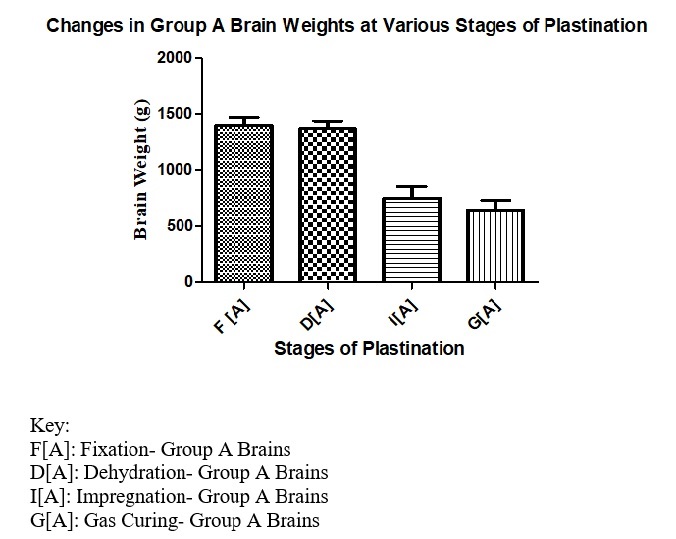 Figure 2.1 Bar chart showing the changes in the weight of the group A brains from the fixation stage to the gas curing stage. The weight of the brains reduced across the stages with the most significant and drastic change in weights being between dehydration and impregnation. There was observable drastic and significant change in weights between the original fixed brain and the final products of the plastination process [p≤0.05]. |
 Figure 2.2 Bar chart showing the changes in weight of the group B brains from the fixation stage to the gas curing stage. The weight of the brains reduced steadily across the stages without any rapid and drastic change in weights. There was only a relatively moderate change between the original fixed brain weight and the final plastinates [p>0.05]. |
 Figure 2.3 Bar chart showing the percentage changes in the volume of the brain after plastination and the percentage changes in the weights of the brains after plastination in an attempt to compare the changes in volume and weights between the two groups. There was a relatively moderate change in the volume of the group B compared to A. Changes in brain weights and volumes were statistically significant [p≤0.05]. |
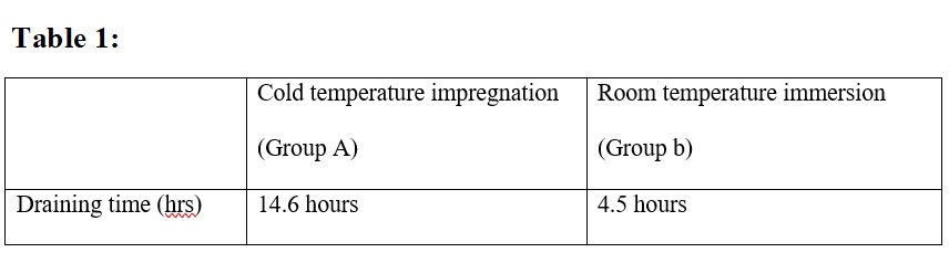 Table 1: Average time of draining excess polymer. Draining excess silicone after room temperature immersion took 4.5 hrs on average, which was 3.24 times faster than at cold temperature. |
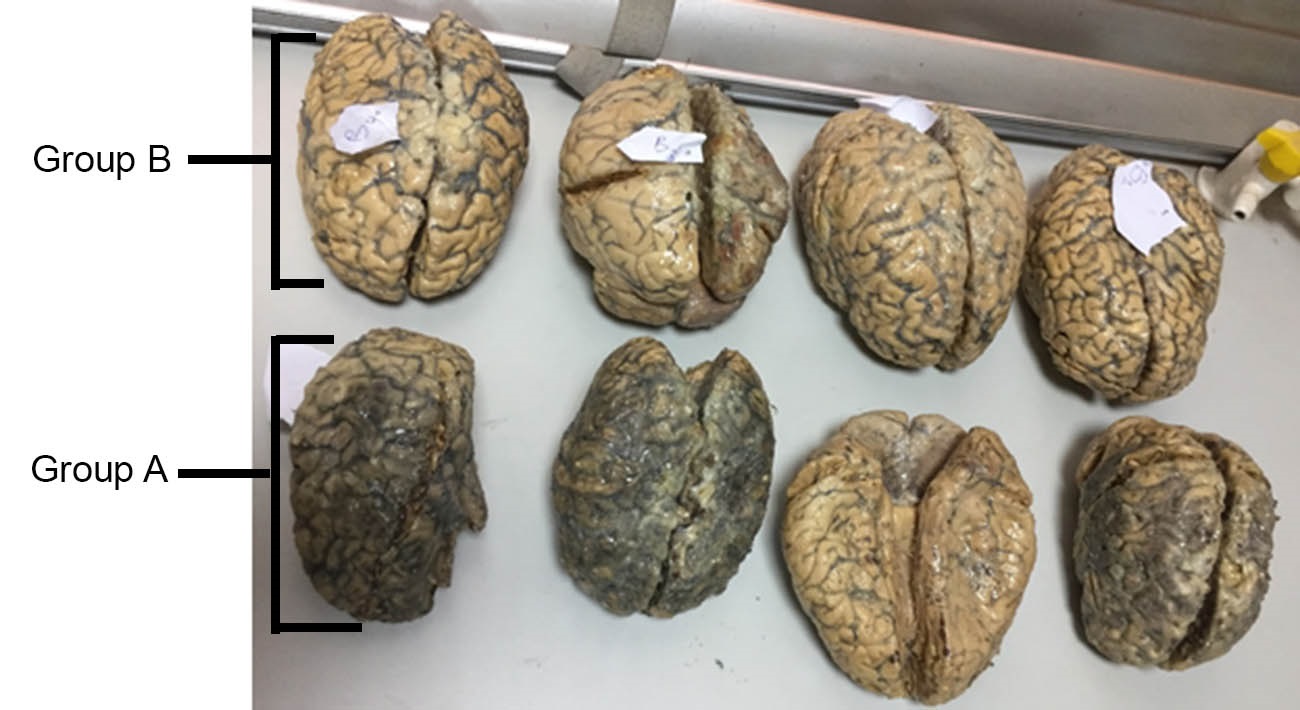 Figure 3. Images showing morphological features of plastinated brains of group A at cold temperature (-25° C) and group B brains processed at room temperature (25° C). Physical appearance of the Group B brains retained the resemblance of the original brain features better than the Group A specimens; thus, the Group B specimens present a better plastination outcome based on physical appearance and presentation of brain morphological features. |
Draining time
After impregnation, the excess polymer was drained from the brain specimens and the specimens remained at room temperature until excess polymer was completely drained off (Table 1).
Quality of the brain plastinates
The quality of brain specimens in each group was examined for color changes and morphological changes such as the prominence of gyri and sulci. Both cold-temperature and room-temperature impregnation techniques yielded brain specimens that were dry, durable, odorless and non- toxic. However, the Group B brains that were processed at room temperature had a better physical appearance by retaining better the original brain colors, better sulci and gyri prominence, and had undergone less shrinkage (Fig. 3).
General observable changes
The present study investigated and compared the standard S10 cold-temperature technique of plastination with a room-temperature immersion process using human brain samples. Both yielded specimens that were dry, durable, odorless and non- toxic. There were observable variations in the outcomes based on the differences in temperature; it was this effect that was under investigation. There were general changes, such as reductions in the weight and volume, thus resulting in physical shrinkage, irrespective of temperature variation. Also, there were changes in the color of the plastinates. There were, then, observable changes in the outcome of the process, irrespective of temperature, and the final outcome is not an absolutely perfect representation of the original mass, volume and color. The results also provide insights into how this knowledge can help understanding and appreciating the original forms of plastinates. The quantification of proportional changes in morphological attributes might also be used as reliable parameters for evaluating the outcomes of plastination.
Changes attributable to temperature variations and treatments
The processed brain samples appeared to have changed colour from the original fixed brain colour to a brownish shade of this color as shown in Figure 3. Also, this brownish shade was darker for the brains plastinated at cold temperature, than for the brains processed at room temperature.
Generally, there was a decrease in weight of the brain specimens after each step of both procedures, which is in agreement with Ameko et al. (2013). The observed decrease in weight was most significant in brain specimens plastinated at cold temperature (-25° C) when compared to room temperature (25° C). This decrease in weight is attributable to the shrinkage experienced in each procedural step of plastination. Decrease in weight was most noticeable during impregnation. The difference in weight after impregnation of plastinated specimens at cold temperature compared to room temperature was 376 g to 599.20 g. The texture of the plastinated brains when felt after the plastination procedure has occurred, appeared to have become more turgid. All specimens (both cold-temperature and room-temperature) became turgid, but the brains processed at room temperature showed better surface clarity, with the anatomical surface markings on the brain clearly visible; when compared to the brain samples plastinated at cold temperature, where the surface markings were not very clear. This result is in agreement with Anant and Madhavi (2015).
The brains produced by both processes were odorless, life-like, and durable, which is in agreement with Wadood et al. (2001). According to Sora et al. (2015), silicone impregnation at room temperature is possible using Biodur silicone. Nelson (1990) and de Jong and Henry (2007), report that plastination of nervous tissue at room temperature using silicone might be challenging, especially with respect to degreasing; however, findings from this study have shown that whole brain specimens can be preserved via this novel S10 technique at room temperature, without the process being compromised by the challenge. Many scholars have recommended the use of the following plastination techniques: P35, P40, and P45, in the study of nervous tissue, while the S10 technique (the gold standard for plastination of anatomical specimens) is not considered ideal for nervous tissues like the brain (Tiedmann and von Hagens, 1982). However, we report here a modified S10 plastination technique at room temperature which is suitable for whole brain study.
It is therefore important to state that more experimentation could be done with the various techniques to further improve the outcomes of plastination, especially under different conditions. While this will enrich the wealth of literature on this technique, it might also help to adapt their uses to various conditions and various organs. It is also expected that organ-specific experimentation might help in identifying the best plastination techniques for different body tissues and organs.
It is evident from this study that the room-temperature S10 technique described here can be used for whole brain sample plastination, and can yield high-quality specimens, particular in terms of morphology, relative weights, texture, and color. This might also come with reduced cost implications, and reduced risk hazards due to the use of room temperature. Both processes, irrespective of the variations in temperature (cold or room), still yielded specimens that were life-like, durable, and odourless, but processing at room temperature yielded a better anatomical brain specimen. It is, therefore, recommended that more research should be done to further the use of plastination as a normal routine for producing teaching aids, and as a means of preservation. Also, processing at room temperature appeared to be a better alternative to plastinating at cold temperature, which requires expensive facilities and power to sustain the required temperature; a factor that might make the procedure less feasible in the developing world.
Ameko E, Achio S, Alhassan S, Sackey M, Danful L. 2013: Room temperature plastination of whole and dissected guinea pigs in Ghana. Int J Pure Appl Sci Technol 19(2): 66-75
http://www.ijopaasat.in/yahoo_site_admin/assets/docs/7_IJPAST-697-V19N2.32215919.pdf.
Anant D, Madhavi D. 2015: Plastination - A boon to medical teaching and research. IJSR 4(5): 1550-1553
https://pdfs.semanticscholar.org/ff16/fe28046ad49dae9c176250892ddc665e13fd.pdf?_ga=2.99310557.2111426504.1546093112-587226869.1545667868.
Azu O, Peter A, Aquaisua N, Ekandem G. 2013: Plastination technology for anatomical studies in Nigeria. Opinion of teachers in medical institutions. Health SA Gesondheld 18(1): 664-666
https://doi.org/10.4102/hsag.v18i1.664
de Jong K, Henry W. 2007: Silicone plastination of biological tissue: cold-temperature technique Biodur S10/S15 technique and products. J Int Soc Plastination 22:2-14.
https://doi.org/10.56507/ZLMJ7068
Jones D, Whitaker I. 2009: Engaging with plastination and the Body Worlds phenomenon: a cultural and intellectual challenge for anatomists. Clin Anat 22(6): 770-776.
https://doi.org/10.1002/ca.20824
Klaus R, Royer D, Stabio M. 2018: Use and perception of plastination among medical anatomy educators in the United States. Clin Anat 31(2): 282-292
https://doi.org/10.1002/ca.23025
Manzoli A, Maraidi M, Capitani S. 2011: In memoriam of Prof. Giovanni Mazzotti. Eur J Histochem 55:175.
https://doi.org/10.4081/1897
Singh O, Mishra B, Pandit S, Maheshwari P, Hasan S. 2013: Plastination: a promising method for preserving biological specimens. Int J Sci Res Pub (IJSRP) 3(6): 1-3
http://www.ijsrp.org/research-paper-0613/ijsrp-p1850.pdf.
Sora M-C and Genser-Strobl B. 2005: The sectional anatomy of the carpal tunnel and its related neurovascular structures studied by using plastination. Eur J Neurol 12(5): 380-384.
https://doi.org/10.1111/j.1468-1331.2004.01034.x
Sora C, Boia M, Banciu CD. 2015: Silicone (Biodur) viscosity and impregnation in plastination. Mat Plas 52(4): 593-595.
Tiedmann K and von Hagens G 1982: The technique of heart plastination. Anat Rec 204(3): 295-299.
https://doi.org/10.1002/ar.1092040315
von Hagens G 1979: Impregnation of soft and biological specimens with thermosetting resins and elastomers. Anat Rec 194: 247-252.
https://doi.org/10.1002/ar.1091940206
Wadood A, Jabbar A, Das N. 2001: Plastination of whole brain specimen and brain slices, J Ayub Med Coll Abbottabad 13(1): 11-13