The National College of Chiropractic, Lombard, IL, USA
For our beginning plastination laboratory the attraction for us of COR-TECH PR-10 silicone was its easily availability for U.S.A. customers as it is locally formulated and supplied and, especially, that its claimed ability to impregnate objects could be done at room temperature, rather than at the -25°C required for standard S10 technique. We could find nothing in the published literature about this new silicone. In order to obtain some indication of its utility for a wide selection of tissues, we first plastinated with COR-TECH PR-10 portions of several organs (kidney, pancreas, gall bladder, brain cortex, muscle, and bone) following verbal instructions from the supplier. In a second trial with the same silicone we plastinated a brain stem and heart. With the cautious, slow plastination of our first specimens no detectable shrinkage had been found (linear, measured between two pins). In our second trial, in spite of fast impregnation at maximum vacuum throughout, the brain stem and heart appeared unchanged after plastination, and shrinkage for the brain stem was only on the order of 3%, compared with as high as 10% in the literature on standard technique. The heart demonstrated 1% shrinkage by the same method of measurement. All of the specimens were usable. Discussion compares and contrasts this silicone process with standard S10, and describes pertinent aspects of our procedures, errors and successes. Future plans are noted. Although the described examples are few, we conclude that this new polymer can easily be used at room temperature. It appears to be faster than standard technique, has minimal shrinkage-even for brain tissue, and is worthy of further exploration.
Presented in part at the 9th International Conference on Plastination, Trois-Rivieres, Quebec, Canada, July 5-10, 1998.
Room temperature impregnation, Human tissue preservation, COR-TECH PR-10 silicone
James Arnold Baker, D.C., The National College of Chiropractic, 200 East Roosevelt Road, Lombard, IL 60148-4583, U.S.A. Tel: 630 889 6852 / Fax: 630 495 6664. Email: JBAKER@NATIONAL.CHIROPRACTIC.EDU

![]()


This report covers two trials of COR-TECH PR-10, each with multiple tissues. First, to gain experience with a wide selection of tissues, we began by plastinating several specimens, viz. a half kidney, a gall bladder, articulated carpal bones, a parasagittal section of lumbar spine, and portions of brain cortex, pancreas, and gastrocnemius muscle. Second, although our plastination laboratory was still under creation, we were implored to plastinate an excellent dissection of a human brain stem in time for the next teaching trimester. The specimen included the diencephalon, pons, and medulla. In addition, the internal capsule and corona radiata were displayed on the left side, and the insula was displayed on the right side. The vasculature, including the circle of Willis, and all cranial nerves were also preserved. We included a heart to be plastinated under the same conditions.
Our primary interest was to try the fast room-temperature impregnation described as possible with the new Corcoran silicone polymers (Corcoran Laboratories Inc., 1405 North Johnson Street, Bay City, MI 48708-5487, U.S.A.). These products were attractive because they were locally available and if they proved usable at room temperature would make low temperature equipment unnecessary for the impregnation step of plastination.
We wanted to examine shrinkage of items plastinated with this silicone because verbal discussions with experienced plastinators warned that brain tissue, especially, suffers in this regard. Long, carefully monitored periods of impregnation time were considered necessary to prevent major shrinkage (von Hagens, 1986; von Hagens et al., 1987). Shrinkage measurement by a linear process has been reported before (Suriyaprapadilok and Withyachumnarnkul, 1997; Sora et al., 1999). To gain an estimate of shrinkage we elected to fix two pins in specimens at a known distance and measure changes in this distance.
Equipment
With the room temperature plastination possible with the Corcoran polymer we were able to use simple, inexpensive equipment. We modified a small counter-height home-type refrigerator by enclosing it in 4 1/2 inches (11.4cm) of polystyrene insulation (Owens Corning Foamular®) (figures 1 and 2), removing all spark-producing contacts to the outside of the refrigerator, and installing an external industrial thermostat with its eight-foot (2.4m) capillary tube leading to the sensing bulb inside (Johnson Controls, Inc. Model A19ABC-24C). A household indoor- outdoor thermometer gave temperature comparisons. An externally controlled 13 watt box-type fan from an old computer circulated internal air. With these modifications we were able to achieve a temperature of -20°C. We rebuilt an existing, but broken, vacuum pump (Gast Mfg. Co. 755 Edgewood Wood Dale, IL 6091-1254. New equivalent pump model 2565V2A). Our vacuum vessel was a four-quart Presto® pressure cooker with a gasketed 3/8 inch (9.5mm) thick Lexan® plastic lid (Baker, 1998). We had a large Bourdon-tube-type vacuum gauge to judge applied vacuum.
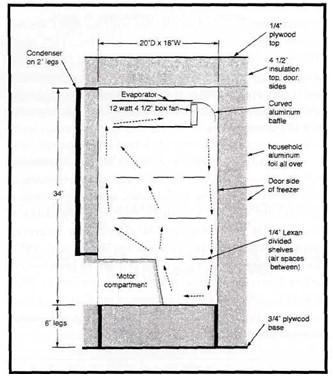 Figure 1. Side View of freezer showing air circulation and construction. |
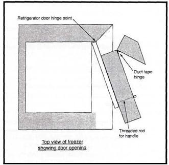 Figure 2. Top view of freezer showing door opening. |
Fixation
All our specimens were from cadavers from our gross anatomy teaching laboratory which had been fixed in a standard anatomic embalming solution (6% formalin, 4% phenol, <2% glycerin, and water), then stored in 2.5% formalin for varying periods up to approximately eight months. After dissection the specimens were stored in 2.5% formalin for one week, until preparations for plastination were completed.
Dehydration
Specimens were drained then placed in cold 100% acetone. All specimens were dehydrated by the freeze substitution method (von Hagens et al., 1987). In an attempt to estimate shrinkage, pins were placed in the specimens and measured prior to dehydration. On each specimen two pins were located as far apart as possible on the largest available reasonably flat surface.
Plastination
Corcoran silicones and associated crosslinkers are supplied in several varieties. At the recommendation of the supplier, we chose to begin with COR-TECH PR-10 and COR-TECH CR-22 crosslinker, said to be easy to use, transparent, of near-water consistency, and producing a reasonably flexible product (Dan Corcoran, personal communication). We used the recommended "preservation catalyst CT-30," a somewhat viscous clear liquid.
Impregnation:
Impregnations were all performed at ambient temperature (24°C).
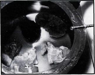
Figure 3. Trial 1 specimens in vacuum chamber.
Trial 1. The half kidney, a gall bladder, articulated carpal bones, a parasagittal section of lumbar spine, and portions of brain cortex, pancreas, and gastrocnemius muscle were all impregnated simultaneously in our one vacuum chamber shown in figure 3. Specimens were kept immersed by an overlying bronze window screen (folded into several thicknesses to increase its weight) in a mixture of 97% COR- TECH PR-10 silicone polymer and 3% COR-TECH CR-22 crosslinker (this ratio was initially recommended by the supplier). We very slowly began to apply the vacuum, just enough to keep a small amount of bubbles flowing from the specimens. Bubbles were easy to see due to the clarity of the polymer-crosslinker mixture. Bubbling stopped when we reached a pressure of 50mm Hg inside the impregnation chamber. We do not yet have a Bennert type manometer so the pressure was calculated from the stable maximum vacuum applied. Impregnation was then considered complete and took 12 days. The specimens were then taken out to drain for one more day, ready for curing.
Trial 2a. The heart was placed in the same vacuum chamber as used in trial 1. However, we had added sufficient COR-TECH CR-22 crosslinker to the mixture to increase its percentage from 3% (as in trial 1) to 5% because the supplier informed us that this would decrease impregnation time (Dan Corcoran, personal communication). Since the specimen floated, a screen was again placed on top of the specimen to hold it below the surface of the liquid.
As opposed to our first experiment, the vacuum was immediately turned up to the maximum. Furious bubbles appeared immediately, both large (air) and small (acetone). Within eighteen hours, bubbling had settled down to minor, regular fine bubbles; this condition persisted with decreasing activity over time, until no more bubbles appeared, at which point the specimen was removed to have its surface wiped off, then it was drained for a day. The final pressure was calculated to be 50mm Hg. Total impregnation time was fifty- four and one half hours.
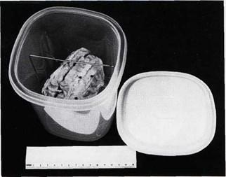
Figure 4. Method of suspending brain stem for drainage.
Trial 2b. The brain stem was impregnated immediately after the heart in the same 95%/5% silicone-crosslinker solution as the heart. As with the heart, vacuum was immediately turned up to the maximum, with frantic bubbling. Residual end-point pressure was 50mm Hg. Total impregnation time was forty-two and one quarter hours. The longer impregnation time of the heart than the brain stem represented interruptions due to other duties rather than the needs of the specimen. The brain stem was drained for two and one quarter days before curing. Because of the delicate structure its surface was not wiped before draining. To suspend the brain stem in a closed polyethylene container for draining, we made a U-shaped wire frame from one sixteenth inch diameter (1.6mm) type 304 stainless steel gas- type welding rod and attached it by a small copper wire looped around a straight pin inserted through the specimen (figure 4).
Curing:
Curing was also done at ambient temperature (24°C).
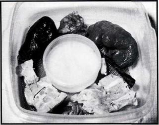
Figure 5. Trial 1 specimens in unsatisfactory curing attempt.
Trial 1. When we completed the first batch of specimens we had understood that curing was effected by placing them in a closed container with an open dish of COR-TECH CT- 30 catalyst so the vapor would begin the curing process (Dan Corcoran, personal communication). We did this for several days, using a closed Rubbermaid™ container, shown in figure 5. Nothing happened. While we were trying to sort out the curing process, we performed an experiment with a few drops of polymer in open petri dishes together with an open petri dish of catalyst, all in a closed container. Each day for 10 days we removed a dish of polymer to discover whether it had cured. None did, so we abandoned trying to cure the polymer with only vapor contact. We phoned the supplier again and found that we had missed the fact that an initial topical application of curing agent was necessary. We applied catalyst with a cloth, being very careful not to contaminate our catalyst with polymer, and waited. Finally, at eleven days the specimens appeared to be cured, but they still had a greasy surface residue which we wiped off with acetone. After eight more days, curing was deemed to be complete, as determined by dry, odorless surfaces.
Trial 2a. Subsequent to trial 1 we were informed that a good way to apply catalyst without the danger of contamination of catalyst by contact with silicone was to spray it on from a common hand sprayer (Dan Corcoran, personal communication). So, after draining was complete, we attempted, without success, to spray COR-TECH CT-30 catalyst on the specimen with a common hand sprayer with an adjustable nozzle. We tried with five different sprayers. The problem was that the catalyst came out of sprayers in a solid stream. We finally squirted catalyst on and carefully wiped off the excess catalyst liquid and droplets using paper towels and light tissue.
We were also told to place the specimen in a common self-seal plastic freezer bag. When air is excluded the process is said to occur much more rapidly (Dan Corcoran, personal communication). So, after wiping off the excess catalyst we placed the heart in a freezer bag, squeezing out as much air as possible before sealing the bag. After forty-one hours we opened it to find polymer cured on the surface of the heart in many spots where the bag had contacted the heart. With care and persistence, it was possible to remove these white spots. Further phone conversation made it clear that we had misunderstood the instructions by leaving it in the bag too long. Twenty four hours is the maximum recommended time (Dan Corcoran, personal communication).
Trial 2b. The brain stem was cured by the same procedure as the heart (in a plastic freezer bag with most of the air squeezed out before closing, as noted above, except that, learning from our experience with the heart, we removed the brain stem from the bag after twenty hours. The bag was opened and the surface wiped. Polymer that had been loosely catalyzed on the surface where the bag contacted it was easily removed. A sheen of polymer showed on a few surfaces where the plastic bag had contacted it. Most of these were easily wiped off. Finally, the brain stem was left suspended in open air (in the container, but with the lid removed) at room temperature. In five days, the surface was dry and the catalyzing process was considered complete enough so the brain stem could be handled.
Shrinkage of our samples by the two-pin method we used is shown in Table 1. Beginning and ending values are probably the most accurate. For the first batch, plastinated in a relatively long procedure (12 days of impregnation), there was essentially no shrinkage. The increase for the brain tissue and carpal bones is probably measurement error. For the heart and brain stem, fast-as-possible plastination produced 1% and 3% shrinkage, respectively. It would appear that, for these two, most of the shrinkage took place during dehydration. The only difference in time between all the various trials was in impregnation.
| Before | >C3H60 | >PR-10 | End | % | |
| Trial 1 |
19 |
18.5 |
19 |
19 |
0 |
| Half kidney | |||||
| Pancreas | 16 | 16 | 16 | 17 | +6 |
| Brain cortex | 18 | 18 | 17.5 | 18 | 0 |
| Carpal bones | 11 | 12 | 12 | 11.5 | +5 |
| Lumbar spine | 14* | - | - | - | |
| Gastroc. | 11 | 11 | 11 | 11 | 0 |
| Gall bladder | 12 | 11 | 12 | 11 | 0 |
| Trial 2a
Heart |
46.5 |
45 |
45 |
46 |
1 |
| Trial 2b
Brain stem |
37.5 |
36 |
36 |
36.5 |
3 |
Figures 6a to e show some of the finished specimens from our first trial. The plastinated heart is shown in figure 7, and figures 8 and 9 show the plastinated brain stem. None of the specimens were unusable, or significantly altered in color, shape or general appearance. The brain stem was immediately put into use for teaching purposes where it still is being used. The heart was put aside for the time being . We had no air to expand the heart chambers or vessels, so the great vessels were somewhat folded. They were fairly rigid, but the heart muscle was not. The brain cortex sample was somewhat brittle . The pancreas tissue and half kidney were barely flexible. The gall bladder was highly flexible, as was the gastrocnemius muscle . The carpal bones were moveable, approximating their state before plastination. Samples of the brain cortex and kidney were taken for microscopic examination, with initial promising results (e.g. cellular organelles and capillaries were easily seen).
 |
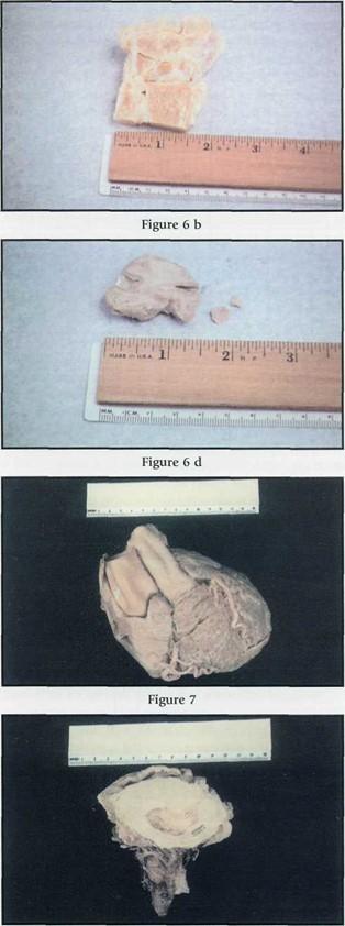 |
| Figures 6 a - e: Trial 1 finished specimens, a. External surface of coronally sectioned kidney b: Articulated carpal bones c: Parasagittal section of lumbar spine d. Brain cortex (with fragments broken off in assessing flexibility) e. Portion of gastrocnemius muscle.
Figure 7. Plastinated heart. Figure 8 and 9. Plastinated brain stem. |
|
The two-pin method we used for measuring shrinkage is admittedly not rigorous, and should be considered an approximation only. Many of the first specimens were too small to use multiple measurement points. For example, three pairs of pins placed in lines orthogonal to each other would give more accurate information. Volumetric procedures, where applicable, could be used to good advantage.
Dehydration is not different from that used for the S 10 technique. Impregnation is the major difference from standard SlO technique. Impregnation with the COR-1ECH-xx series of silicones may be done at room temperature, whereas S10 requires minus 25°C for impregnation (von Hagens, 1986). Room temperature impregnation means that low temperature facilities are needed only for dehydration, thus reducing equipment cost.
A second difference is that with the one we have used, COR-1ECH PR-10, rising acetone bubbles are easy to see, as the polymer is crystal clear. As well, the bubbles rise quickly, since this polymer is near water consistency, whereas S10 polymer is "highly viscous" (von Hagens, 1986). This facilitates faster impregnation time.
Curing for COR-1ECH PR-10 appears to be a gas-cure, similar to S10, however an initial topical application of catalyst must be made, followed by enclosing the specimen in a plastic bag with as much air as possible excluded for a maximum of twenty-four hours. Initially excluding air in the curing process is said to allow a high concentration of catalyst vapor pressure to "drive" the catalyst deep into the specimen (Dan Corcoran, personal communication) .
Surface curing proceeds rapidly. If there is any remaining polymer on the surface, this will cure against the bag or in droplets, leaving white cured drops or a sheen of cured plastic where the bag touched. If the specimen is taken out soon enough, these surfaces may be wiped clean before complete curing has set in. After removing the item from the bag and wiping off any excess, it should be left to air cure at ambient temperature .
We had wondered how inaccessible crevices in the highly convoluted brain stem would cure when they could not be directly painted with catalyst. This turned out not to be a problem because curing seems to proceed throughout the specimen, once started. The end point of curing may be considered to be when the surface of the specimen is dry.
Any problems we had were due to our newness to the process of plastination and to our misunderstanding of the verbal instructions. We made several procedural errors, notably not making an initial topical application of catalyst and keeping it in the plastic bag for longer than 24 hours. Since we processed the above samples, a summary set of instructions has been made available (Corcoran Laboratories, Inc., 1998). This describes the polymer we used as well as the several others which are available.
After phone discussion with the supplier we chose to start with COR-1ECH PR-10 for all the described specimens. It is stated to have "easy penetration with some flexibility, fastest impregnation time" (Corcoran Laboratories, Inc., 1998). The great vessels of the heart we plastinated are more stiff than we had expected. This may be due to reported firmness of some tissues, as intestine, if left too long in acetone (von Hagens, 1986). We plan to experiment with COR-1ECH PR-12 polymer, which is stated to have more flexibility "easy penetration with more flexibility that COR- 1ECH PR-10. A little longer penetration time." (Corcoran Laboratories, Inc., 1998). With the above noted reservation, the plastinated heart is otherwise excellent.
From this limited trial it is unclear exactly what results may be expected from varying the silicone crosslinker ratio. We noticed no difference that could be specifically attributed to the two concentrations we used. We intend to experiment with varying amounts of crosslinker, and varying amounts of directly applied catalyst to obtain a better conception of appropriate curing parameters. Sheet plastination with this silicone remains to be explored.
It is imperative not to contaminate either the catalyst or polymer, such that one's stock of raw material begins to catalyze. To_this end it is suggested to spray ("mist") catalyst onto the surface of the specimen, thus avoiding physical contact. We understand that catalyst COR-1ECH CT-32 can be sprayed from a common hand sprayer with an adjustable nozzle (Dan Corcoran, personal communication). We intend to try this on a future specimen.
The plastination materials (polymer, crosslinker and catalyst) are Dow Corning products (Midland, MI), and labels list multiple constituents. Reasonable precautions need to be taken with regard to handling the three components, e.g. gloves and proper ventilation, especially when spraying catalyst. Of these three components, the crosslinker is the only one which was shipped with a "flammable liquid" label and a "poison" warning, possibly because it contains methanol.
In spite of our inexperience and the dearth of information about the new silicone, we had no disasters with the above- noted specimens. Our initial specimens remain unchanged in appearance or consistency after a year. All were usable as planned. Rapid application of the vacuum for impregnation of the heart and brain stem appeared to cause no problems. The small amount of shrinkage might have been reduced even further by increasing the vacuum gradually, but the shrinkage was well within our expectations. We did not have any difficulty with handling silicone, cross-linker or catalyst. We conclude that COR-TEC PR-10 is an excellent, easy-to-use silicone for plastination of the materials for which we have used it, including brain stem tissue. It may be used at room temperature and, importantly, allows quick preparation of specimens. We realize that the limited examples cited do not compose a rigorous examination of the use of this silicone for plastination, but we hope that our experience may benefit those who also wish to explore using this material.
Acknowledgements
Baker JA: An Economical Plastination Pilot Project and Trial of the COR-PR-10 Polymer. 9th Int Conf Plast, Trois- Rivieres, Quebec, Canada, 1998. Abstract in J Int Soc Plastination 13 (2): 32-33, 1998.
Corcoran Laboratories, Inc.: Room Temperature Process: Suggested Use for Best Results. [Note. This five page, undated combination material description and instruction booklet was received in July 1998 at the 9th International Conference on Plastination at Trois-Rivieres, Quebec]
Sora MC, Brugger P, Traxler H: P40 Plastination of Human Brain Slices: Comparison between Different Immersion and Impregnation Conditions. J Int Soc Plastination 14 (1): 22-24, 1999.
https://doi.org/10.56507/XLSJ5724
Suriyaprapadilok L, Withyachumnarnkul B: Plastination of Stained Sections of the Human Brain: Comparison between Different Staining Methods. J Int Soc Plastination 12 (1): 27-32, 1997.
https://doi.org/10.56507/YISQ6047
von Hagens G: Heidelberg Plastination Folder. Anatomisches Institut, Universitat Heidelberg, Heidelberg, Germany, 2nd ed, sec. 3.3.4, p. 3.8, 1986.
von Hagens G, Tiedemann K, Kriz W: The current potential of plastination. AnatEmbryol 175 (4): 411-421, 1987.
https://doi.org/10.1007/BF00309677