Orthopaedic University Hospital and Department of Anatomy¹, University of Heidelberg, Germany
In order to determine the yascularization of cranial (anterior) cruciate ligament (ACL) allografts, six white New Zealand rabbits underwent ACL allotransplantation. The animals were sacrificed at 6, 12 and 24 weeks postoperatively, and their hind limb vessels were injected with an epoxy resin and cut with a cryomicrotome. The blood supply of the ACL allograft was shown to arise mainly from the infrapatellar fat pad and the hypertrophied synovial membrane.
ACL allograft, Blood supply, Animal model, Plastination, Cryomicrotome.
B. Fromm, Orthopaedic University Hospital University of Heidelberg, Germany

![]()


The blood supply of the undamaged cranial cruciate ligament is mainly derived from the surrounding soft tissue structures (infrapatellar fat pad and synovial membrane), which form a f ine paraligamentous envelope surrounding the ligament (Arnotzky et al., 1979). These vessels penetrate the ACL and arborize around the collagen fibers, forming an endoligamentous network of vessels which course in a longitudinal plane both proxirnally and distally (Clancy et al., 1981). Other authors found the endosteal vessels of the tibia and the femur to be a pathway for delivery of nutrients to the cruciate ligaments (Whiteside and Sweeny, 1980).
This study was undertaken to determine the blood supply of the cranial cruciate ligament allografts in an animal model.
The cranial cruciate ligament with its femoral and tibial bony attachments was harvested from six mature New Zealand white rabbits (weight 3,500 ± 200 gms) under aseptic conditions, frozen at -96 "C for 72 hours, and then transplanted into the ipsilateral knee joint of the rabbits, whose own ACLs were removed during the surgical transplant procedure (similar weight as donor animal). No immobilization was performed. The animals were sacrificed after 6,12 and 24 weeks and the blood vessels of their hind limbs injected with an epoxy resin using a continuous pressure infusion (120 mm Hg) into the abdominal aorta as described by von Hagens (1987). A mixture of 100 parts Biodur E20, 30 parts methyl ethyl ketone and 45 parts of Biodur E2 was injected. During the injection procedure, the caudal (inferior) vena cava was opened. The injection was stopped when epoxy resin appeared in the transected vena cava. The dissected knee specimen was frozen and sliced using a cryomicrotome at -70 °C. No further preparation was necessary. Sequential close up photographs of the frozen knee sections were taken after the application of a thin film of paraffin which enhanced visualization.
No blood supply was seen to originate from the endosteal vessels of the tibial or femoral cavities. A gradual increase in numbers of blood vessels from the surrounding infrapatellar fat pad and the thickly hypertrophied synovial membrane grew from the tibial and femoral attachments of the transplanted ACLs towards the mid-thirds of the ligaments (Figs. 1, 2a, 2b). Twenty four weeks after transplantation, the middle third still showed a zone of hypovascularization (Fig. 3). No signs of rejection were observed.
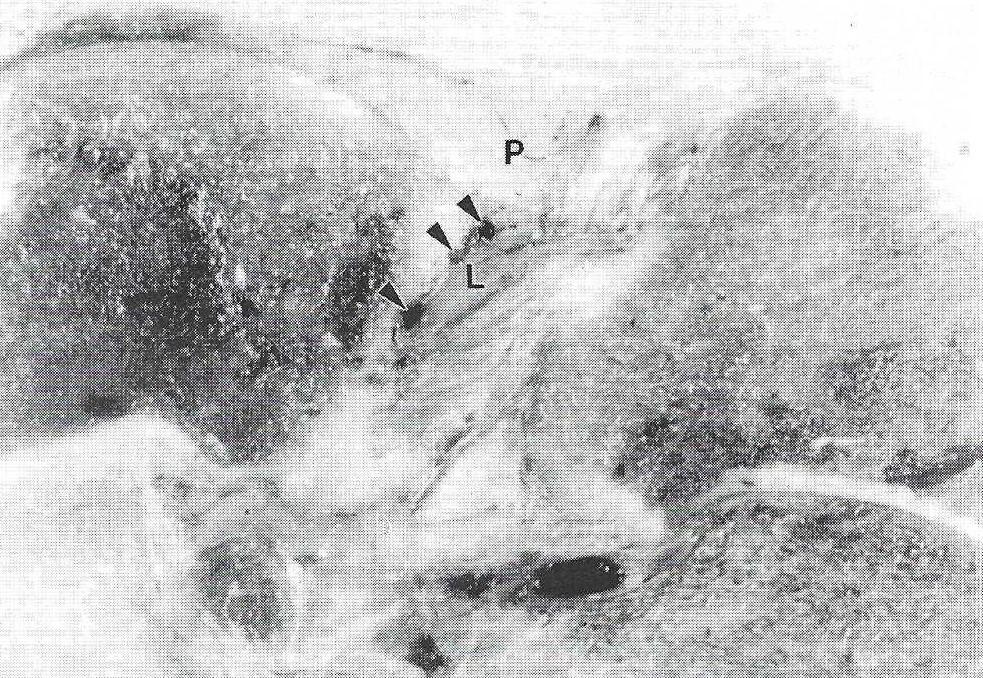 Figure 1. Six weeks after homologous ACL transplantation, the cranial cruciate ligament (L) is ensheathed with blood vessels (arrowheads) derived from the infrapatellar fat pad (P). |
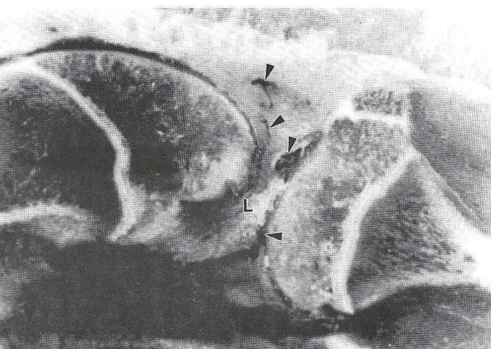 Figure 2a. Twelve weeks after surgery, the cranial cruciate ligament (L) has a well-developed vascular supply (arrowheads) at both its proximal and distal ends. |
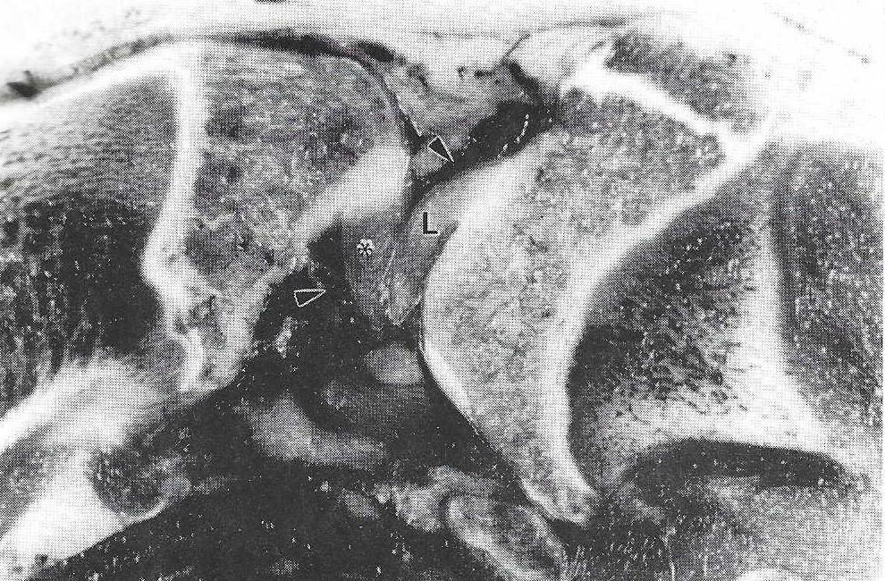 Figure 2b. Twelve weeks after surgery, the cranial cruciate ligament (L) has a well-developed vascular supply (arrowheads). Caudal (posterior) cruciate ligament (*). |
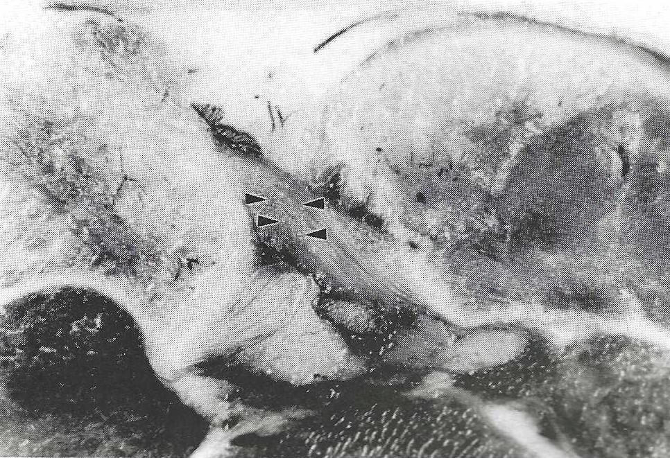 Figure 3. Twenty four weeks after surgery, a zone of hypovascularization remains (outlined by arrowheads) in the middle third of the transplanted ligament. |
The ACL allografts were incorporated into the hosts knee joint and vascularized in a similar way as autologous transplants. The time period for a complete revascularization to occur was prolonged as compared to autologous transplants. In experiments with rhesus monkeys, Clancy and co-workers (1981) found no difference in vascularization after the eighth postoperative week in autologous tendon transplants. The incomplete vascularization, still seen after the 24th week in our series, could be explained by the prolonged substitution period needed for the incorporation of the allografts. The vascular injection of epoxy (von Hagens, 1977, 1981, 1985, 1986, 1987) was simple and easy to perform. The injected specimens offered excellent demonstration of the microangiography of the rabbit's knee joint.
Arnotzky SP, RM Rubin, JL Marshall: Microvasculature of the anterior cruciate ligaments and its response to injury. An experimental study in dogs. J Bone and Joint Surg 61 A: 1221-1229, December 1979.
https://doi.org/10.2106/00004623-197961080-00013
Clancy WG, RG Narechania, TD Rosenberg, JG Gmeiner, DD Wisnefske, TA Lange: Anterior and posterior cruciate ligament reconstruction in Rhesus Monkeys. A histological, micorangi- ographic, and biomechanical analysis. J Bone Joint Surg 63A: 1270-1284, October 1981.
https://doi.org/10.2106/00004623-198163080-00008
Whiteside LA, RE Sweeny: Nutrient pathways of the anterior cruciate ligaments. An experimental study using the hydrogen wash-out technique. J Bone Joint Surg 62A:1176-1180, October 1980.
https://doi.org/10.2106/00004623-198062070-00019
von Hagens G: DB Patent 27 10 147 (1977), USA Patent 4, 205 059 (1981).
von Hagens G: Heidelberg Plastination Folder. Anatomisches Institut I, Universitat Heidelberg, 6900 Heidelberg, Germany, 1985,1986
von Hagens G: The current potential of plastination. Anat Emryol 175:411-421, 1987 and personal communication.
https://doi.org/10.1007/BF00309677