Division of Anatomical Sciences, Office of Medical Education, The University of Michigan Ann Arbor, USA
Plastination has enhanced the way students study human gross anatomy by providing them with three-dimensional specimens that they can hold and manipulate. These specimens allow students to learn gross anatomy, especially difficult areas, more efficiently. However, the intricacies of organ function in life are is still difficult to understand from dissected specimens. At the University of Michigan Medical School, innovative approaches to enhance the quality of plastinated specimens have been implemented to demonstrate complex anatomical features. The heart is a particularly difficult organ for students to,visualise because of the unique changes it undergoes during systolic and diastolic phases of the cardiac cycle. The aim was to develop a plastinated heart model that demonstrates how cardiac valves function during the systolic and diastolic phases. Five hearts were collected from cadavers, dissected and plastinated. Various incisions in the heart were made to reveal the cardiac valves. Corks, sutures, and hinges were used to position and to hold the valves in place either in its contracted state (systole) (2 out of 5 hearts) or in its relaxed state (diastole) (3 out of 5 hearts). A pilot survey was administered to get students’ feedback on these plastinated models. The results indicate that a majority of students favor this novel animated model as it displays both systolic and diastolic phases while keeping superficial structures of the heart intact.
plastination, heart, valves, systole, diastole
Dr. Ameed Raoof, Office of Medical Education, The University of Michigan Medical School, 3740 Med. Sci. II Bldg., Ann Arbor, MI, 48109-5608, USA; Telephone: 734-615-2597; Fax: 734-615 -8191; E-mail: ameedr@umich.edu.

![]()


The advent of the plastination process in gross anatomy laboratories provided students with a learning tool that transforms two-dimensional textbook images into three-dimensional models that can be examined and manipulated (von Hagens et al., 1987). One of the most challenging organs to understand for medical students is the human heart, with its complex physiology and unique two-stroke mechanism. Knowledge of the basic anatomy and physiology of the heart is an essential component of any medical or health science curriculum. Demonstrating how heart valves open and close during systolic and diastolic phases of a cardiac cycle is difficult with cadaveric specimens. The aim of this study was to develop a plastinated heart model that demonstrates how cardiac valves function during the systolic and diastolic phases. Plastinated models of hearts in both systole and diastole would allow students to identify each valve and its role in each phase of the heartbeat, as well as provide them with an intact three-dimensional tool for learning the fine details of heart anatomy and how its structure is related to its function.
Five embalmed human heart specimens were harvested from cadavers donated to the University of Michigan’s Anatomical Donations Program, age ranged between 70-80 years (average 75). The hearts were kept in a water bath throughout the duration of the dissection to preserve their hydration and to enhance clot removal from the heart. At first the pericardium was removed, as well as fat from around veins and arteries.
To remove the superior aspect of the heart, the semilunar cusps were located and an incision was made just above the pulmonary and aortic valves. This incision was extended around the heart superior to the coronary sulcus for a full transverse section of the atria and aorta and pulmonary trunk. To mimic the contracted state of the heart (systole), the mitral and tricuspid valves were sutured closed while the aortic and pulmonary semilunar valves were positioned open using corks of a suitable size in two of the hearts.. The other three hearts were used to demonstrate the heart in its relaxed state (diastole). The mitral and tricuspid valves were positioned open using corks and the aortic and pulmonary semilunar valves were sutured closed. After the heart valves were prepared, the two portions of the heart were sutured to align and preserve the original external anatomy (Raoof, 2001).
After flushing the hearts with water, they were dehydrated in cold acetone. The prepared hearts were plastinated using the room temperature method (Raoof 2001, Raoof et al., 2007). The impregnation polymer was mixed with 8% cross-linker (CR 22- Dow Corning). After forced impregnation, all sutures and corks were removed. A small brass hinge was attached with pins and glue to keep the basal and apical portions of the specimens as one unit. The hinges were attached with sutures, fine screws, and rubber silicone (Figure 2).
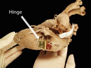
Figure 2: Transected human heart with hinge
After the hinge had been securely attached to the heart, the specimen was cured using a catalyst (CT 32- Dow Corning) (Raoof, 2001, Raoof et al., 2007).
The impregnated and cured hearts were used by undergraduate students in Human Anatomy. The course introduced the students to the basic concepts of systemic anatomy and included visits to the lab where pertinent plastinated specimens are displayed. To evaluate student perception after using the plastinated hearts, a pilot survey was developed and administered to these 199 undergraduate students. Questionnaires were distributed and students were asked to express their opinions to three questions on a 5-point Likert scale. The questions were (1) “whether the heart specimens have been useful in demonstrating structural relationships”, (2) “whether the specimens have been beneficial to learning the heart’s anatomy”, and (3) “whether the specimens have been beneficial to understanding function in correlation with structure”. Data from the survey was compiled and analyzed using Microsoft Excel.
Careful preparation of the hearts permitted production of heart specimens for demonstration of systole and diastole of the cardiac cycle while preserving the relationship of superficial anatomy of the heart
(Figure 3).
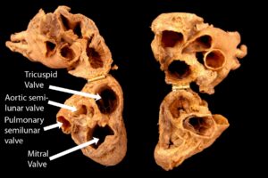
Figure 3: Plastinated human heart displays the systolic (left) and diastolic (right) forms.
The valves were misshaped during closure and opening but could be made anatomically correct prior to hardening/application of the catalyst.
The value of these models for undergraduate study of heart anatomy and physiology was assessed by conducting a pilot survey among undergraduate anatomy students. Students were allowed to spend time studying and manipulating the heart, after which they were asked if they felt a model of this type was beneficial to learning anatomy. Sixty out of 199 students responded to the survey. A majority of students felt that these specimens were useful in demonstrating structural relationships (Figure 4) and were helpful to their understanding of details of heart anatomy (Figure 5) and how structure correlates with function (Figure 6).
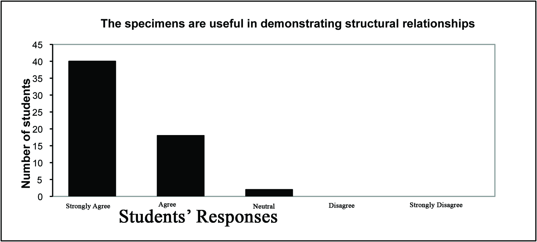 Figure 4: Student response, pilot survey, question 1 - whether the heart specimens have been useful in demonstrating structural relationships. |
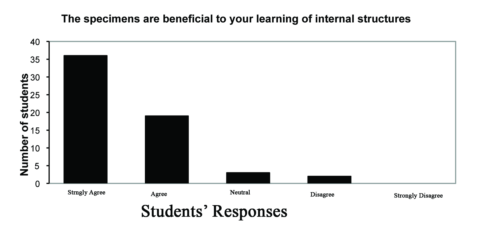 Figure 5: Student response, pilot survey, question 2 - whether the specimens have been beneficial to learning the heart’s anatomy. |
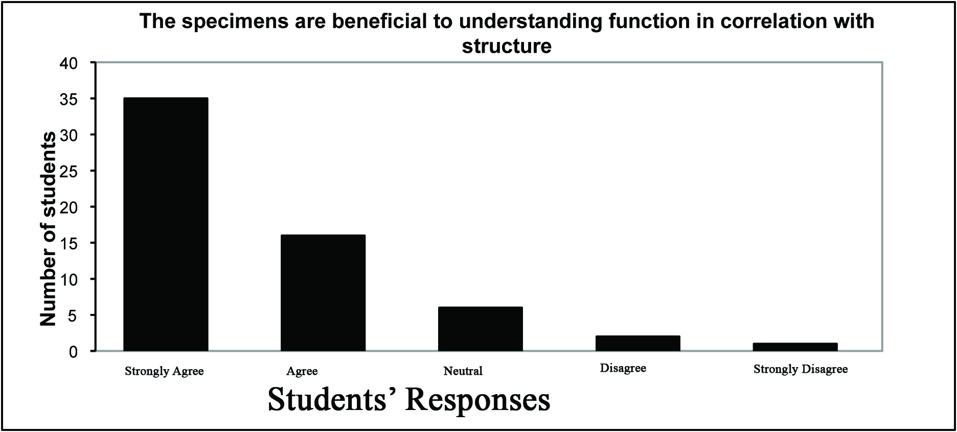 Figure 6: Student response, pilot survey, question 3 - whether the specimens have been beneficial to understanding function in correlation with structure. |
Metal hinges were used to create a simple animated model of the heart that would permit students not only to study the external features of the heart, but also observe the internal structures and to gain a better understanding of the correlation between valve function and blood flow during various stages of the cardiac cycle. These systolic and diastolic models help students comprehend the sequence of events that make up a cardiac cycle. Technically, it is essential to modify valve contours which are misshaped due to the pressure of the cork and/or suture to their final perceived anatomical shape after silicone impregnation and before adding the catalyst. During preparation of the cardiac valves an incision was made above the pulmonary and aortic valves to view the semilunar valves. The incision around the heart superior to the coronary sulcus permitted the view of the atrioventricular valves. After plastination the two portions of the heart were sutured to align and preserve the original external anatomy (Figure 1).
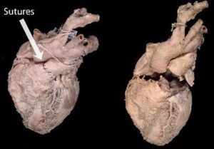
Figure 1: Left specimen: heart sutured at the pulmonary trunk. Right specimen: base of the heart transected from the apex.
Baptista and Conran, 1989, prepared the cardiac valves prior to dehydration by anchoring the leaflets of the right and left atrioventricular valves with small pieces of moistened cotton introduced through a small incision in the right and left ventricles. Small pieces of cotton were also positioned in the cusps of the semilunar valves. After plastination, the incision was widened reviewing the internal structures of the ventricles such as chordae tendineae and papillary muscles, and their relationship with the cardiac valves. Gomez et al., 2011, in an attempt to correlate plastinated heart slices with echocardiographic images, used 13 dog hearts fixed by dilation. The plastinated slices corresponded accurately with the echocardiographic images revealing the internal anatomy with great details. The dilated cavities of the plastinates (due to fixation) made comparison with echocardiographic images of systole difficult.
A current project will further enhance understanding of heart anatomy. The heart is being sectioned along a sagittal plane through the septum and a second hinge attached, giving another view into the heart. Hinges are rather obtrusive, therefore, magnets will be used instead of hinges.
Baptista, C.A.C. and Conran P.B.1989: Plastination of the heart: Preparation for the study of the cardiac valves. J Int. Soc. Plastination 3: 3-7
https://doi.org/10.56507/PEXL8984
Gomez A., Del Palacio J. F., Latorre R., Henry R. W., Sarria R., Arbors O. L. 2011: Plastinated heart slices aid echocardiographic interpretation in the dog. Vet Radiol Ultrasound 53 (2): 197-203
https://doi.org/10.1111/j.1740-8261.2011.01880.x
Raoof, A. 2001: Using a room-temperature plastination technique in assessing prenatal changes in the human spinal cord. J Int Soc Plastination 16: 5-8
https://doi.org/10.56507/UIHR4575
Raoof A, Henry RW, Reed RB 2007: Silicone plastination of biological tissue: room temperature plastination technique - DowTM /Corcoran technique and products. J Int Soc Plastination. 22: 21-25.
https://doi.org/10.56507/AWAC9285
von Hagens G, Tiedemann K, Kriz W. 1987: The current potential of plastination. Anat Embryol 175(4): 411-421.
https://doi.org/10.1007/BF00309677