Department of Diagnostic and Biomedical Sciences, The University of Texas Health Science Center at Houston School of Dentistry, Houston, TX 77054, USA
For centuries, cadavers have been the traditional model of teaching gross anatomy. However, with the large time investment and high cost of maintenance associated with cadavers, studying anatomy via plastinated specimens has become increasingly attractive. Plastination is a novel technique for preservation of the human body by replacing water content with polymers, creating specimens that are dry, odorless, durable, and nontoxic. In 2016, The University of Texas Health Science Center at Houston School of Dentistry (UTSD) underwent a significant curriculum reform by replacing cadaveric specimens with plastinated prosections. Objectives: The aim of this study was to investigate our dental students’ and residents’ perceptions of learning anatomy from cadavers or plastinated specimens, and to determine if student performance in anatomy lab exams have changed since this reform. Methods: A survey was administered to dental students and residents who studied anatomy via cadavers and/or plastinated specimens. Lab exam scores from the past six years were compared in these two models of anatomy instruction. Results: More than half of students in all cohorts believed that plastinated prosections can effectively replace the need for dissection. ANOVA analysis revealed that the switch from cadavers to plastinated specimens had a significant improvement of lab exam scores. Conclusions: At UTSD, the shift in learning anatomy from cadavers to plastinated specimens increased student satisfaction with anatomy instruction and improved student performance in the course.
anatomy; cadavers; prosections; dissections; plastination
Dr. Vuvi H. Nguyen, Department of Diagnostic and Biomedical Sciences, The University of Texas Health Science Center at Houston School of Dentistry, 7500 Cambridge Street Suite 5371, Houston, TX 77054. Tel: 713-486-4554; Fax: 713-486-4416; E-mail: vuvi.h.nguyen@uth.tmc.edu

![]()


Anatomy is one of the most important and clinically relevant curricular necessities in dental and medical education. Historically, dissection and didactic lectures were its sole pedagogy (Sugand et al., 2010). Over the past few decades, anatomy education has undergone significant changes in order to meet the demands and evolution of curriculum design (Drake 1998; Drake et al., 2009; Pyle 2012). Despite many anatomists who still favor the use of dissection over other teaching tools, there has been on-going debate whether or not cadaveric dissections is still suitable in anatomy education (Patel and Moxham, 2006; Korf et al., 2008; Estai and Bunt, 2016). For example, in place of cadaveric dissections, institutions across North America and Europe have implemented prosected (already-dissected) cadavers along with other teaching modalities (e.g., computer-based learning) in their anatomy curricula. As a result, time required for dissection from both students and faculty has dramatically reduced (Reidenberg and Laitman, 2002; Estai and Bunt, 2016; Rizzolo et al., 2010). In addition, the maintenance of cadavers is associated with high costs such as preservation fluid, ventilation equipment, and lab space (Sugand et al., 2010; Rizzolo et al., 2010; Estai and Bunt, 2016). One particular challenge many dental and medical education programs often face is finding time in their curricula for new content in other courses, while upholding the numerous hours typically allocated to a traditional anatomy course (Rowland and Joy, 2015; Estai and Bunt, 2016). According to the American Dental Association (ADA), anatomy instruction in lecture and lab takes up nearly twice the amount of time compared to other disciplines in the biomedical sciences in dental education (Baker et al., 2013).
In 2016, The University of Texas Health Science Center at Houston School of Dentistry (UTSD) underwent a significant reform in their Head and Neck anatomy curriculum by implementing the use of plastinated prosections in place of cadaveric specimens. Plastination is a novel technique for preservation of the human body by replacing water content with polymers. As a result, these specimens are dry, odorless, durable, and nontoxic, which allows for easy storage and handling (Bickley et al., 1981; von Hagens et al., 1987). This reform eliminates the time needed for dissection, including cleanup and maintenance of specimens, and therefore reduces the many hours required for students and faculty to spend in the gross anatomy lab. Previous studies report that students believed plastinated specimens to be very useful during their anatomy coursework (Latorre et al., 2007; Fruhstorfer et al., 2011; Baker et al., 2013). For example, students from Cambridge University felt that the plastinated specimens allowed them to see certain details (i.e., nerves) that were often more difficult to identify in their own dissections (Latorre et al., 2016). Students at Warwick Medical School reported that plastinated prosections were “very useful” and provided opportunities for students to learn anatomy in a short period of time (Fruhstorfer et al., 2011). The New York University College of Dentistry (NYUCD) was the first reported institution in the United States to use plastinated specimens exclusively as an educational model in their anatomy curriculum. Since their switch from cadavers to plastinated prosections in 2005, NYUCD reported improvement in student satisfaction of their anatomy course as well as improvement in board scores (Baker et al., 2013).
Although the use of plastinated specimens has been fairly well received by medical institutions nationally and internationally, currently, there are no reports from other dental schools in the United States as to the measurable outcome of student performance in lab exams for cohorts who studied anatomy via plastinated specimens versus cadavers. The purpose of this study was to investigate dental students’ and residents’ opinions in regards to the change in UTSD’s anatomy curriculum from cadaveric dissections to plastinated prosections, and to determine if student performances in lab practical exams have significantly improved since this change.
This study took place during the 2017-2018 academic year. A survey was administered to 2nd, 3rd, and 4th year dental students from the UTSD graduating classes of 2018-2020 who took anatomy during their first year of dental school. The dental class of 2018 (4th year students) was the last cohort to have studied anatomy via cadavers, whereas the dental classes of 2019 and 2020 (2nd and 3rd year students) are the first two cohorts to have studied anatomy via plastinated specimens. Dental residents were also surveyed, because they studied anatomy via cadavers at their respective dental schools and plastination during residency at UTSD. The survey contained items asking whether or not students had previous educational experience with cadavers, students’ views on learning anatomy via cadaveric dissection or plastinated specimen, and their general experience in the anatomy course. Dental students and residents filled out the anonymous digital survey administered through Qualtrics. The survey was approved by the University Institutional Review Board (IRB).
Using a Likert scale (with answer choices: agree, neutral and disagree), dental students and residents responded to the following statements:
In the survey, students and residents answered questions that pertained to their cohort. For example, only students who studied anatomy via plastination answered statement #6, whereas only the class of 2018 and residents as well as any students with prior dissection experience answered statement #5.
This study also gathered data from lab exam grades from the past six years. Specifically, lab practical scores from the dental graduating classes of 2016-2021 were assessed in order to compare performances between those who have studied anatomy via cadavers vs. the plastinated specimen. The dental graduating classes of 2016-2018 studied anatomy via cadaveric dissections whereas the classes of 2019-2021 learned anatomy using the plastinated prosections. All statistical analyses were performed using R statistical software (R Core Team 2017).
One hundred and thirty-five dental students (N= 43, 33, and 59 students, respectively, from the graduating classes of 2018-2020) responded to the survey. There were approximately 100 dental students enrolled in each class, giving an overall response rate of 45%. Out of 40 residents in the graduate anatomy course, 10 responded to the survey. At UTSD, the dental class of 2018 learned anatomy from cadaveric dissections, whereas the classes of 2019 and 2020 were the first two cohorts to learn anatomy from the plastinated prosections. Residents enrolled in the Advanced Head and Neck anatomy graduate course learned anatomy via the plastinated prosections.
When asked if students had previous experience with cadaver dissections, only 15% of students in the classes of 2018 and 2020 had human dissection experience prior to attending UTSD. However, for an unknown reason, 40% of students from the class of 2019 had previous human dissection experience. All resident respondents studied anatomy via cadavers prior to residency.
At least half of respondents from all cohorts believed that dissection is an essential part of learning human anatomy (Fig. 1). Cohorts who learned anatomy by cadaver dissection were more likely to agree that dissection is essential to learning anatomy. About 60% of students from the graduating classes of 2018 and 2019, and residents, had this classical view of anatomy education. In the graduating class of 2020, however, 49% of students believed dissection was important to learn anatomy. More students in this cohort who might have viewed dissection as essential had they had dissection experience were swayed to answer neutral because they had education exclusively with plastination. Even though half or more of each cohort viewed dissection as essential to learning human anatomy, approximately a third or less of respondents from each cohort actually preferred doing cadaveric dissection (Fig. 2). Specifically, only 30% of residents and 40% of students from the graduating class of 2018 preferred dissection, whereas less than 20% of dental students from the graduating classes of 2019 and 2020 preferred dissection. The graduating classes of 2019 and 2020 had the most “neutral” responses (about 30%), which may have been due to their lack of experience working with cadavers. If dissected for them, nearly two-thirds of respondents from each cohort were willing to learn anatomy from cadavers (Fig. 3). Interestingly, even through dental residents had experience in dissection and the hand skills of a trained dentist, 70% preferred to learn anatomy from prosected cadavers.
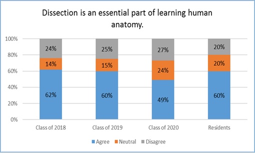 Figure 1. Cohorts who learned anatomy by cadaver dissection were more likely to agree that dissection is essential to learning anatomy. |
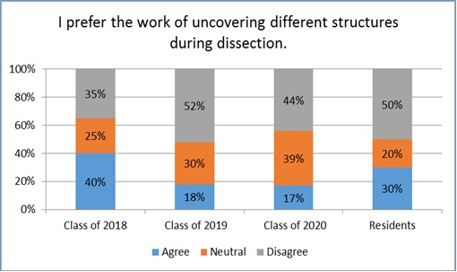 Figure 2. In every cohort, the majority of respondents did not prefer or did not care if they performed the dissection. |
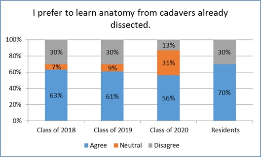 Figure 3. If prosected for them, students are willing to learn from cadavers. |
Respondents were also surveyed on their confidence in identifying important anatomical structures on cadavers (Fig. 4) compared to plastinated specimens (Fig. 5). Because all residents, as well as some respondents from the graduating classes of 2019 and 2020, had human dissection experience prior to UTSD, they were also asked of their confidence in identifying structures on cadavers and the plastinated specimen. Of the three cohorts who had learned from plastinated specimen, at least 96% of respondents believed that they could easily identify structures from plastinated specimens. In contrast, only about one-third of respondents who had dissection experience believed they could easily identify structures on cadavers (23% from the classes of 2018 and 2019, 33% from the class of 2020, and 40% of residents). Lastly, when asked if respondents believed plastinated prosections can effectively replace the need for dissection (Fig. 6), 85%, 92%, and 70% of respondents from the classes of 2019, 2020, and residents (respectively) agreed with the statement. Only 51% of students from the class of 2018, who were without plastinated experience, believed dissection can be replaced by plastinated prosections. Dunn-Bonferroni (DB) post hoc analysis revealed 2018 < 2020: DB= -22.28, p<0.032).
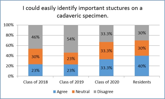 Figure 4. Of students with prior cadaver experience, only about 25% had confidence in identifying structures. |
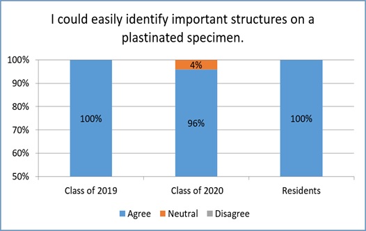 Figure 5. Of students with experience of plastinates, virtually all students had confidence to identify structures. |
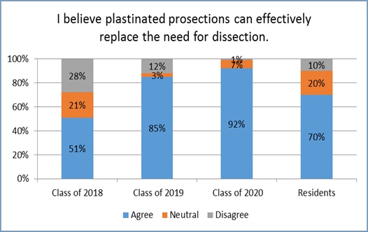 Figure 6. Only the class of 2018, (without experience of plastinates), are divided in their opinion on whether plastination can replace dissection or not (p<0.032). |
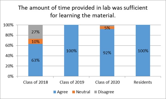 Figure 7. Over 90% of the class of 2019, 2020, and residents, believed that there was enough time in lab to learn anatomy from the plastinated specimens, compared to only about a 2/3rd of the class of 2018 (cadaveric cohort) (p<0.001). |
When asked if the amount of time in lab was sufficient for learning anatomy (Fig. 7), over 90% of students from the graduating classes of 2019, 2020, and residents, agreed with the statement. In the graduating class of 2018 (the cadaver cohort), about 63% agreed with the statement. This is remarkable, as the cadaver dissection lab was double the duration of plastination labs. This indicates that those who disagree believed that dissections require more time to learn. Dunn-Bonferroni (DB) post hoc analysis revealed 2018< 2019: DB= -44.78, p<0.001 and 2018< 2020, DB= -34.18, p<0.001).
Interestingly, the commonality amongst all the cohorts is that over 96% of all respondents did not prefer to learn head and neck anatomy exclusively through textbooks and computer software. This indicates that anatomical specimens (whether dissection or prosection) is crucial for students to see and understand the structural relationships in anatomy.
Although this report revealed that more than half of all cohorts believed that plastinated prosections can effectively replace the need for dissection, students do recognize the advantages of dissection. For example, our survey also asked if the unique tactile feel of tissues on the specimens were helpful in learning and differentiating structures. There was a clear agreement among respondents from the class of 2018 who agreed (63%) that the unique tactile feel of tissues from the cadaver helped them learn. In contrast, less than 44% of students and dental residents who learned anatomy via the plastinated models agreed with this statement. In fact, the graduating classes of 2019, 2020, and dental residents who learned anatomy from the plastinated models had the highest percentage of responding neutral (34-40%). This may be because touching the plastinated specimens was discouraged in order to preserve their pristine condition. In addition, the tactility of plastinated models is rather homogenous, whereas when learning with cadavers, students can touch and squeeze (for example) nerves vs. arteries. These differences in textures can help students differentiate these structures. Our study also included comments from students on the pros and cons of learning anatomy from cadavers compared to the plastinated models. Here are some comments from the respondents:
In addition to assessing students’ opinions and comments of their experiences in learning anatomy via cadavers or plastinated specimens, we compared laboratory exam scores amongst the cohorts to investigate if student performances have significantly improved since this change.
We compared student performances in anatomy lab exams within the past 6 years from the graduating dental classes of 2016 to 2021. The cohorts who studied anatomy via cadavers are from the graduating classes of 2016-2018 and those who studied anatomy via the plastinated specimens are from the graduating classes of 2019-2021. Four anatomy laboratory exams are administered in the Head and Neck Anatomy course. For each cohort, the class average for each anatomy laboratory exam was determined. Then an overall average of the class averages from the four exams was calculated, giving one laboratory exam score average for each cohort. The overall lab exam averages for the cohorts that studied anatomy via cadavers were 83%, 84%, and 83% from the graduating classes of 2016-2018, respectively. For the cohorts that studied anatomy via the plastinated specimens, the class averages were 90%, 88%, and 91% from the graduating classes of 2019-2021, respectively. This indicates vast improvement in lab exam performances since the implementation of the plastinated models in our anatomy curriculum.
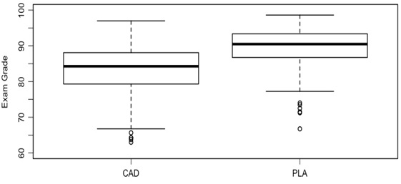 Figure 8. Anatomy laboratory exam means for all cohorts who studied anatomy via cadavers (CAD) versus plastinated (PLA) specimens. |
The Box-and-Whisker plot (Fig. 8) shows the anatomy lab exam averages for cohorts who studied anatomy via cadavers (CAD) versus the plastinated models (PLA). ANOVA test (F1, 598= 161.81, p<0.0001).
Following the switch of teaching anatomy from cadaveric dissections to plastinated prosections, our data revealed that student satisfaction and laboratory performance in the Head and Neck Anatomy laboratory curriculum have significantly improved.
At UTSD, Head and Neck Anatomy is a 16-week course offered to first year dental students. One-hour lectures are held twice a week with a 1.5-hour laboratory session following every lecture. Four laboratory exams are administered in the course. Prior to spring 2016, laboratory sessions consisted of cadaveric dissections. There were 20 groups consisting of 5-6 students per group with their assigned cadaver. Students spent approximately 6 hours per week working on their dissections. In spring of 2016, plastinated prosections replaced cadaveric dissections. The plastinated specimens were purchased from the Institute for Plastination in Heidelberg, Germany. Laboratory time was reduced by half in that each 3-hour lab session is divided into two 90 minute sessions with about 50 dental students assigned in each session. Within each session, students are divided into 4 groups consisting of 12-13 students per set of plastinated specimen group along with an anatomy instructor in each group. Students then divide themselves into pairs or groups of threes to discuss and identify structures.
Similar to NYUCD’s anatomy curriculum, frequent, low-stakes quizzing is an integral part of the course. Unlike previous laboratory sessions with cadavers, after each lab session with the plastinated models, students take an online quiz consisting of five questions relating to the anatomical structures discussed in lab that day. The practical exams, however, are similar to those given in the cadaver dissection course; structures are pinned and students must identify the structure by writing out the answer. These low-stakes quizzes not only encouraged attendance in lab, but also motivated students to spend their time in lab wisely learning and discussing the anatomical structures with their colleagues.
The use of plastinated prosections in studying anatomy is advantageous in terms of presenting detailed anatomy in a clear and efficient manner. Previously, students spent an average of 6-8 hours a week in the anatomy lab working on their dissection with some assistance from faculty and teaching assistants. In addition, students often attended lab outside of class time in order to finish their dissection or review structures from other cadaveric specimens. Because head and neck anatomy is one of the most complex and difficult areas to dissect, for novice dissectors, it is difficult to do a quality dissection and preserve structures of interest. The plastinated prosections allow students to locate muscles, nerves, and vasculature without the added pressure of dissecting during a limited amount of class time. The dissections on the plastinated specimens are flawless and have allowed students to see structures and relationships that are often destroyed in a traditional lab setting where student do the dissections.
Although plastinated specimens are not widely used, there are numerous health professional schools that, similarly, utilize cadavers that are already dissected (prosected) to teach anatomy to students (Ashdown et al., 2013). Interestingly, a study performed by Williams et al. (2019) from the University of Mississippi Medical Center, compared the pedagogical approaches of whether prosection versus dissection was best in learning the human anatomy of the hand and foot. By comparing students’ lab exam performances, they found that students who studied anatomy via prosected cadavers performed better than their classmates who studied anatomy via dissections. Nnodim et al. (1996) also investigated students’ retention by comparing cohorts who learned anatomy through dissection versus prosected cadavers. Not only did prosected cadavers take up less time in their curriculum, students’ anatomical retention 5 years after the course was comparable or slightly better than students who studied anatomy via dissections (Baker et al., 2013). Unlike cadavers, the advantage of the plastinated specimens is that they are odorless, durable, and easy to store. Critically, they save faculty many hours of dissection, as the prosection is done once by the supplier and saved for perpetuity. Therefore, the specimens are available not to only first-year dental students, but also to advanced students preparing for their boards, and residents enrolled in the Advanced Head and Neck anatomy course.
During the course of this study, we encountered several limitations. For example, not all respondents could directly compare cadaveric dissections to plastinated prosections in anatomy education in dental school. The only cohort that was able to do this was the residents. As a result, the dental students who have had exposure to either the cadaveric dissections or plastinated prosections may have answered the survey statements based on their limited to no experiences with other modalities. Ideally, we would like to have had participation from more residents in regards to their attitude about dissection and studying anatomy from plastination. Another limitation was that the overall improvement in anatomy lab grades could be due to fewer specimens for students to study. Previously, students were responsible for recognizing head and neck structures from 20 full-bodied cadavers that were variable in size, shape, anatomical anomalies, and quality of dissection. Our collection of plastinated specimens consists of four sets of 7 hemisected head and neck models, ranging from superficial to deep dissections. Therefore, it could be possible that students had an easier time navigating through the few pristine prosected specimens as opposed to deciphering through cadavers, in which structures are more fragile and are typically torn or missing.
In conclusion, a great majority of dental students and residents believed plastinated prosections can effectively replace instruction with dissection. Of interest to programs continuing with cadaveric instruction, we found that, if taught by cadaveric dissections, students prefer prosected specimens, saving them time and frustration with indiscernible structures. Further, student performance in anatomy laboratory examinations significantly improved since our change to studying anatomy via plastinated models. These facts could possibly ease the decision at schools to transition to teaching anatomy with plastinated specimens.
Acknowledgements
Special thanks go to Drs. Charles Streckfus and J. Nathaniel Holland for their assistance in the statistical analysis of our work. We also give many thanks to the UTSD dental students and residents for their input and participation of our survey.
Ashdown L, Lewis E, Hincke M, Jalali A. 2013: Learning anatomy: can dissection and peer-mediated teaching offer added benefits over prosection alone? ISRN Anat 2013: 873-825.
https://doi.org/10.5402/2013/873825
Baker EW, Slott PA, Terracio L, Cunningham EP. 2013: An innovative method for teaching anatomy in the predoctoral dental curriculum. J Dent Educ 77: 1498-507.
https://doi.org/10.1002/j.0022-0337.2013.77.11.tb05626.x
Bickley HC, von Hagens G, Townsend FM. 1981: An improved method for the preservation of teaching specimens. Arch Pathol Lab Med 105: 674-6.
Drake RL. 1998: Anatomy education in a changing medical curriculum. Anat Rec 253: 28-31.
https://doi.org/10.1002/(SICI)1097-0185(199802)253:1<28::AID-AR11>3.0.CO;2-E
Drake RL, McBride JM, Lachman N, Pawlina W. 2009: Medical education in the anatomical sciences: the winds of change continue to blow. Anat Sci Educ 2: 253-9.
https://doi.org/10.1002/ase.117
Estai M, Bunt S. 2016: Best teaching practices in anatomy education: A critical review. Ann Anat 208: 151-57.
https://doi.org/10.1016/j.aanat.2016.02.010
Fruhstorfer BH, Palmer J, Brydges S, Abrahams PH. 2011: The use of plastinated prosections for teaching anatomy--the view of medical students on the value of this learning resource. Clin Anat 24: 246-52.
https://doi.org/10.1002/ca.21107
Korf HW, Wicht H, Snipes RL, Timmermans JP, Paulsen F, Rune G, Baumgart-Vogt E. 2008: The dissection course - necessary and indispensable for teaching anatomy to medical students. Ann Anat 190: 16-22.
https://doi.org/10.1016/j.aanat.2007.10.001
Latorre R, Bainbridge D, Tavernor A, Lopez Albors O. 2016: Plastination in anatomy learning: an experience at cambridge university. J Vet Med Educ 43: 226-34.
https://doi.org/10.3138/jvme.0715-113R1
Latorre RM, Garcia-Sanz MP, Moreno M, Hernandez F, Gil F, Lopez O, Ayala MD, Ramirez G, Vazquez JM, Arencibia A, Henry RW. 2007: How useful is plastination in learning anatomy? J Vet Med Educ 34: 172-6.
https://doi.org/10.3138/jvme.34.2.172
Nnodim JO, Ohanaka EC, Osuji CU. 1996: A follow-up comparative study of two modes of learning human anatomy: by dissection and from prosections. Clin Anat 9: 258-62.
https://doi.org/10.1002/(SICI)1098-2353(1996)9:4<258::AID-CA8>3.0.CO;2-A
Patel KM, Moxham BJ. 2006: Attitudes of professional anatomists to curricular change. Clin Anat 19: 132-41.
https://doi.org/10.1002/ca.20249
Pyle MA. 2012: New models of dental education and curricular change: their potential impact on dental education. J Dent Educ. 76: 89-97.
https://doi.org/10.1002/j.0022-0337.2012.76.1.tb05237.x
R Core Team 2017: R: A language and environment for statistical computing. R Foundation for Statistical Computing, Vienna, Austria. URL https://www.R-project.org
Reidenberg JS, Laitman JT. 2002: The new face of gross anatomy. Anat Rec 269: 81-8.
https://doi.org/10.1002/ar.10076
Rizzolo LJ, Rando WC, O'Brien MK, Haims AH, Abrahams JJ, Stewart WB. 2010: Design, implementation, and evaluation of an innovative anatomy course. Anat Sci Educ 3: 109-20.
https://doi.org/10.1002/ase.152
Rowland KC, Joy A. 2015: The gross anatomy laboratory: a novel venue for critical thinking and interdisciplinary teaching in dental education. J Dent Educ 79: 295-300.
https://doi.org/10.1002/j.0022-0337.2015.79.3.tb05884.x
Sugand K, Abrahams P, Khurana A. 2010: The anatomy of anatomy: a review for its modernization. Anat Sci Educ 3: 83-93.
https://doi.org/10.1002/ase.139
von Hagens G, Tiedemann K, Kriz W. 1987: The current potential of plastination. Anat Embryol (Berl) 175: 411-21.
https://doi.org/10.1007/BF00309677
Williams SR, Thompson KL, Notebaert AJ, Sinning AR. 2019: Prosection or dissection: which is best for teaching the anatomy of the hand and foot? Anat Sci Educ 12: 173-80.
https://doi.org/10.1002/ase.1808