Department of Anatomy and Cell Biology, University of Melbourne, Parkville, Victoria 3052, Australia
This paper documents the procedures followed in establishing an anatomical technologies laboratory for the preparation of anatomical and plastinated specimens, in the Department of Anatomy and Cell Biology at the University of Melbourne. Funding for the laboratory was obtained through a Faculty of Medicine teaching development and innovation grant, supplemented by funds from the Department. Next to the embalming room, an existing space, which was formerly used for the storage of cadavers, was converted into two suitable work areas. One a preparation room and the other a spark-proof area housing the freezer with an isolated vacuum pump and refrigeration unit. Part of the funds obtained from the Faculty have been used to employ a technical officer who has dedicated responsibility to the facility and its day-to-day operation. The laboratory has been fully operational for over a year and a large number of plastinated specimens have been prepared during that time. We are in the process of developing 'sheet plastination' and have a project under-way with radiologists from the Royal Melbourne Hospital correlating cross-sectional plastinated specimens with CT-scans and magnetic resonance images. It is our intention to direct most of our work towards the Department's teaching requirements, however we also intend using the laboratory to train technicians in anatomical techniques and encourage honours and higher degree students in the area.
Preservation, Plastination, Laboratory design, Cost, Equipment
Dr Christopher A. Briggs, Associate Professor of Anatomy, Department of Anatomy and Cell Biology, The University of Melbourne, Parkville, Victoria 3052, Australia. Telephone: 61 3 9344 5776 / Fax: 61 3 9347 5219. Email: C.Briggs@anatomy.unimelb.edu.au

![]()


The Department of Anatomy and Cell Biology at the University of Melbourne, like Anatomy departments universally, has extensive teaching commitments. In any given year we provide instruction to over 1500 undergraduate students in medicine, physiotherapy, dentistry, optometry and science, as well as to a variety of postgraduate groups and professional organizations. Our undergraduate teaching pro- grams continue to run along traditional lines, comprising lectures, demonstrations using prosected wet specimens, and student dissection. Students receive practical demonstration of material presented in lectures through a program known as 'wet specimen workshops'. The term 'wet' refers to the manner of storage of such specimens - immersed in a bathing solution of 99% ethanol: water (1:3). Prior to such work- shops, specimens are removed from a number of storage tanks and displayed on tables in the laboratory, where stu- dents preview, under supervision, the anatomy of a structure or region before proceeding to dissect it for themselves. We also have a large and well-stocked Anatomy museum but unfortunately specimens are mounted, unlabeled, in Perspex containers behind glass cabinets. Structures are of- ten difficult to see and identify and as a result our museum has been progressively underutilized in recent years.
Due to large student numbers, increased demand on the Department's limited stock of suitable prosected material has meant greater wear and tear and an increased rate of attrition of such specimens in recent years. Ideally we should be producing greater numbers of specimens to increase the variety and range of those already available. However, at present we are unable to produce enough to maintain our existing stock. As S10 silicone fully cured specimens are durable (Weiglein and Henry, 1993) and do not require 'wet' storage, plastination provides us with the opportunity both to extend the life of this material, and additionally maintain effective student/specimen interaction. Therefore in 1994, following a visit by one of the authors (CB) to Heidelberg in Germany to examine Von Hagen's plastinated materials, as well as visits to anatomy departments in North America and the United Kingdom (CB and BK) to thoroughly research the approaches to anatomy teaching taken by those departments, we decided to investigate means of extending the life of cadaveric material by plastinating our own anatomical specimens.
The S10 technique (Von Hagens, 1986) is highly reproducible and nearly always results in a satisfactory, durable specimen. The methodology can be applied to older formalin fixed specimens (Cannas and Fuda, 1991) which effectively extends the life of our museum stored specimens, and has the added bonus of making them much more user friendly as they can now be handled by both teachers and students. Our initial challenge was to find the funds to establish a laboratory, as well as the space in an already crowded building to house one. With the assistance of a Teaching Development and Innovations Grant from the Faculty of Medicine at the University and additional funds from the Department we were able, at the end of 1994, to start planning the facility.
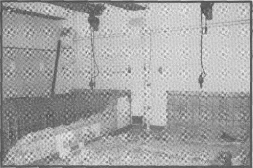
Figure 1. Preparation of Anatomical Technologies Laboratory. Existing space during demolition.
Laboratory Construction
In addition to the traditional prosected anatomical specimens, techniques are available to cast anatomical cavities, viscera and blood vessels, assemble articulated skeletons and display joints and their ligaments (Tompsett, 1970). New methods of preparing anatomical specimens are also appearing in the literature (Ocello, 1997). Because we intend to experiment with a number of these techniques the laboratory was named the Anatomical Technologies Laboratory rather than merely Plastination Laboratory. However, plastination has become the major focus. The laboratory was constructed within an area occupying approximately 45 square meters of floor space in the 'tank room', a room in the basement of the Medical Centre adjacent to our embalming room, where cadavers were formerly stored immersed in large tanks of alcohol, water and formaldehyde. The tanks were made of reinforced concrete, lined with stainless steel and connected to the floor. It was a major undertaking to remove the tanks and make good the floor (figure 1).
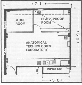
Figure 2. Plan of the Anatomical Technologies Laboratory (all measurements in metres).
This task and the overall refurbishment of the space, including sealing-off a 3-phase electronic lifting system, upgrading the air conditioning, making selected power out- lets and light fittings spark proof and altering the space to conform to safety standards, cost over $Aus.40 000. The final plan of the laboratory is illustrated in figure 2.
By early 1995 this task was achieved and we then started to acquire equipment to get the plastination process under way. This phase of the project could not have happened as quickly as it did without the appointment, again funded from the grant, of a Technical Officer, a science graduate with training in human anatomy, who has taken on responsibility for day-to-day operation of the laboratory. Our initial objective was to quickly produce S10 specimens and to this end we sought a freezer sufficiently large to have several specimens simultaneously under preparation, a vacuum system for acetone removal and impregnation, and a curing chamber. With considerable help and ad- vice from Mr Robbie Boyes at the University of Queens- land we found local producers for all major items of equipment (details available on request). The overall cost of fit- ting out the laboratory was approximately $Aus. 10 500, the freezer including remote unit (2000x700x1000 cm) and vacuum pumps being the most expensive items ($Aus.5 000 and $Aus.2 600 respectively). The freezer has been especially built for our purposes and is capable of fitting four large barrels for dehydration. It possesses an easily removable lid and has no ignition sources. The walls contain an extensive coiling system allowing rapid cooling, and they are insulated in a fashion that allows the freezer to remain below zero (Celsius) for a period of 24-48 hours in the event of a power failure. Using a normal household unit the freezer will draw and hold -40° Celsius. The vacuum pump is a RV3 rotary van pump, 220-240V, 50/60 Hz single phase Edwards pump (Edwards High Vacuum International, Manor Royal, Crawley, West Sussex, RH102W, U.K.) which has warranty certificates and good servicing facilities. Maintenance is simple and requires minimal time. The pump is capable of holding a vacuum in many large chambers and is therefore suitable for current or future needs should we further expand the laboratory. We did not investigate the use of reconditioned parts but they are undoubtedly available.
To get us started we required S10, hardener and other products from Biodur, which significantly increased costs (by an additional $Aus.5 600 in the first year). The initial purchase consisted of Biodur S10: 15 kg ($Aus.915 / DM1005), Biodur Hardener S3: 0.15 kg ($Aus.l6.14/ DM16.50), Biodur Hardener S6: 1L ($Aus.65.52 / DM72), acetonometer: 90-100% ($Aus.lOO / DM106), Biodur Fix A: 10.4 kg ($Aus.327 / DM360), Biodur Fix T20: 2L ($Aus.61.88 /DM68), Pine Oil: 2L ($Aus.72.80 / DM80).
To the above will shortly be added a purchase of Biodur S10: 50 kg ($Aus.2 935.42 / DM 3 000), Biodur Hardener S3:500ml ($Aus.50 / DM55) and Biodur S6:1L ($Aus.70.45 /DM72). The total cost of these purchases was $Aus.4614.21 and the freight cost $Aus.999.43. As may be seen, approximately 20% of our funds are spent on freight charges. We are currently developing sheet plastination and because of the above cost factors are experimenting with local products. We are also examining various combinations of curing time with these local products.
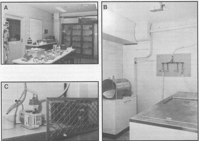
Figure 3. Laboratory in current use: A. Preparation room. B. Spark proof room with vacuum distribution apparatus. C. External bench with remote units.
During our first full year of operation we have man- aged to produce approximately 60 quality plastinated specimens, such as dissected hearts with display windows cut into the chambers, a series of dissections of the foot to re- veal ligamentous attachments, the entire small and large bowel, an axilla, popliteal fossa, superficial dissection of the face as well as brain sections in a variety of planes. These are essentially for our own teaching purposes, however the laboratory has been assisting a neighboring institution by plastinating a number of their prosections. In formulating the successful funding proposal for this innovation, we costed the preparation of a wet specimen (including academic and technical time) at between $Aus.350-500, depending on size and complexity of the structure or region. With an average life span for a wet specimen of only three to five years, we anticipate making significant cost savings after three years, through the use of plastinated rather than wet specimens. The added benefit will be a large and diverse number of durable plastinated specimens which we anticipate using for many years to come. In addition, we have instituted a research project with radiologists from the neighboring Royal Melbourne Hospital to develop correlated MRI and CT-scans for teaching purposes. We also in- tend using the laboratory to train technicians in anatomical techniques and encourage honours and higher degree students in the area. Student and teacher reaction to the avail- ability of plastinated specimens has been favorable. We are currently preparing plastinated preparations of both nor- mal and pathological abdominal and pelvic viscera. These will also be photographed and the images stored in CD-ROM format with appropriate labelling and text which the students may access in the dissecting room and in the museum. In addition, neurosciences teaching staff have requested multiple sets of sagittal, coronal and transverse views of the brain for neuroanatomy teaching purposes.
Cannas M, Fuda P: Plastination of old formalin fixed specimens. J Int Soc Plastination, 5(1): 11-15, 1991. https://doi.org/10.56507/IYFR4714
Ocello PJ: A new process for preserving biological tissue. http://cvm.msu.edu/pare/docs/silyophilization.htm Tompsett DH: Anatomical Techniques, 2nd ed. E. & S. Livingstone, Edinburgh, 1970.
von Hagens G: Heidelberg Plastination Folder: Collection of all technical leaflets for plastination, 2nd Ed. Anatomische Institut 1, Universitat Heidelberg, Heidelberg, Germany, 1986.
Weiglein AH, Henry RW: Curing (hardening, polymerisation) of the Polymer - Biodur S10. J Int Soc Plastination, 7(1): 32-35, 1993. https://doi.org/10.56507/ABNZ7085