Departement de Chimie-Biologie, Universite du Quebec a Trois-Rivieres, Trois-Rivieres, Quebec, Canada.
The aim of this study was to produce a plastinated specimen of the human cerebral dura mater with the base of skull, showing its morphological and topographical features. The authors give a detailed account of the dissection procedure to obtain a very helpful specimen in neuroanatomical and neurological curricula. Plastination of the specimen preserved the consistency of the meninges, and allowed a good understanding of its structure and different septa.
Presented in part at the 8th International Conference on Plastination, Brisbane, Australia, July 14-19, 1996.
Meninges, Cranial Dura Mater, Falx Cerebri, Tentorial Incisure, Polymer S10
G. Grondin, Departement de Chimie-Biologie, Universite du Quebec a Trois-Rivieres, C.P.500, Trois-Rivieres, Quebec, Canada G9A 5H7. Telephone: 819 376 5053 / Fax: 819 376 5084. Email: Gilles_Grondin@uqtr.uquebec.ca

![]()


The place of plastination in neuroanatomical pedagogics is well documented (Holladay and Hudson, 1989; Resch, 1989; Purinton, 1991; Weiglein, 1993, 1997; Magiros et al., 1997). However, the morphology and relationships of the meninges are also of outstanding importance in the teaching in medicine and allied sciences: circulation of the cerebrospinal fluid from the apertures of the fourth ventricle to the arachnoid granulations, location of the main cranial dural venous sinuses, infra or supratentorial topography of the main components of the central nervous system in relation with the tentorium cerebelli, distinction between extradural and subdural hemorrhages, transtentorial herniations. Specimens of the cerebral dura mater are therefore very helpful to neuroanatomical and neurological curricula. Such specimens were previously produced using the freeze-drying method, but were described as «fragile» and «would not stand up to rough handling* (Romero-Sierra et al., 1983). To our knowledge, the dissection and plastination procedure of this kind of specimen never was the subject of a full-length publication (Grondin and Olry, 1996a, 1996b). We describe in this paper the dissection and plastination procedures to obtain a specimen of the human cerebral dura mater with the base of the skull.
Removal and fixation of the specimen
An unfixed body, free of any obvious pathological or surgical history (fracture of the skull, meningioma, extradural or subdural hemorrhage) was chosen for this project. Its head was removed through the intervertebral disc between the sixth and seventh cervical vertebrae.
A solution of 10% formalin was injected via both com- mon carotid arteries, and the specimen was immerged in the same solution at 4°C for 24 hours. The specimen was then kept in the solution during its dissection which lasted about three weeks.
Dissection of the specimen
Soft tissues of the scalp (skin, subcutaneous fibro-adipose tissue, epicranial musculature and pericranium) were removed, and a hole (2x2 cm) was pierced in the left parietal bone about 2 cm inferior to the sagittal suture with a Lipshaw autopsy saw (Shandon Lipshaw, Pittsburg, USA). Care was taken to avoid perforation of the underlying cerebral dura mater during this procedure. After removal of the
piece of bone, a blunt probe was introduced through the cranial aperture to carefully release the dura mater from the in- ner aspect of the skull. The cranial vault could then be cut piece by piece, in order to make the gradual detachment of the dura mater more easy. The cranial vault was removed down to a line passing inferior to the groove for transverse sinus of occipital bone, above the external acoustic meatus and zygomatic arch, at mid-level of the frontal process of zygomatic bone, and about 1 cm superior to the nasofrontal suture. The base of skull was also dissected, and cervical vertebrae were carefully removed so that the spinal dura mater became visible.
A hole (5x5 cm) was cut in the convexity of the left half of the cerebral dura mater, and the whole brain (prosencephalon, brain stem and cerebellum) could then be pulled out in small pieces through this aperture.
All soft tissues of the face, including eye balls and accessory visual apparatus, were also removed, but the dural sheath of the optic nerve was minutely dissected and preserved.
Plastination of the specimen
The specimen was then dehydrated and impregnated according to the standard S10 procedure (von Hagens, 1985). After impregnation, the whole dural cavity was filled with absorbent paper in order to restore the normal shape of the cerebral dura mater and its septa, and the specimen was fast cured (Weiglein and Henry, 1993).
After complete curing, the opening on the left side was enlarged to permit a full view of the falx cerebri with its inner free margin, as well as the tentorium cerebelli with its tentorial incisure.
The convexity of the cerebral dura mater (figures 1-2) shows arterial ramifications (middle meningeal artery) and the location of the superior sagittal and transverse sinuses. The sheath of the optic nerve appears through the optic canal of the orbital cavity (figure 3).
The aperture of the left side of the dura mater gives a full view of the intradural cavity and its partitioning (figures 4-5). The anterior part of the falx cerebri is attached to the crista galli of the ethmoid bone and is perforated by numerous apertures, as usual (Williams et al., 1989). Many fibrous tracts can be observed at the junction of the falx cerebri with the convexity of the dura mater (Renard et al., 1967). The straight sinus is visible along the attachment of the posterior part of the falx to the tentorium cerebelli. The free border of the tentorium cerebelli and the dorsum sellae of the sphenoid bone delimit the tentorial incisure, and the anterior attachments of the tentorium cerebelli can be followed to the anterior clinoid processes.
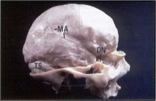 Figure 1. Lateral aspect of the specimen (Norma latera- lis). MA - ramifications of the middle cerebral artery, ON - sheath of the optic nerve, TS - transverse sinus. |
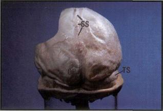 Figure 2. Posterior aspect of the specimen (Norma oc- cipitalis). OP - occipital poles, SS - superior sagittal sinus, TS - right transverse sinus. |
 Figure 3. Close view of the orbital region. FS - frontal sinus, NG - nasolacrymal groove, ON - sheath of the optic nerve, S - lesser wing of sphenoid bone, SS - superior sagital sinus. |
|
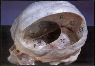 Figure 4. Anterolateral view of the specimen. CF - mid- dle cranial fossa, F - hypophysial fossa, FC - falx cerebri, FT |
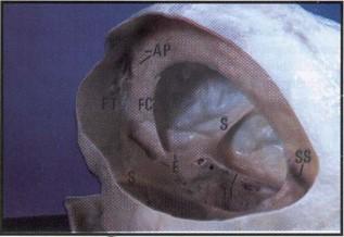 Figure 5. Posterolateral view of the specimen. AP - apertures of the anterior part of the falx cerebri, E - cribriform plate of the ethmoid bone, FC - anterior part of the falx cerebri fixed to the crista galli, FT - fibrous dural tract, S - lesser wings of the sphenoid bone, SS - straight sinus, TI - tentorial incisure. |
The aim of this study was to provide students with a plastinated specimen of the cerebral dura mater showing all its morphological and topographical features. The base of skull and parts of bones of the face were preserved, so that topographical relationships between the meninges and its osseous envelope could be better understood.
Unlike the specimen of the same kind produced by using the freeze-drying method (Romero-Sierra et al., 1983), our plastinated specimen can be handled without particular caution. Moreover, the consistency of the cerebral dura mater is comparable with those of fresh, i.e. non plastinated specimens.
Grondin G, Olry R: Current Plastination Index. Trois- Rivieres, Publication of the International Society for Plastination, 1996a.
Grondin G, Olry R: Preparation and plastination of the cerebral dura mater with the skull base. Presented at The 8th International Conference on Plastination - University of Queensland, Brisbane, Australia. July 1996. J Int Soc Plastination 11 (1): 4, 1996b.
Holladay SD, Hudson LC: Use of plastinated brains in teach- ing neuroanatomy at the North Carolina State Univer- sity, College of Veterinary Medicine. J Int Soc Plastination 3 (1): 15-17,1989.
https://doi.org/10.56507/FEKB4686
Magiros M, Kekic M, Doran GA: Learning Relational Anatomy by Correlating Thin Plastinated Sections and Magnetic Resonance Images: Preparation of Specimens. Acta Anat 158: 37-43, 1997.
https://doi.org/10.1159/000147908
Purinton PT: Plastinated brains used with computer assisted learning modules for teaching veterinary neuroanatomy laboratories. J Int Soc Plastination 5 (1): 16-19, 1991. https://doi.org/10.56507/WDQQ3260
Renard M, Poisson P, Cayotte JL: Structure de la faux du cerveau: Orientation et signification des faisceaux fibreux communs a la faux du cerveau et a la dure-mere de la voute cranienne. Trav Lab Anat Nancy 1: 44-53, 1967.
Resch KDM: Plastinated specimens for demonstration of microsurgical approaches to the base of the cranium. J Int Soc Plastination 3 (1): 29-33, 1989. https://doi.org/10.56507/FUGF5217
Romero-Sierra C, Driscoll J, Lyons W, Lane P: A technique for the preparation of specimens for the meninges of the brain. Syllogeus 44: 45-48, 1983.
von Hagens G: Heidelberg Plastination Folder. Anatomisches Institut, Universitat Heidelberg, Heidelberg, Germany, 1985.
Weiglein A: Plastinated brain specimens in the anatomical curriculum at Graz University. J Int Soc Plastination 7 (1): 3-7, 1993. https://doi.org/10.56507/EHRX7749
Weiglein A: Plastination in the Neurosciences. Acta Anat 158:6-9, 1997. https://doi.org/10.1159/000147902
Weiglein A, Henry RW: Curing (Hardening, Polymerization) of the polymer Biodur S10. J Int Soc Plastination 7 (1): 32-35, 1993. https://doi.org/10.56507/ABNZ7085
Williams PL, Warwick R, Dyson M, Bannister LH: Gray's Anatomy, 37th Ed. Edinburgh, London, Melbourne and New York: Churchill Livingstone, 1989.