1Department of Anatomy, Faculty of Medicine, Masaryk University, Brno, 625 00, Czech Republic
2Faculty of Education, Faculty of Arts, Masaryk University, Brno, 602 00, Czech Republic
3Section of Investment Development, Technology and Operation, St. Ann´s University Hospital in Brno, 656 91, Czech Republic
4Department of Animals Anatomy, Histology and Pathomorphology, National University of Life and Environmental Sciences of Ukraine, Kiev, 03041, Ukraine
5 Department of Human Anatomy, National Pirogov Memorial Medical University, The Ministry of Healthcare of Ukraine, Vinnytsya, 21018, Ukraine
6 Department of Morphology, Sumy State University, 40000, Ukraine
There are only a few methods of preserving bodies that keep them in excellent condition for decades. These methods were developed mostly to sustain the cult of a political leader. Of the long-term embalmed bodies, only V.I. Lenin, Ho Chi Minh, Mao Zedong, Kim Il-sung and Kim Jong-il, the body of Professor Pyrogov, Rosalia Lombardo, and the corpse of a man in the Omsk Anatomical Museum, remain on display. The other bodies were subsequently buried in a tomb. Mention may also be made of the embalmed body of Philippine dictator Ferdinand Marcos, exhibited 1989-2012, which was embalmed by Philippine embalmer Frank Malabed. The body was original with a wax mask on his face. The embalming of the Vatican popes is carried out by the funeral home of Signoracci. Embalming solutions contain formaldehyde, ethanol, glycerine, sodium acetate, potassium acetate and thymol, or use paraffin impregnation. In most cases, it is also necessary to maintain standard environmental conditions such as humidity and air temperature. This paper provides an overview of the major and most successful embalming techniques, with a focus on the Soviet-Russian method and its development.
embalming; Lenin; Lombardo; paraffinization; Perón; Pyrogov
Jan Frišhons, Department of Anatomy, Faculty of Medicine, Masaryk University, Brno, 625 00, Czech Republic. Tel.: +420 54949 6418; E-mail: 203563@mail.muni.cz

![]()


The individual components of solutions for long-term embalming each fulfill a certain function. Formaldehyde consolidates proteins and adipose tissue, and inhibits most enzymes and microorganisms, while reducing hemoglobin to methemoglobin. Ethanol stabilizes tissue and converts methemoglobin to pigment, removes epidermal lipids, and increases skin permeability. Glycerine and potassium acetate preserve the pigments and enhance the effects of ethanol. Sodium acetate is used as a pH regulator and preservative. In addition, glycerine maintains tissue elasticity, facilitates distribution of the embalming agent, and draws moisture from the surrounding air to the body. During long-term embalming, i.e. long-term stabilization, water is expelled from the tissues, being gradually replaced with the embalming solution, which results in a hydrothermal equilibrium between the body and the surrounding atmosphere. In most cases, it is also necessary to maintain specific humidity and temperature (Lopukhin, 1997; Frišhons and Vacín, 2014a). Carrying out long-term embalming and research into tissue changes (Fig. 1, Fig. 2) is a very complex process, and for the purposes of this article, the issue has been considerably simplified. With the use of industrially produced solutions, e.g. by Dodge Chemicals (Slocum, 1947), there is no evidence of long-term monitoring of the body for more than a year after the embalming (Labush, 2014).
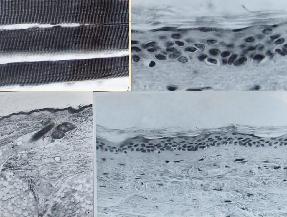 Figure 1. Well-preserved microscopic structure of the skin, subcutaneous tissue, and muscle fibers in Klement Gottwald’s tissues as of March 22, 1960. Top left: longitudinal section of muscle fibers; top right and bottom right: skin with papillary layer; bottom left: skin, subcutaneous tissue with follicles, and adipose tissue. Source: NA, f. 100/24, unprocessed - Mausoleum, photoalbum No. 4. |
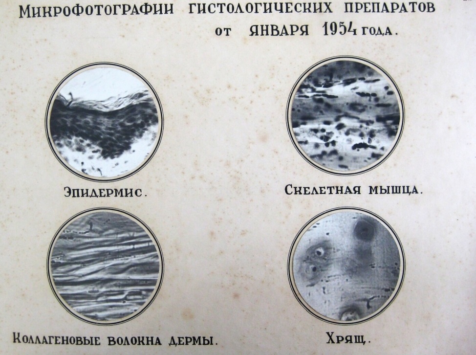 Figure 2. Well-preserved microscopic structure of Georgi Dimitrov’s tissues as of January 1954. Top left: skin with subcutaneous tissue; top right: skeletal muscle; bottom left: collagen fibers; bottom right: cartilage. Source: Dimitrov Mausoleum Lab. Central State Archives (CDA), ChP 190B. |
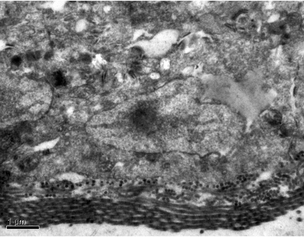 Figure 3. Tissue structure in samples taken from the body of Prof Pirogov during periodic embalming in 2018. Basal cells of the skin epidermis of the anterior abdominal wall. Intercellular connections; basement membrane. SEM (scanning electron microscope), Enlargement x 6000. Source: archive of the authors. |
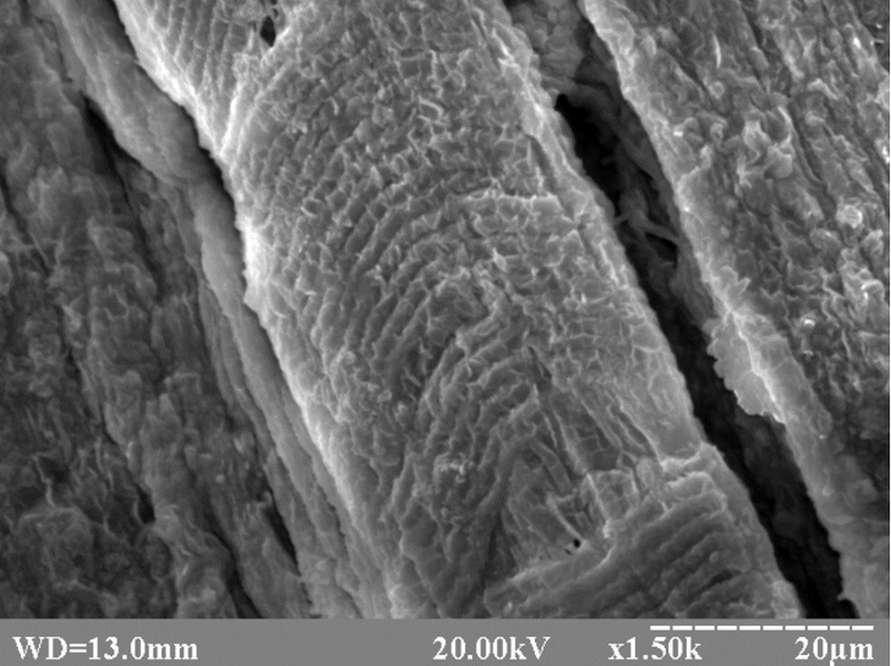 Figure 4. Tissue structure in samples taken from the body of Prof Pirogov during periodic embalming in 2018. Muscle fibers. Note the presence of sarcomeres responsible for the striated appearance. Disorganization of connective tissue layers. SEM. Enlargement x 1500. Source: archive of the authors. |
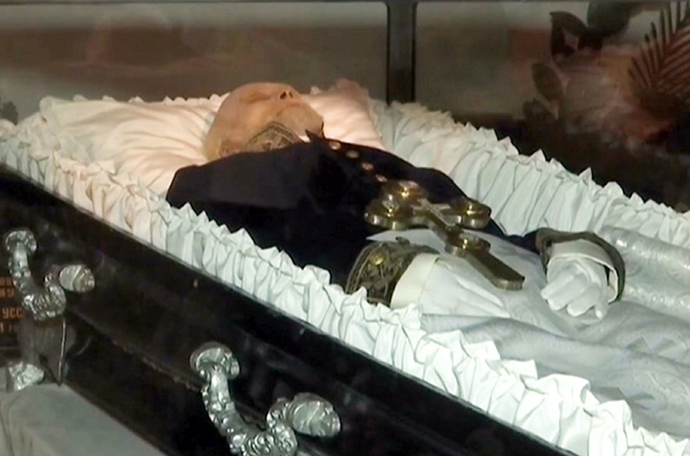 Figure 5. The body of Prof Pirogov stored in a sarcophagus after periodic embalming in 2018. Source: archive of the authors. |
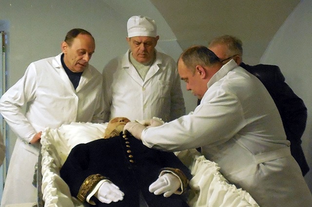 Figure .6 Adjustment of the clothing on Prof Pirogov’s body after periodic embalming in 2018, performed by Prof Melnyk. Source: archive of the authors. |
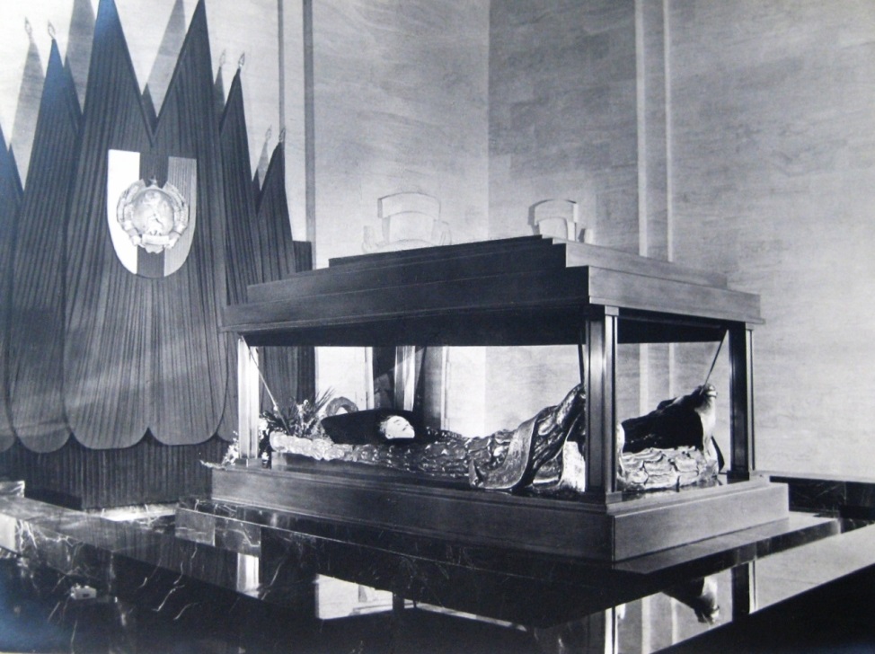 Figure 7. Sarcophagus with the body of Georgi Dimitrov in the mourning hall of his Mausoleum. Source: Photo album to reports on the embalming of the body of GM Dimitrov (1949-54). Central State Archives (CDA), ChP 190B. |
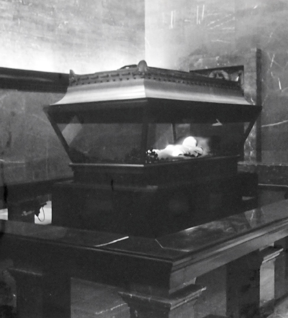 Figure 8. Sarcophagus with the body of Klement Gottwald in the mourning hall of his Mausoleum. Source: fond NA, f. 100/24, unprocessed - mausoleum, photo album from January – February 1955. |
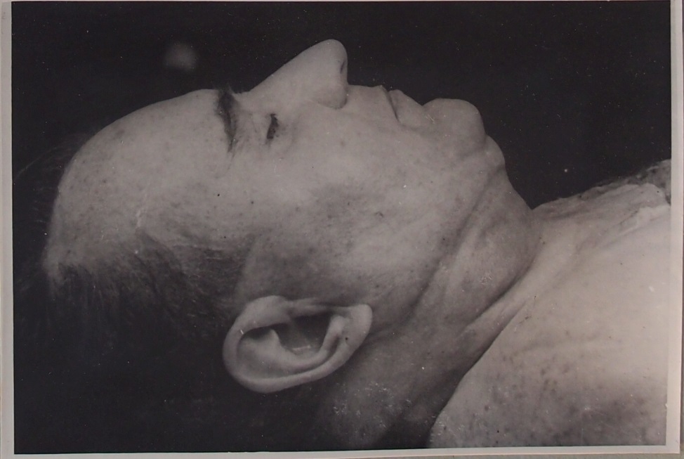 Figure 9. A close-up of Klement Gottwald's right profile during the body’s stabilization. Source: NA, f. 100/24, unprocessed - mausoleum, photo album from July 22, 1953 in Russian. |
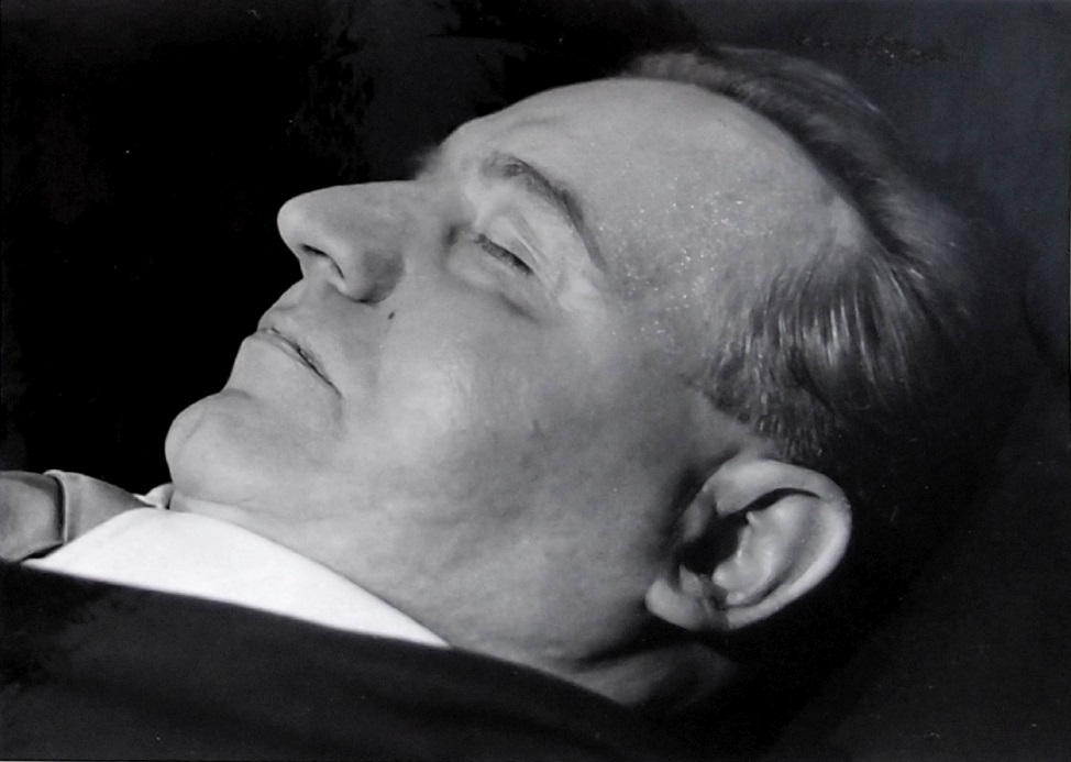 Figure 10. A view of Klement Gottwald's left profile on the lift pedestal in the laboratory. Source: NA f. 100/24, unprocessed - mausoleum, photo album from January – February 1955 |
 Figure 11. Detail of the fingers of Klement Gottwald's right hand on the lift pedestal in the laboratory. Source: NA f. 100/24, unprocessed - mausoleum, photo album from January – February 1955. |
At the beginning of this method was the embalming of V.I. Lenin, initially performed by Vorobyov, Abrikosov, and others. A 1%, and then a 2%, formaldehyde solution was first applied to the face, hands, and the front of the body, covered with cotton swabs, as well as injected into certain areas. The body cavities were washed with acetic acid. Subsequently, a 3% formaldehyde solution was applied to the hands and head, and injected into the skull cavity, which had been trepanated. For the whitening of facial and hand tissues, hydrogen peroxide and ammonia were used after intradermal and muscular application of acetic acid. The whole body was then immersed in a 3% formaldehyde solution for several days. Incisions were made in the abdominal wall, shoulders, forearms, thighs, calves and along the major muscles of the back and in the gluteal region, the skin of the palm, and the lower surfaces of the fingers on both hands. After a week, the body was immersed in a 20% ethanol solution, with the head and hands placed separately in 30 to 35% ethanol, for 6 days. After some time, 20% glycerine was added to the bathtub with 30% ethanol. Following this 14-day ethanol-glycerine bath, the body was immersed in an aqueous solution of glycerine, in May 1924. Eye prostheses were placed in the eye sockets and the eyes were then closed with 2–3 sutures on the edges of the eyelids. The head and hands were regularly soaked in a 1% formaldehyde solution. By the end of June 1924, the body was immersed in 240 l of glycerin, 110 kg of potassium acetate and 150 liters of water, with the addition of a 1–2% solution of quinine and hydrochloric acid. The body was wrapped in rubber elastic bandages, gloves were put on the hands, the body was dressed and placed in a sarcophagus. A petri dish containing an aqueous solution of thymol was placed under the pedestal (Lauer 1924; Melnikov-Razvedenkov 1930; Lopukhin 1997; Zbarskii 1998).
This experimental method has been refined over the decades by further experiments, and the growing experience of the scientists. The resulting methodology is now patented. First, an autopsy is performed to remove the visceral organs while preserving the main blood vessels. This is followed by perfusion of the vascular bed through the arteries with a solution of up to 20% formalin (according to tissue condition, and post mortem interval), with the possible addition of 5% ethanol, 10% glycerin, and 15% potassium acetate. The solution may also be injected into the subcutaneous tissue and muscles. Subsequently, the body is immersed in a bath of the solution for 1 to 2 months. The ratio of body volume to the fixative solution in a triplex silicate glass vessel is 1:10. In the first phase of stabilization of the body by impregnation, a solution is used with a volume of components as follows: glycerin 3–12%, potassium acetate 3–15%, sodium acetate 5%, and up to 100% with distilled water, for 45 days. The wide range of some of the components is because it is an experimental method, and the volume of substances is determined many factors, such as skin pigment, the rate of tissue impregnation, and other variables, known only to the Russian scientists who have been dealing with the issue for decades.
In the second phase, the impregnation lasts 45 days, in a solution consisting of glycerin 12–20%, potassium acetate 15–25%, sodium acetate 5%, up to 100% with distilled water. The third phase lasts 60–75 days, in a solution of glycerin 20–50%, potassium acetate 25–40%, sodium acetate 5%, excipient thymol 0.02% and up to 100% with distilled water. The pH of the solutions must be 8.2–8.4. The ratio of ingredients was determined experimentally, depending on the condition of the body before embalming, skin color, hair color, skin pigment, and subcutaneous fat. Stability of proteolytic enzymes was 64% (Bykov 2002). The procedures are performed by a group of 3 to 5 experts.
The body is stored in a sarcophagus in disposable overalls (jacket, collar, pants, shoe covers, sleeves, drawstrings and drainage valve) made of non-woven hydrophobic material. Sealing tapes at the cuffs and neck are sealed with a 15–30% solution of animal glue gelatin, technical casein, bone glue, and skin glue (Bykov 2013). The overalls are filled with about 10 L of the embalming solution. Clothes are placed on the overalls (Fig. 12). The exact position of the body in the sarcophagus is set by the fixed coordinates (Fig. 13), which ensures perfect adjustment of the lighting system.
 Figure 12. The placement of the body on the lift pedestal in the sarcophagus. Graphics by Kateřina Vacínová. |
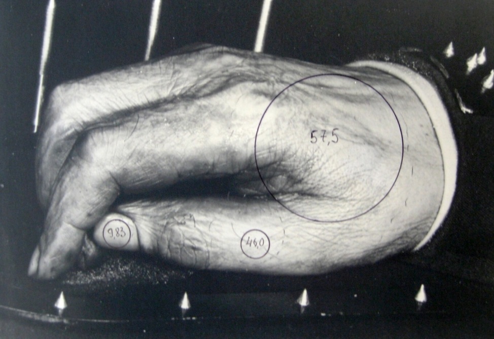 Figure 13. Georgi Dimitrov's right hand placed on the coordinates during the measurement of the surface brightness of the monitored body parts. Source: Dimitrov Mausoleum Lab. Central State Archives (CDA), ChP 190B. |
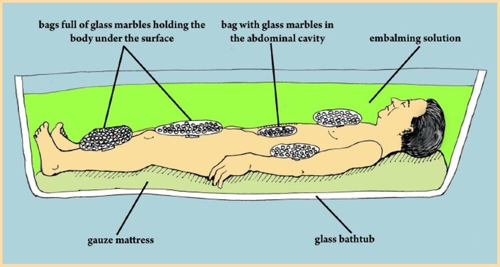 Figure 14. The placement of the body in an embalming tank with the embalming solution during the stabilization and periodic re-embalming. Graphics by Kateřina Vacínová. |
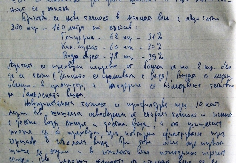 Figure 15. The composition of the maintenance fluid for the body of Georgi Dimitrov in 1957. Source: Dimitrov Mausoleum Lab. Central State Archives (CDA), ChP 190B. Embalming book No. 1 (1955–1957), p. 49. |
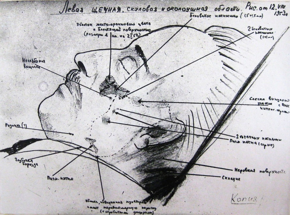 Figure 16. A sketch of the relief of Georgi Dimitrov’s left profile. Source: Dimitrov Mausoleum Lab. Central State Archives (CDA), ChP 190B, а.е. 9. |
In addition to the technical background for body maintenance, the method also includes daily monitoring of the condition of the body, and possible correction of the humidity of the visible parts of the body, as well as the temperature and humidity of the surrounding atmosphere. Periodic re-embalming after 18 months entails 4 weeks of impregnation in a tank with 200 L of a solution made up according to the condition of the body (Fig. 14). For instance, the body of Georgi Dimitrov was immersed in glycerin 62 liters (31% by weight), potassium acetate 60 kg (30%), and distilled water 78 liters (39%), at a temperature of 14 to 15 °C (CDA, 1957) (Fig. 15). In case of Klement Gottwald, it was a solution of 31% glycerin, 31% potassium acetate and 38% distilled water (National Archive Prague, 1955). The body was weighed before and after impregnation, with an average weight gain of 2 kg. Bonding and sealing of skin incisions was made by rubbing in 15% gelatin dissolved in 10% chloralhydrate. These spots were then covered with silk and another layer of gelatin, and fixed with cotton soaked in 10% formalin. Any soft tissue relief adjustments were made using subcutaneous injections of a liquid mixture of paraffin with white vaseline (8 parts to 2 parts), with wax and gelatin added, at a temperature of 44 °C. The respective spots were then cooled and shaped by hand (National Archive Prague, 1956). Another mixture for correcting the volume of wrinkles and folds is glycerin and potassium acetate 20-40%, in a ratio of 1: 1, thymol 0.02%, hydroxyapatite or bone mineral with a size of 50µ 40-70%, and distilled water up to 100% (Matvejchuk 2019), or Celladamm (a type of dammar resin) 16-20%, white beeswax 55-70%, paraffin 6-19%, and petroleum jelly 6-12% (Avramov, 2013; Abramov, 2016;). Embalming fluid samples were taken for spectrometric examination, and histological specimens were extracted, e.g., from the thighs, for a complex examination of the body’s overall condition. Monitoring and evaluation of the shape and volume of the soft tissues of the head and hands (Fig. 16) were performed at an interval of 4 to 5 years, using reliefometry (a system of relief mapping). As for an assessment of skin color, photoelectric colorimetry was used (Figs. 17, & 18) (Vasilevskii, 1988; Ryabtsev, 1997; Vasilevskii, 2004; Litvinov, 2015; Litvinov, 2016; Matvejchuk 2016). Subsequently, the new results were compared with previous measurements. Moreover, the brightness of the illuminated parts of the body in the sarcophagus, i.e., the head and hands, was measured from the right, left, and front, using a photometer.
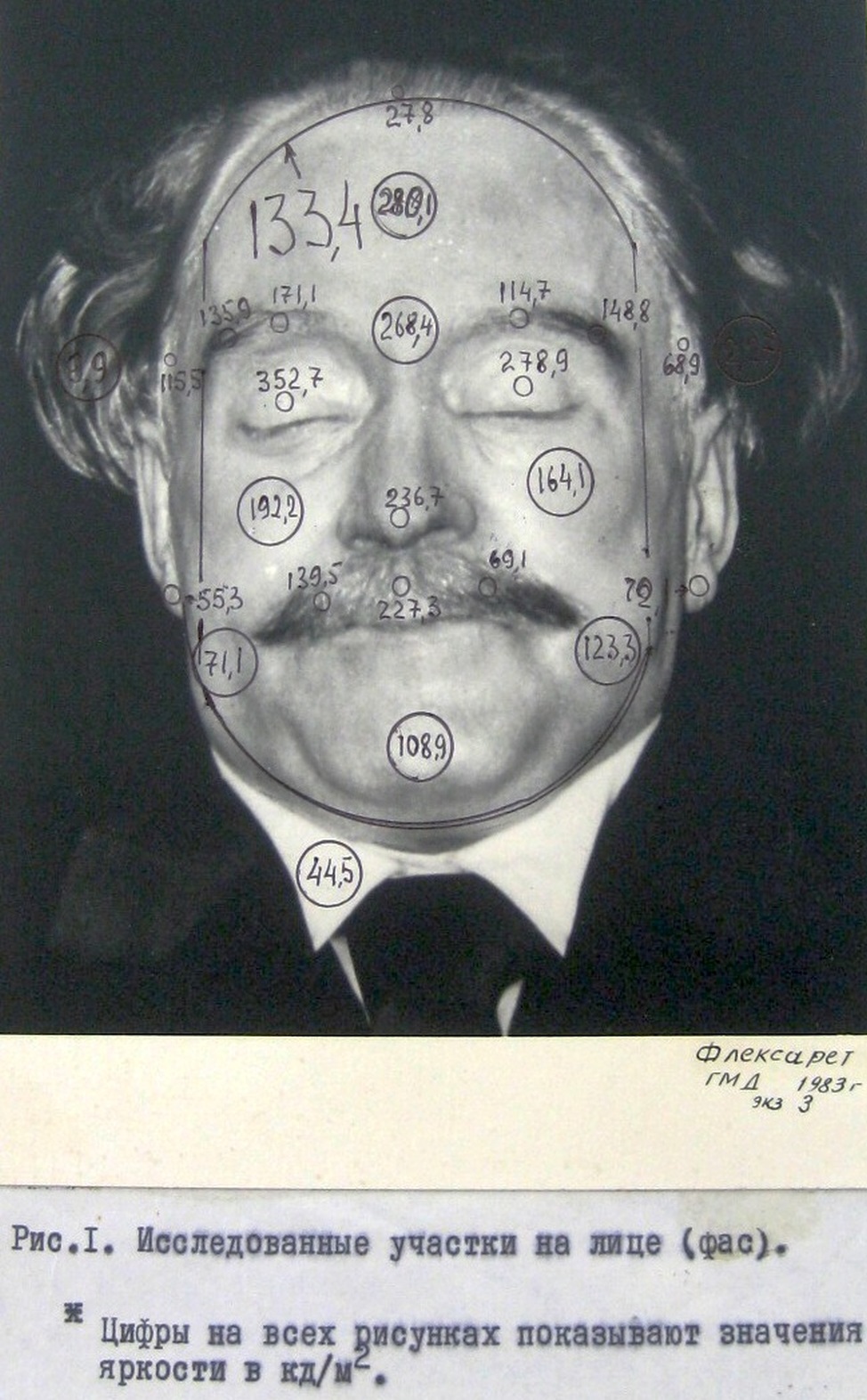 Figure 17. Brightness measurement values in candela units per square meter on Georgi Dimitrov’s face in 1983. Source: Dimitrov Mausoleum Lab. Central State Archives (CDA), ChP 190B, а.е. 9. |
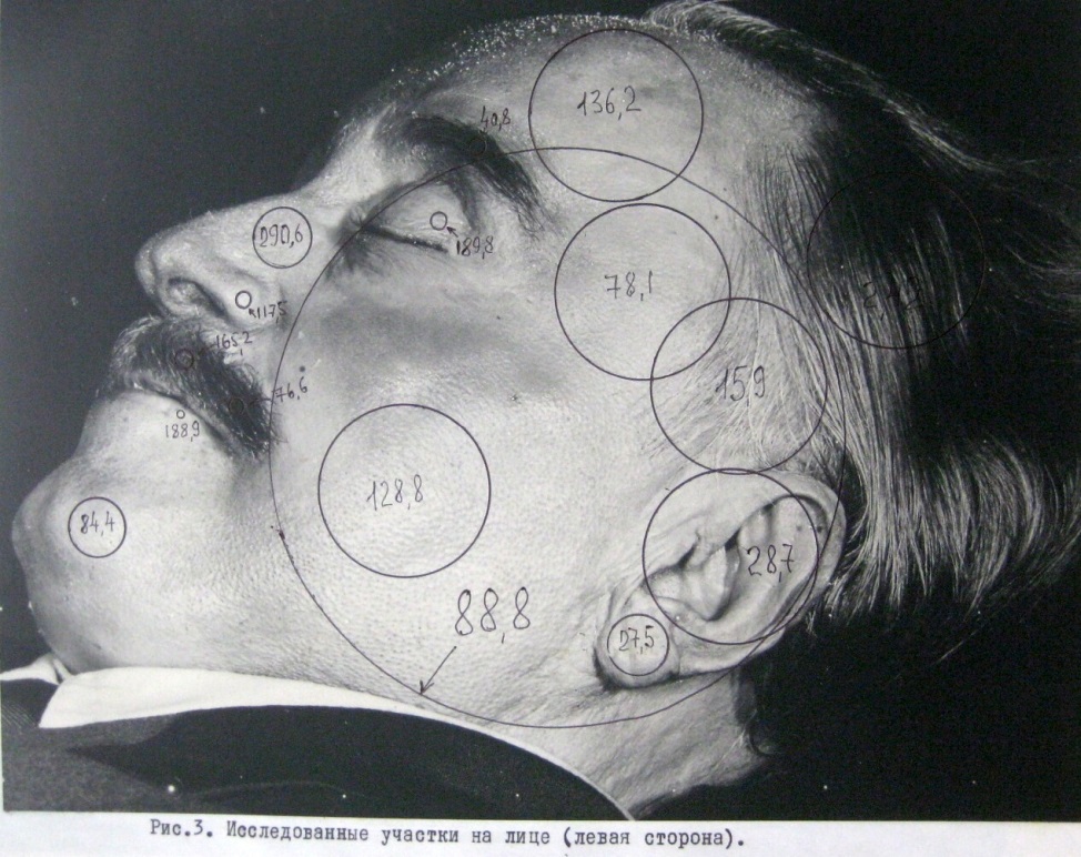 Figure 18. Brightness measurement values in candela units per square meter on Georgi Dimitrov’s left cheek in 1983. Source: Dimitrov Mausoleum Lab. Central State Archives (CDA), ChP 190B, а.е. 9. |
Air conditioning system and the technical background of a mausoleum
According to the Soviet-Russian method, the technical facilities of a mausoleum and the disinfection of the laboratory premises are additional preconditions for the successful long-term preservation of embalmed bodies. The equipment of the individual mausolea is similar. Over the decades, improvements have been made to the technology of monitoring and control of the air conditioning system. The facilities consist of a mourning hall with the sarcophagus, a laboratory for observation and body care, and some adjacent spaces. Furthermore, there is a control room and air conditioning machine room. Below, we provide a simplified description of the principles of the air conditioning system in the mausoleum of Klement Gottwald in Prague (early 1960s) and its technical background (Fig. 19). The air conditioning system in the mourning hall was designed to maintain a temperature of 16 °C ± 1 °C and relative humidity of 70% ± 5% in the presence of 80 visitors. The outdoor intake air was filtered and led through the supply channel to the mixing chamber, where it was mixed with the air already discharged from the mourning hall. Through a box dust filter, the air flowed into a pre-heater and a washing device, where it was cooled and saturated to 100% humidity. The air was then led to a fan which drove it into individual distribution pipes with heaters, which took the air to the required temperature and humidity. The warmed and humidified air entered the mourning hall through the front and side vents. The upper part of the sarcophagus was in a constant stream of air. The air was then sucked out of the mourning hall by a fan separate from the system, and drawn into the mixing chamber, where it was treated again.
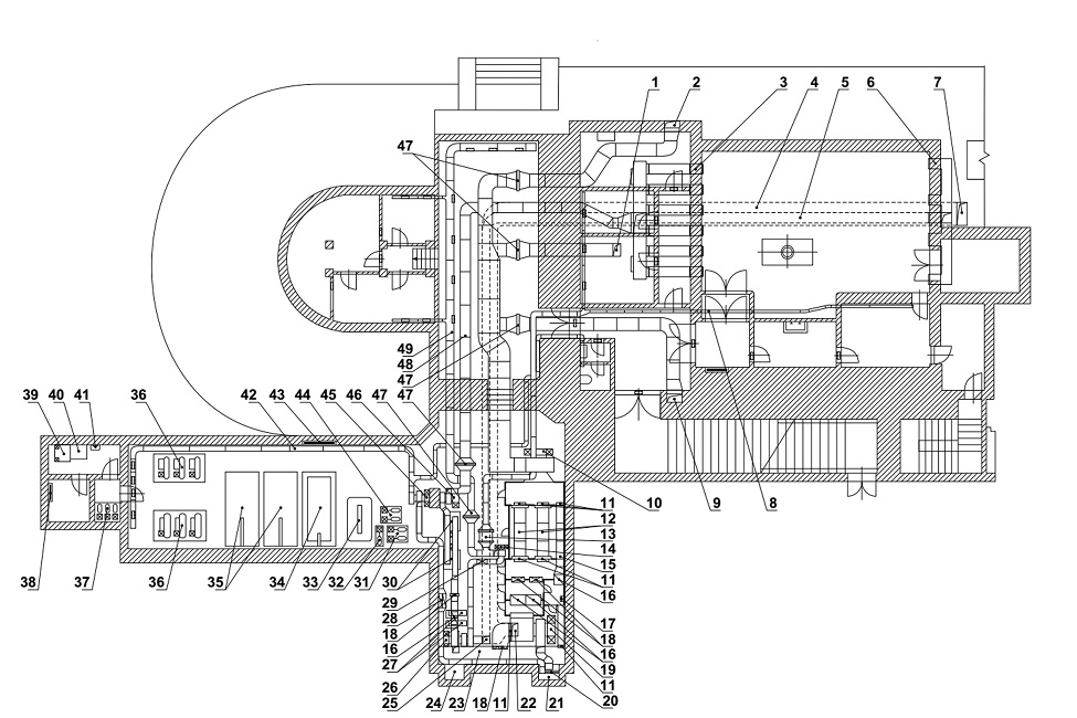 Figure 19. Ground plan of the mausoleum premises including the design of the air-conditioning system. Source: VHA, f. MKG 1953–1955, inv. no. 10, box no. 7. Reconstruction © Jiří Tauš. 1 air supply to the mourning hall - front wall; 2 air supply to the mourning hall - right side; 3 vents to the laboratory; 4 return channel mourning hall; 5 return channel laboratory; 6 exhaust air from the laboratory; 7 air extraction from the mourning hall; 8 air supply to auxiliary spaces; 9 air supply to the mourning hall - left side; 10 device fan no. 1 - output; 11 shut-off valves; 12 air conditioning equipment no. 1 for the mourning hall; 13 dry air cooler; 14 device fan no. 3 for auxiliary spaces; 15 bypass; 16 cell plate filter; 17 ladder; 18 heater; 19 device fan no. 1 - input; 20 venetian blind; 21 discharge shaft; 22 return channel mourning hall; 23 common channel; 24 suction shaft; 25 return channel laboratory; 26 device fan no. 2 – input; 27 compressed air station for automatic regulation; 28 cabinet plate filter; 29 metal oil filter; 30 air conditioning device no. 2 for laboratory; 31 cooling pumps no. 2; 32 hot water pump; 33 electric hot water boiler; 34 buffer tank; 35 cooling tanks; 6 refrigeration compressors; 37 cooling water pumps; 38 switchboard; 39 filter washer; 40 shower; 41 wash-basin; 42 three switchboards for pumps; 43 exhaust air; 44 cooling pumps equipment no. 2; 45 exhaust fan; 46 device fan no. 2 – output; 47 air heaters; 48 air supply to the laboratory; 49 air supply to the control room. |
Automatic control of temperature and relative humidity was based on a sensor, controlled by a thermostat set to the desired temperature. The automatic temperature and relative humidity control for ventilation and air conditioning in the mourning hall consisted of four parts: thermostatic control of electric outdoor air pre-heater, dew point regulation, signaling, and hall temperature regulation with regulation of the minimum amount of outdoor air.
The sarcophagus in the mourning hall was hermetically sealed. The internal temperature of the sarcophagus was maintained at 16° C ± 1°C under the lights, which was controlled by a remote device with an accuracy of ± 0.1 °C. The body was illuminated by a system of 12-volt, 35-watt light tubes with apertures and color filters (Fig. 20). The need for thermal isolation necessitated the placement of dethermal glass under the bulbs, which prevented overheating but did not change the color of the emitted light.
The air conditioning system for the laboratory maintained a temperature of 16° C ± 0.75° C with a relative humidity of 75–85% ± 2.5%. The system contained the same elements, and worked on the same principle, as the system for the mourning hall, except that the air conditioning chambers with pre-heaters were made of sheet metal, and placed upright. Another difference, intended to reduce the occurrence of dust particles and microorganisms, was the inclusion of cotton filters installed on the supply pipe, which cleaned the air before it entered the laboratory. A disinfectant solution was added to the water in the washing device. The walls, ceiling, and floor of the laboratory were lined with washable and disinfectable material. Ethanol (70%) was used as disinfectant. All equipment had to be made of materials that resisted corrosion and minimized the nutrient surface for microorganisms. A special hydraulic lift with a platform, of a maximum height of 4 meters and a load capacity of 1000 kg, was installed in the laboratory, below the mausoleum. A metal pedestal was mounted on the platform, and a glass casket containing the embalmed body was placed on the pedestal (Fig. 21). An electrically driven, hermetically sealable board was installed in the ceiling. It formed the bottom of the sarcophagus and was closed only when the platform was lowered into the laboratory for a treatment of the body (Fig. 22).
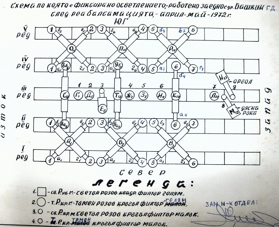 Figure 20. Lighting system with tubes and apertures in the ceiling of Georgi Dimitrov’s sarcophagus in 1972. State Archive Sofia: DA-Sofia, f. 3868, op. 1 and 2, a.e. 1M-86. |
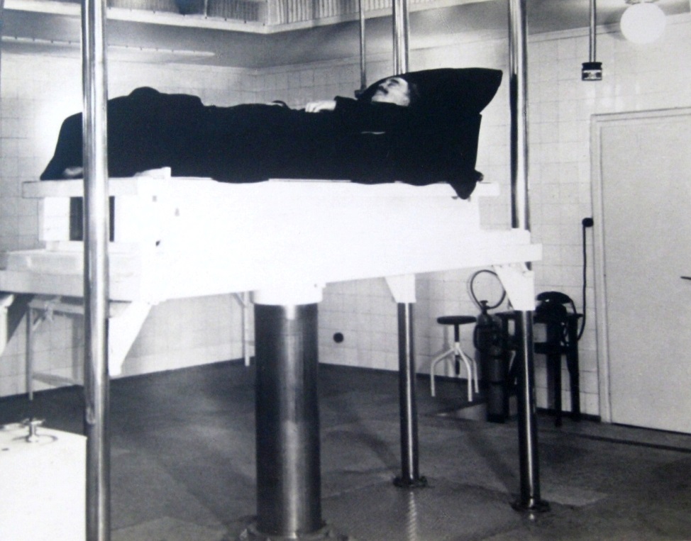 Figure 21. Georgi Dimitrov’s body on the pedestal of the lift platform. Source: Dimitrov Mausoleum Lab. Central State Archives (CDA), ChP 190B, а.е. 9. |
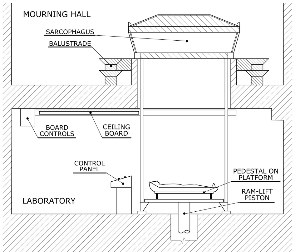 Figure 22. Location of the sarcophagus in the mourning hall and the lifting device in the laboratory. Graphics |
The laboratory's air conditioning system operated in two modes. In the first, standard mode, the main and backup laboratories were air conditioned in parallel. Ventilation vents and doors in the hermetically sealable wall were opened. Thus, the same temperature and humidity was maintained in both laboratories. The second mode was used for maintenance and additional embalming of the body. The orifices and doors in the airtight wall would be sealed, and the reserve laboratory would be cooled by an evaporator. Only the required temperature would be maintained in the laboratory, while humidity was not controlled. Rooms adjacent to the laboratory were a cloakroom, a doctors' office, an autoclave room, a lab technicians’ room, and a hallway. Air conditioning of all auxiliary spaces, i.e. the ventilation of the physicians’ office and room for lab technicians, the storeroom, the autoclave room, the ventilation engine rooms, the controller’s room, workshops and toilets, was provided by a separate unit. Air was drawn into the ventilation system of the auxiliary spaces from a common duct, it was then passed through an oil filter, heated, and piped through vents into the individual rooms. An automatically controlled, dry air cooler was installed in the duct for the doctors' office, lab technicians’ room, storeroom and autoclave room. The air supply to the individual rooms could be regulated by dampers (Frišhons and Vacín 2014b).
The paraffinization embalming method was developed by Frédéricq, Hoschetter and Schmeidel, and improved by Professor Pedro Ara Sarrìa (1891–1973) (Ara, 1934b) who performed the embalming of Eva Perón’s body from July 26, 1952 until November 24, 1955. Initial conservation was carried out with a solution of 10% formaldehyde, 96% ethanol, and 1:1000 solution of water and thymol, injected through the carotid artery, as well as into the visceral cavities and subcutaneous tissue. Subsequently, the body was impregnated with a solution of ethanol, glycerin, potassium acetate, nitrate, and thymol in a 150 L tank. The same solution was used for moistening the face and hands with cotton swabs. The fingers were left wrapped in bandages impregnated with trichlorethylene. Subsequently, the face and hands were treated with cotton swabs soaked in an aqueous solution of 3–5% hydrogen hydroxide. This was followed by a gradual dehydration with 50%, 70%, then 96% ethanol, replacing the tissue water with ethanol. It was later replaced again with a paraffinic organic solvent, turpentine oil, and gasoline, binding the ethanol until the preparation became translucent, which took several months. Excess paraffin was then removed from the body with the use of filter paper at 56° C. Finally, the body was impregnated in a paraffin tank immersed in a water bath, as well as by vascular infusion. First, paraffin with a low melting point of 36 to 48 °C was used, then a higher temperature of 56 °C was set using a thermoregulator. This procedure made the solvent evaporate by bubble formation, while the paraffin subsequently hardened in air at room temperature. The body was stored in a sarcophagus with a temperature of up to 25° C. It was protected against mechanical, chemical, and thermal damage, and monitored (Ara 1934a; Ara 1996). Two incisions made to facilitate arterial injection, as well as an incision on the head, were sealed with the impregnating agent. After the repatriation of the body from Spain back to Argentina, it was restored from November 22 to 29, 1974, by Professor Domingo Isaac Tellechea (1935-) (Tellechea, 1975). The paint covering the neck and shoulders was removed with a solution of petroleum ether. The right shoulder and breasts were restored. A strong insecticidal mixture was applied to a deep incision in the neck, followed by covering the open defects of the neck with hard wax. Synthetic wax was used to replace the small missing part of the left ear. Next, a very fine transparent wax with red cadmium and black pigmentation was applied to adjust the skin color on the head, hands, feet, and the rest of the body. The fingers were repositioned. Lime encrustations and fragments of oxidized iron were removed from the back. At the level of the knee, a wide crack with a large amount of adipose tissue was found. The hair was cleaned and treated. The feet were treated against mold and moisture with an insecticide. The fingernails were treated with a polyester solution in order to conceal the yellowish skin, and to clean and stain the nails. A cast of the missing part of a finger from the right hand was made and put in place. Eyelashes made of natural hair were applied with a needle. Deep defects of the soft tissues of the left anterior shin and right ankle were fixed. An X-ray examination of the body was performed, showing dehydration of the visceral organs. Furthermore, dactyloscopic examination, and a histological examination of the tissue from the right ear was performed (Tellechea 1975). Subsequently, the body was placed in a coffin and interred in a tomb.
Preserved embalmed bodies prove that long-term embalming of a human body is possible under certain conditions. Researchers face problems, such as changes in skin color, brought about by changes in melanin due to UV radiation or formaldehyde, which reduces hemoglobin in tissues, and other factors. Changes in tissue volume, drying out, hydrolysis and oxidation of fats, decalcification of bones, or the possible presence of microorganisms in the form of molds or fungi are major problems to deal with. In the initial stages, preservation with formaldehyde, bleaching of brown skin spots with hydrogen peroxide, or treatment of parchment-like spots with water, dilute acetic acid, and hydrogen peroxide is necessary (Lopukhin, 1997). Changes in tissue volume, e.g. swelling, are treated with concentrated ethanol. Constant monitoring and automatic regulation of temperature and humidity in the sarcophagus is necessary (Kozeltsev and Romakov, 2000). Further, non-contact methods for the control of skin condition and color, as well as relief and volume of soft tissues, or the application of advanced imaging methods for monitoring internal structures, have been developed and improved. Last but not least, the continuous analysis of the embalming solution from the overalls, and the collection of tissue specimen to assess the state of cytological and histological structure are of paramount importance. For example, in a specific physiochemical analysis of the skin and epidermis, diffusion parameters of tissue impregnation were found. The histological structure of the skin had been preserved with a number of structural changes in the stratum corneum, cell layers, and dermis, such as disorders of karyolysis, decreased volume of nuclei, decreased DNA content, histones, or RNA concentration. The extent of the changes is determined by the duration of the embalming and the properties of the skin. Substances released during UV absorption, or morphological damage to cells or threshold levels of structural integration of the cell organelle system were also detected (Tomashevich, 1997). A major disadvantage of the Soviet-Russian embalming method is the enormous financial cost of the establishment and operation of the technical facilities.
Postscript
In the 1990s, the staff of the Lenin Mausoleum laboratory studied the effects of the embalming process on the state of epidermal nucleoproteins, using histochemical detection of nucleic acids and proteins in embalmed materials. They also explored the UV absorbers passing into the fluid during the embalming of skin and muscle tissues, using methods of mathematical physics in order to solve problems with the diffusion of preservative components, or using mathematical models of the process of preservative component diffusion in various tissues, and many other topics (Tomashevich and Nikitina, 1989). No other institution in the world has achieved such knowledge in the study of thanatochemical and microscopic changes in long-term embalmed tissues. Currently, the same team are working on the development of new methods for a non-destructive quantitative assessment of the main components in aqueous solutions containing potassium acetate and glycerin, with the use of Quantitative 1H-NMR spectroscopy (Proton Nuclear Magnetic Resonance) (Abramov 2018; Agrafenin, 2018) or thermogravimetry (Polakov, 2019). While there are a number of research perspectives concerning the long-term preservation of embalmed tissues, it may be assumed that no other laboratory will address these issues at such a level of excellence in the foreseeable future.
Agrafenin AV, Abramov Yu V, Denisov-Nikolsky Yu I, Blinova GI. 2014: Simultaneous quantitative determination of glycerin and potassium acetate in aqueous solution by Raman spectroscopy. Trace Elem Med 15(3): 48−51. [In Russian]
Abramov YV. 2013: Razrabotka nauchno-metodicheskih podhodov k provedeniju restavracionnyh rabot na biologicheskih objektah [Development of scientific and methodological approaches to the restoration work at biological sites]. Voprosy biologiceskoj i medicinskoj i farmacevticeskoj chimii [Problems of Biological, Medical and Pharmaceutical Chemistry] (11): 149-153.
Abramov YV, Matveichuk IV, Nakoskin AN, Krasnov VV, Litvinov YY, Rozanov VV, Logachev SE, Bykov VA, Sidelnikov NI, Astakhov YY. 2016: Restavracionnaja plasticheskaja massa [Restoration plastic mass]. Patent RF № 2576820 C1.
Abramov YV, Abramov YV, Sheichenko VI, Blinova GI. 2019: Simultaneous quantitative determination of glycerine and potassium acetate in aqueous solution by 1H-NMR spectroscopy. Patent RF № 2690186 C1.
Ara P. 1996: "Eva Perón" (La verdadera historia contada por el médico que preservó su cuerpo) [The true story told by the doctor who preserved her body]. Buenos Aires: Ediciones Sudamericana.
Ara P. 1934a: A New process of embalming. Journal of Technical Methods 13 (3):36.
Ara P. 1934b: Zur Geschichte und den heutigen Ergebnissen der Paraffinierungsmethode [On the history and today's results of the paraffin wax method]. Anat Anzeiger 78:117-127.
Bykov VA, Vasilevskii VK, Denisov-Nikolsky YI, Matveichuk IV, Banin VV, Abramov YV, Astakhov YY, Kukushkina EN, Kuznetsova LI. 2002: Sposob balzamirovaniya tela [Body embalming method]. Patent RF №2185060.
Bykov VA, Denisov-Nikolsky YI, Matveichuk IV, Banin VV, Abramov YV, Astakhov YY, Kukushkina EN, Kuznetsova LI. 2013: Odnorazovyi kombinezon dlya khraneniya zabalzamirovannogo tela [Disposable jumpsuit for storing the embalmed body]. Patent RF №127313U1.
Bykov VA, Denisov-Nikolsky YI, Matveychuk IV, Abramov YV, Astakhov YY, Kukushkina EN, Kuznetsova LI. 2013: Kombinezon dlya khraneniya zabalzamirovannogo tela [Jumpsuit for storing the embalmed body]. Patent RF №135493U1.
CDA: Centralen drzhaven arkhiv Sofia, Bulgaria, Collection ChP 190 B (Mavzolei na Georgi Dimitrov). Embalming log book No. 1, 1955–1957 20.6.-24.4.1957, p. 49.
Frišhons J, Vacín, L. 2014a: "Aby byla zachována tvář soudruha Gottwalda": Postup a průběh balzamace těla Klementa Gottwalda [“In order to preserve the face of Comrade Gottwald": The procedure and course of embalming the body of Klement Gottwald]. Acta Musei Nationalis Pragae. Series A - Historia Praha: Národní muzeum. 68(1-2):3-31.
Frišhons J, Vacín, L. 2014b: "Zařízení, která se v ČSR dosud nikdy nevyráběla": K technickému zázemí mauzolea Klementa Gottwalda ["Equipment that has never been produced in the Czechoslovak Republic": On the technical background of the Klement Gottwald Mausoleum]. Acta Musei Nationalis Pragae. Series A - Historia Praha: Národní muzeum. 68 (1-2): 69-90.
Hunko P, Haidukov V, Martynova Z. 2019: Long-term preserving of M. I. Pyrogov’s embalmed body as a unique scientific experiment. Scientific Bulletin of the National Museum of the History of Ukraine 4: 625-639.
Kozeltsev VL, Romakov JA. 2000: Novyi sposob sokhraneniya chelovecheskikh mumii [A new way to preserve human mummies]. Arkheologia, etnologia i antropologia Evrazii [Archeology, Ethnology and Anthropology of Eurasia] 4:103-106.
Kuznetsov LE, Khokhlov VV, Fadeev SP, Shigaev VB. 1999: Balzamirovanie i vosstanovlenie trupov: rukovodstvo [Embalming and restoring corpses: a guide]. Lekarstvo [Medicine], 496 pp.
Lauer VV. 1924: O sposobach, primenennykh pri balzamirovanii tela V. I. Lenina [On the methods used for embalming the body of V.I. Lenin]. Kubanskii nauchno-medicinskii vestnik [Kuban Scientific Medical Bulletin] 4: 23–24.
Labush S. 2014: Long-term embalming. Dodge Magazine 206 (3):16-23.
Litvinov YY, Rozanov VV, Matveichuk IV, Chernyaev AP, Nikolaeva AA. 2016: Innovacionnye podhody k objektivnoj registracii sostojanija poverhnosti i objemov obrazcov rastenij i biologicheskih tkanej [Innovative proposals for the objective registration of surface condition and volumes of plant and animal specimens]. Sbornik nauchnyh trudov mezhdunarodnoj konferencii «biologicheskie osobennosti lekarstvennyh i aromaticheskih rastenij i ih roli v medicine» [Digest of scientific papers of the international conference "Biological characteristics of medicinal and aromatic plants and their role in medicine"].
Posvjashhennoj 85-letiju vilar 23-25 ijunja 2016g., FGBNU Vilar, Moscow, Russia. 395-399.
Litvinov YY, Matveichuk IV, Rozanov VV. 2015: Ispolzovanie beskontaktnogo metoda objektivnoj registracii sostojanija poverhnostej biologicheskih objektov s primeneniem stereoscan 3D [Implementation of a contactless method of objective registration of the composition of biological objects with a 3D stereoscan]. Nauchno-tehnicheskaja konferencija “Mediko-tehnicheskie tehnologii na strazhe zdorovija”. Sbornik dokladov 18-25 sentjabrja 2015, Krym [Scientific and technical conference on medical and technical technologies guarding health. Collected papers 18-25 September 2015, Crimea].
Lopukhin JM. 1997: Bolezn, smert i balzamirovanie V.I. Lenina: Pravda i mify [Illness, death and embalming of V.I. Lenin: Truth and myths]. Moscow: Respublika. 246 pp.
Malyshev BF. 1955: Metod rekonservacii balzamirovannogo trupa (sokhranenie ostankov N.I. Pirogova) [Repeated Embalming of a corpse (preservation of the body of N.I. Pirogov)]. Sov. zdravookhranenie Kirgizii [Sov. Healthcare in Kyrgyzstan] 6: 63-65.
Matvejchuk IV, Denisov-Nikolsky YI, Gunko PM, Gaidukov VV, Martynova ZS, Abramov YV. 2013: Sovremennye dostizhenija v konservacii i dlitel'nom sohranenii biologicheskih struktur i objektov, NI Pirogov [Modern achievements in conservation and long-term preservation of biological structures and objects, NI Pirogov]. Voprosy biologiceskoj i medicinskoj i farmacevticeskoj chimii [Problems of Biological, Medical and Pharmaceutical Chemistry] 11:143-147.
Matvejchuk IV, Litvinov YY, Rozanov VV. 2016: Nauchno-metodicheskie osnovy objektivnoj registracii sostojanija poverhnostej biologicheskih objektov s ispol'zovaniem innovacionnyh metodov [Scientific and methodological foundations of objective registration of surface condition of biological objects using innovative methods]. Morfologija [Morphology] 149(3):134.
Matvejchuk IV, Nakoskin AN, Krasnov VV, Litvinov JJ, Rozanov VV, Logachev SE, Bykov VA, Sidel'nikov NI, Astahov JJ, Abramov JV. 2019: Pestavracionnaja massa dlja korrekcii objemov i ustranenija vnutrennih defektov pokrovnyh tkanej biologicheskih objektov [Restoring mass for volume correction and elimination of internal defects of integumentary tissues of biological objects]. Patent RF № 2683313 C1.
Melnikow-Raswedenkow NF. 1898: Ueber die Herstellung anatomischer Präparate nach der Formalin-Alkohol-Glycerin-essigsauren-Salz-Methode [On the production of anatomical specimens by the formalin-alcohol-glycerin-acetic acid-salt method]. Zentralblatt für allgemeine Pathologie und pathologische Anatomie 9/8 (9): 299–301.
Melnikov-Razvedenkov NF. 1930: Pro naukovi sposobi permanentnogo zberezhenya tila V. I. Lenina [On the scientific methods of permanent preservation of V. I. Lenin’s body]. Ukrainskii medichnii arkhiv [Ukranian Medical Archive] 5 (2): 148–152.
National Archive Prague (NA): Collection 100/24 (Central Committee of the Communist Party of Czechoslovakia – Klement Gottwald), Book No. 36, 16 Oct 1956, p. 55.
Piombino-Mascali D, Aufderheide AC, Johnson-Williams M, Zink AR. 2009: The Salafia method rediscovered. Virchows Archiv 454:355-357.
https://doi.org/10.1007/s00428-009-0738-6
Piombino-Mascali D, Williams MJ. 2009: Rosalia Lombardo and Her Master Embalmer. American Funeral Directors. 6: 52-55.
Polakov NA, Abramov YV, Blinova GI. 2018: Simultaneous quantitative determination of glycerol and potassium acetate in high-concentrated aqueous solution by thermogravimetry method. Trace Elem Med Moscow 19 (1): 51-55.
https://doi.org/10.19112/2413-6174-2018-19-1-51-55
Ryabtsev V. 1997: Semidesyatitrehletnii eksperiment [A seventy-three-year experiment]. Nauka i tehnika [Science and Technology] 10:18-26.
Sheychenko VI, Abramov YV, Blinova GI. 2019: Quantitative determination of glycerol and potassium acetate in aqueous solution by the method of 1H nuclear magnetic resonance. Voprosy biologiceskoj i medicinskoj i farmacevticeskoj chimii [Problems of Biological, Medical and Pharmaceutical Chemistry 22 (8): 10-14.
https://doi.org/10.29296/25877313-2019-08-02
Slocum RE. 1947: Permanent Embalming. The De-Ce-Co Magazine 37 (1): 15-22.
Tellechea DI. 1975: Informe sobre la conservación y restauración del cadáver momificado de la señora Eva Perón [Report on the conservation and restoration of the mummified body of Mrs. Eva Perón]. State Press.
Tomashevich SV, Nikitina EK. 1989: Ocenka matematicheskikh modelei diffuzii konservantov v myshechnuyu tkan s ispolzovaniem kompleksa biokhimicheskikh parametrov [Assessment of mathematical models of preservatives diffusion in muscular tissue using different biochemical parameters]. VI Vsesoiuznaya konferenciya po biokhimii myshc [6th All-Union Conference of Muscle Biochemistry] Tbilisi, p 240.
Tomashevich SV. 1997: Biomedicinskie aspekty processov transporta razlichnykh veshshestv v kozhu cheloveka [Biomedical aspects of transport of various substances in human skin]. Dissertacii po Nauchno-issledovatel'skom centre biologicheskikh struktur [Dissertations at the Research Center for Biological Structures], Moskva.
Vasilevskii VK. 2004: Opyt kompleksnogo primeneniya obiektivnykh metodov ocenki reliefa i cveta kozhnogo pokrova cheloveka v kosmetologii [Experience in the complex application of objective methods for assessing the relief and color of human skin in cosmetology]. Biomedicinskie tehnologii i radioelektronika [Biomedical Technology and Radioelectronic] 1-2: 68-76.
Vasilevskii VK, Zherebtsov LD. 1988: Cvet i morfologicheskie osobennosti kozhnogo pokrova u lyudei razlichnykh rasovykh grup [Color and morphological features of the skin in people of different racial groups]. Bul. eksperim. biologii i mediciny [Bulletin of Experimental Biology and Medicine] 10: 495-498.
Wiesner J, Baumjohann K, Beneck M, Scheidt J. 2013: In the Catacombs of the Capuchin Monastery in Palermo. Leica Microsystem https://www.leica-microsystems.com/science-lab/in-the-catacombs-of-the-capuchin-monastery-in-palermo/
Zbarskii IB. 1998: Pod "kryshei" Mavzoleya [Under the "roof" of the Mausoleum]. Tver: Polina.