1Department of Veterinary Medicine, Federal University of Viçosa, Viçosa, MG, 36570-000, Brazil.
2Faculty of Health and Science, Univiçosa, 36576-340, Viçosa, MG.
Plastination is a technique for preserving body parts and cadavers, in which tissue water is replaced by polymerizable silicone resin. Currently it is in wide demand due to its many applications in animal and human anatomy laboratories, even being popularized in artistic exhibitions of the human body. The development of this preservation technique requires a laboratory with specific adaptations and a high budget for the acquisition of equipment and reagents. The protocol, originally developed by von Hagens in Germany in 1977, describes the use of acetone as an intermediate solvent in the dehydration stage, which, in addition to being costly, requires a series of safety modifications, thus increasing the production cost significantly. The open vacuum system recommended for impregnation requires a vacuum pump to be running for long periods, considerably increasing its wear and tear, with the need for frequent replacement of the equipment. The adaptations for adopting the silicone plastination technique in the pre-existing laboratories of the Morphology Section of the Veterinary Department of the Federal University of Viçosa (DVT/UFV) prioritized the reduction of the production costs by adaptations in all stages of the technique, especially the substitution of the intermediate solvent by ethyl alcohol, and the development of a closed vacuum system. The proposed modifications and adaptations resulted in low-cost production of good quality specimens for the teaching collection of the veterinary anatomy laboratory.
acetone; anatomy; closed vacuum reservoir; formaldehyde; plastination
Zerlotini, M.F Tel. +55 31 3612-5610; Fax +55 31 3612-5607; Email address: mayfz.mz@gmail.com

![]()


Plastination is a technique for preserving organic tissues, organs, and cadavers, where tissue water is replaced by polymerizable silicone resin. With this technique it is possible to produce anatomical specimens which are odorless, free of moisture and with a realistic appearance, not requiring any maintenance, and constantly available for immediate study. It is, therefore, in great demand, due to its great applicability in animal and human anatomy laboratories, even being popularized in artistic exhibitions of the human body (G1 GLOBO, 2017).
The standard plastination technique proposed by von Hagens (1977) consists of four stages: 1) fixation - in this stage, the samples are fixed in a formaldehyde solution between 3% and 10%, and then washed in running water; 2) dehydration - the stage of replacement of tissue water by a secondary solvent begins. The main solvent used is acetone, and the best results are obtained at freezing temperature (-26° C). Generally, a series of three baths in pure acetone over three weeks is applied. 3) Impregnation - this is the main step in plastination where the specimens dehydrated in acetone are immersed in liquid silicone polymer at freezer temperature, and subjected to an increasing vacuum. Depending on the texture and size of the samples, forced impregnation takes between 4 and 12 weeks; 4) the final step of plastination processing is curing, in which the resin-impregnated body part is hardened by a cross-linking reaction catalyzed by a gaseous agent (Biodur S6 hardener). The specimens are arranged in the desired positions in airtight chambers, where the curing agent is vaporized (von Hagens, 1979; de Jong and Henry, 2007; Raoof, Henry, Reed, 2007; Ottone et al., 2015).
The development of this preservation technique requires a laboratory with specific adaptations and a high budget, around US$61,334 just for the acquisition of equipment and reagents. This is the estimated cost of the plastination laboratory at the Federal University of Rio Grande do Norte, considered to be the largest in Latin America (UFRN, 2019). The main obstacles in implementing the different stages of the plastination technique are the necessary changes for the dehydration and impregnation stages. The use of acetone as an intermediate solvent is widely employed, and constitutes in itself a logistical and economic problem, since its commercial availability is strongly controlled and costly in some South-American countries. Here in Brazil, we have high alcohol production, which makes it cheap, and hence favors its use in plastination. Acetone is extremely volatile and flammable, and must be handled in a closed environment with an efficient exhaust system. In addition, it is not recommended to use electrical equipment and incandescent lighting.
In the impregnation stage, the technical protocol requires, among other things, an increasing vacuum for forced impregnation, since the silicone resin is extremely thick, particularly at low temperatures, which makes tissue penetration difficult. The protocol originally developed by von Hagens describes the use of an open vacuum system, where pressure is controlled from a regulated vent in a chamber connected to a vacuum pump. As the impregnation process can extend for up to three months, the wear and tear and consequent maintenance of the vacuum pump is a major obstacle in a laboratory with a limited budget (Von Hagens, 1979).
In this context, the present work describes the development of a series of structural adaptations and technical modifications for the installation of a plastination laboratory in the Morphology Section of the DVT/UFV, which was already engaged in the production of a collection of anatomical specimens for the disciplines of domestic animal anatomy, and veterinary anatomy. The main objectives were to develop a closed vacuum system, using a reservoir, distribution line and impregnation chambers, from recycled equipment; adapt curing agent vaporizing chambers from recycled flow cabinets, and produce a collection of plastinated anatomical specimens with didactic quality.
 Figure 1: floor plan of the DVT/UFV Morphology Section, before the installation of the plastination laboratory. 1, teachers' rooms; 2, research laboratory; 3, corridor; 4, theoretical classroom; 5, practical classroom; 6, skeleton assembly workshop; 7, first floor storeroom; 8, bathroom; 9, staircase to the second floor of the storeroom 10, storeroom; 11, body storage and fixation room; 12, bone cooking and maceration room |
In order to adopt the plastination technique, the DVT/UFV Plastination Laboratory was established within the morphology section. Some reforms and adaptations were necessary, aiming at safety and efficiency in the production of the plastinated specimens. The Morphology Section of the Veterinary Department of UFV has a total area of 550 m2 (Fig. 1) made up of: theoretical classroom; practical classroom; teachers' rooms, research laboratory, storage area and preparation of cadavers and parts; room for bone cooking and maceration; workshop for assembly of skeletons; and rooms for storage (on two floors). With small structural modifications, part of the skeleton assembly workshop and the lower of the two storage rooms were designated for the installation of the plastination laboratory.
The plastination laboratory was sectorized according to the stages of the plastination process, i.e. fixation, dehydration, impregnation and curing. The fixation sector, also used in the washing of the specimens to be plastinated, was also used for fixation and other routine activities, in the previously-existing area for storage and preparation of cadavers, and no further structural modifications were necessary. The dehydration and impregnation sectors were installed on the lower floor of the old storage area (Fig. 2), and a small area of the workshop, separated by means of office partitions, was also used to accommodate the electrical equipment. As a safety measure, an emergency shower and a fire extinguisher were installed near the access door. The curing section was installed in part of the skeleton assembly workshop, also isolated by office partitions (Fig. 2).
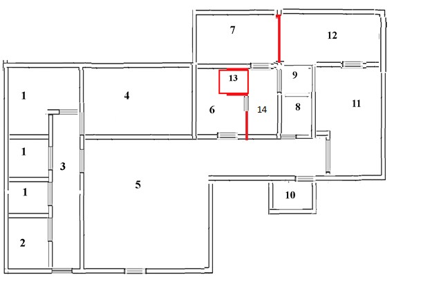 Figure 2: floor plan of the DVT/UFV Morphology Section, after the installation of the plastination laboratory. 1, teachers' rooms; 2, research laboratory; 3, corridor; 4, theoretical classroom; 5, practical classroom; 6, skeleton assembly workshop; 7, dehydration and impregnation room; 8, bathroom; 9, staircase to the second floor of the storeroom; 10, storeroom; 11, storage and fixation room for cadavers; 12, bone cooking and maceration room; 13, machine room; 14, curing room |
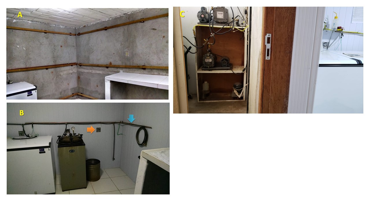 Figure 3: exhaust system production. A, installation of PVC ceiling lining, 5 cm from the walls, interconnected to the exhaust equipment; B, exhaust system distribution valves (orange arrow), earthing system (blue arrow); C, view of the transition between the machine room (left) and the dehydration and impregnation room (right). Freezer inside the dehydration and impregnation room, with the motor and compressor installed in the machine room next to the production and vacuum storage plant. |
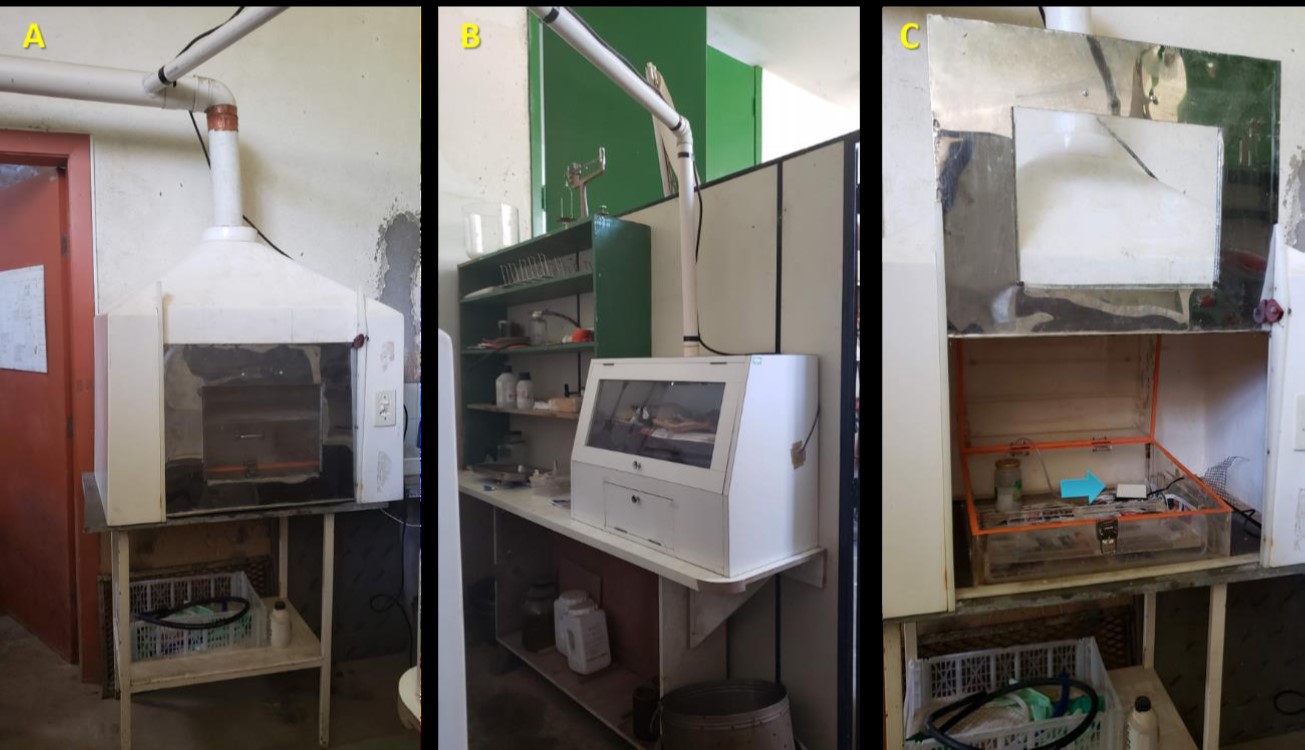 Figure 6: flow cabinets adapted as curing chambers. A and B, flow cabinets interconnected to the exhaust system of the dehydration and impregnation room; C, secondary curing chamber inside the flow cabinet, with an aquarium bubbling pump for the hardening agent. |
In order to reduce production costs, experiments were carried out to replace acetone with ethyl alcohol as an intermediate solvent, although tests indicated that acetone was still recommended as a degreasing agent during the dehydration process (Zerlotini et al., in prep. section 2, 2020). In consequence, the following structural modifications to equipment and electrical installation were necessary:
-Exhaust system installation: the only existing window in the room was isolated with a plywood cover, where a large external exhaust was installed. A PVC sheet lining was installed at about 5 cm on all internal walls of the room, forming a continuous surrounding space interconnected to the exhaust, so that, from openings equipped with valves strategically positioned in the PVC lining, an exhaust system can be directed and distributed (Fig. 3a).
-Changes in equipment and electrical installation: due to the highly flammable nature of acetone, any ignition source was avoided inside the room, so the electrical installation was isolated. The light was replaced by a LED ceiling light, and the switch was installed externally. The freezer used in dehydration and impregnation was adapted by transferring the motor and compressor to the external area of the room (machine room) (Fig. 3c). An over-capacity earthing system was also installed inside the room (Fig. 3b).
A distillation apparatus for recycling the acetone was developed from an electrically heated container, connected to a condenser tube cooled with running water. This produced an acetone distillate of approximately 98% concentration, but with a significant loss of 30%. The whole apparatus was positioned under an air exhaust system (Fig. 4).
For the impregnation stage, a methodology was developed using a closed vacuum system (Zerlotini et al., 2020 (in prep.)). A vacuum production plant and reservoir, outside the room, were connected to the impregnation chambers by a pipeline (Fig. 5). The vacuum production and storage plant were developed using a main vacuum pump, recovered from discarded cryo-centrifuge equipment, and a large cylinder (200 L) purchased at an industrial scrap merchant facility. The connection line interconnects the entire system (vacuum pumps, reservoir, and impregnation chambers) by means of connectors and gas valves and special high-pressure piping. In this way, it is possible to accurately control the vacuum in each unit of the system, by means of industrial analog vacuum meters (Fig. 5).
The impregnation chambers were developed from four disused and discarded autoclaves. Two of them had their internal chambers removed, and were fitted with flat tempered glass lids, connected to the vacuum distribution system, and installed inside the freezer for cold impregnation. The other two autoclaves were adapted for use at room temperature, so the internal chambers were kept, which were connected to the vacuum system, and the lids were adapted for internal visualization by replacing the upper part of the original lid with an inverted tempered glass bowl (Fig. 5 C).
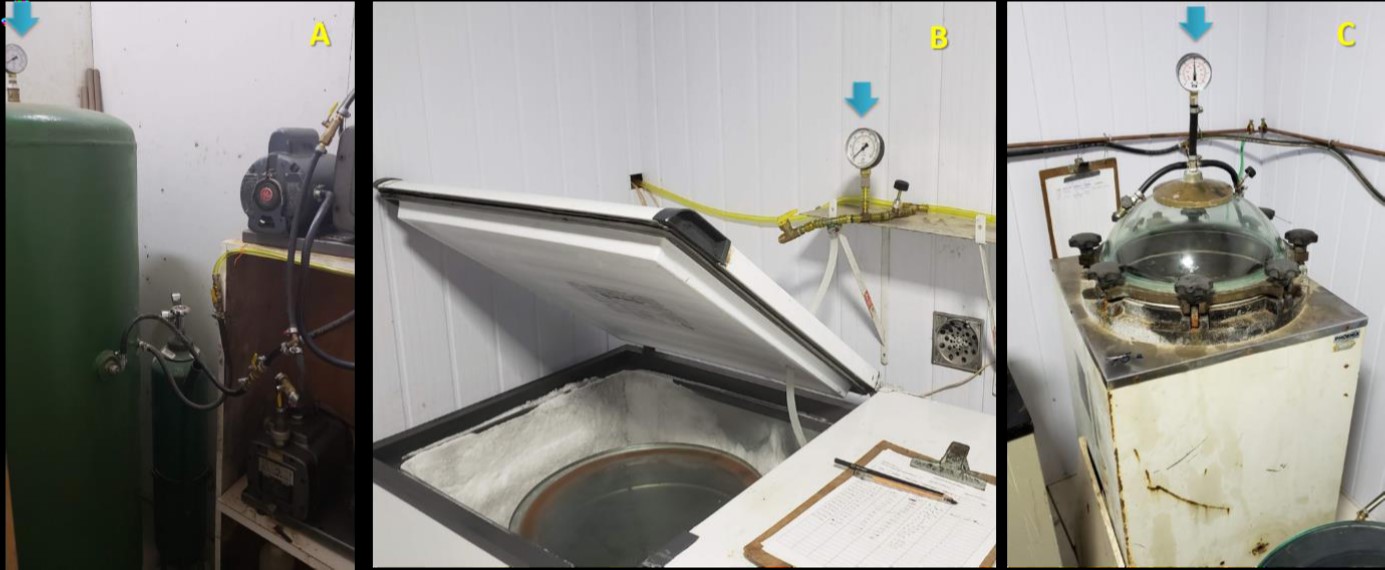 Figure 5: vacuum production and storage plant, distribution line and impregnation chamber. A, cylindrical reservoir interconnected to the pumps and vacuum distribution line; B, impregnation chamber inside the freezer, interconnected to the distribution system; C, room temperature impregnation chamber, adapted from recycled autoclave equipment. Arrows highlight the presence of vacuum gauges in different components of the system. |
Figure 6: flow cabinets adapted as curing chambers. A and B, flow cabinets interconnected to the exhaust system of the dehydration and impregnation room; C, secondary curing chamber inside the flow cabinet, with an aquarium bubbling pump for the hardening agent. |
The curing area, installed in part of the skeleton assembly shop, was equipped with two flow cabinets obtained from the UFV equipment disposal repository (Fig. 6). The cabinets were interconnected to the exhaust system of the dehydration and impregnation room, and were either equipped directly with aquarium pumps, or via a secondary curing chamber, to vaporize the curing agent (Fig. 6 C).
The first records of the study and teaching of anatomy date back to the School of Alexandria where, according to Galen's records, the first public dissections of animals and human bodies were performed (Singer, 1996). In recent decades, anatomy teaching has undergone many changes, in part due to the curricular reforms of health courses, necessary for accommodation of the new curricular guidelines for undergraduate courses (Fornaziero et al., 2010), but also, and mainly, to follow the didactic-pedagogical evolution related, specifically within the context of anatomy, to several factors, such as: advances in digital technology (including advances in digitized images that allow visualization of 3D structures), difficulty in obtaining cadavers for dissection due to a greater control of the origin of bodies, increasing numbers of students in the anatomy subjects, and reduction of the time the student spends working with the same structures, which would facilitate memory consolidation (Silva et al., 2018).
The Section of Morphology of DVT/UFV offers the subject of the anatomy of domestic animals for Agronomy and Zootechny courses, and veterinary anatomy for Veterinary Medicine, both initially covering the locomotor apparatus, and later the other systems, structures and organs. These subjects are taught semi-annually, each comprising fifteen weeks of study in the first and second semesters. The time allocation on the veterinary anatomy course is eight hours per week, with a minimum attendance of 60 students in each subject (UFV Graduation Catalogue, 2020).
The methodology used for the study of the locomotor system is by topographic dissection of cadavers of the different domestic species (bovine, swine, equine, dogs and cats), and this is performed by the students themselves, under the guidance and supervision of the relevant teacher, while systematic anatomy involves the study of hollow organs in loco on the same cadavers, as well as isolated organs.
The working dynamics consists of the formation of groups of four to five students, to whom a cadaver, fixed in a 10 % formaldehyde solution, of any of the domestic species (dog, cat, horse, pig, calf and goat) is assigned. About 60 to 70 % of the class time, especially in the locomotor system, is spent in dissecting the study material. Thus, dissection over the years has become a target and not a means of anatomical study. Dissection still involves the inconvenience of working on wet specimens with strong residual formaldehyde content, and is dependent on the ability and individual dedication of inexperienced students, not always resulting in dissections with acceptable didactic quality, and thus putting descriptive study at risk. Since the classes usually have 30 students there is an overload on the teacher's attention, who often needs to take over the dissection on certain cadavers, which generates delay and disengagement of the other students.
As an experiment, during the last cycle of locomotor system anatomy at DVT/UFV, formalin-fixed animals that had been already dissected were used. This considerably optimized the use of time, which could now be used mainly in the study of previously dissected structures, complying in a much more satisfactory way with the target activity of topographic anatomical study. It was also possible to see the improvement in general performance of the students. This is supported by Weiglein (1996) and Sora et al. (2019), who reported that students of health courses showed better development, and consequently better learning, from the use of previously dissected specimens, and that the use of plastinated specimens additionally encouraged handling and personal study, due to the elimination of moisture and tissue odor.
The fixation of anatomical specimens and cadavers can be performed using several types of fixatives, with formaldehyde being the most common (Ramos, 2018). For plastination there is no specific fixation process, but formaldehyde solutions with concentrations ranging from 1 to 20 % are widely used, 5% being the most common (Oostrom, 1987). The first specimens to be plastinated in the DVT/UFV plastination laboratory were specimens that were already fixed, and had been stored in a 10% formaldehyde solution. Fresh specimens were also used, fixed by forced perfusion with formaldehyde solution in concentrations varying, for experimental purposes, between 3 and 10 %. In all cases, as an initial step in the plastination process, the specimens were washed in running water for one week to remove excess formaldehyde.
The dehydration step is critical for the plastination process, as it involves replacing tissue water with an intermediate solvent, which is essential for later penetration of the silicone resin. Acetone is the solvent of choice as it is a powerful dehydrating agent and also an excellent degreasing agent (Valdés et al., 2010). However, acetone is also highly flammable, and is required in large amounts. It can be very dangerous if not handled correctly; its ignition point is from -18oC, and low temperatures are recommended for its handling (Gubbins, 1990). In addition, the price and bureaucracy involved in the purchase of acetone are important restrictive issues here in Brazil (Santana, 2018).
Besides the fact that acetone is difficult to acquire in Brazil, it is also very expensive: one liter costs an average of US$26.66, compared to fuel alcohol, which has an average cost of US$0.94 per liter. The difference in price of the products was one of the motivating factors in replacing acetone with alcohol during the dehydration stage. For example, in the dehydration of dog and cat hemi-carcasses, 40 liters of alcohol were used in each exchange, and a total of 5 exchanges were performed, giving a total of 200 liters of alcohol used. Using acetone, the cost would have been US$5,334; thus, the replacement by alcohol generated a saving of US$5,147. However, in preliminary tests it was found that dehydration with alcohol alone is not enough to promote total degreasing of the specimen; an acetone bath at the end of the process is also recommended. For that, 40 liters of acetone would be used, with a cost of US$1,067, giving a total a saving at the end of the whole dehydration process of US$4,080. Even though this cost can be reduced with the reuse of acetone, it would still involve the cost of the dehydration apparatus, and the inherent losses of acetone during the distillation process; even so, alcohol is more advantageous. Studies using of commercial detergents and degreasers are being conducted, with the aim of completely replacing acetone in the degreasing stage.
Srisuwatanasagul et al. (2010) also describe the use of ethyl alcohol as an intermediate solvent to replace acetone, concluding that, although acetone presents better results in terms of coloring and tissue shrinkage, alcohol is a safer and economically viable alternative for the production of didactic quality specimens. Most authors recommend using refrigeration for the dehydration stage, to prevent excessive tissue shrinkage (von Hagens, 1986; Gubbins, 1990; Valdés et al., 2010; Pentea et al., 2016); however, Santana (2018) concluded that the dehydration temperature does not compromise the morphological quality of the plastinated specimens. Although there is known shrinkage in tissues such as the brain, Santana (2018), in studies on specific conditions, and in the same region as where this study was carried out, considered that the observed shrinkage was not sufficient to necessitate having to do dehydration under refrigeration. Thus, the protocol used in the DVT/UFV plastination laboratory used room temperature dehydration. With the intention of reducing the production costs even more, the acquisition of locally-produced resources, such as the silicone resin, catalyst and hardener, was the option chosen.
The impregnation stage of the plastination technique is carried out in a vacuum system, with the vacuum applied increasingly throughout the process. This is necessary so that the penetration of silicone resin is concurrent with the exit of the intermediate solvent; the vacuum level, therefore, is adjusted according to the bubbling resulting from evaporation of the intermediate solvent. When the bubbling subsides, it is restored by increasing the vacuum level (Ottone et al., 2015). The widely recommended vacuum application system is the open (active) system, which means the use of a vacuum pump, directly connected to the impregnation chamber, while the vacuum level is controlled by letting air into the system through a bypass valve (von Hagens, 1979; Briggs et al., 1997; de Jong & Henry, 2007; Latorre et al., 2007).
Ottone et al. (2015) developed the concept of a closed (passive) vacuum system, in which, once the desired vacuum level is reached, the chamber is completely isolated and the vacuum pump can be switched off. The same authors recommend the alternate use of the open and closed vacuum systems for impregnation at room temperature, which avoids the continuous operation of the vacuum pump, and allows slower and more uniform penetration of the silicone into the specimen, reducing tissue shrinkage. In the DVT/UFV plastination laboratory, a closed vacuum system was developed which allows vacuum level control from a large reservoir. The different cold- and room-temperature impregnation chambers are thus independently connected to the reservoir, which can be periodically replenished separately, using a vacuum pump. In this system, from previous experiments, it was found that the visualization of the bubbling alone is not a decisive factor for control of impregnation, and consequently the vacuum level used. The vacuum level growth curve adopted in the DVT/UFV plastination laboratory was established from previous experiments (Zerlotini et al., 2020, in prep.), being standardized and applied incrementally in -50 mmHg steps every two days, until the maximum level, which is then maintained until impregnation is completed.
The last stage of the plastination process is the curing of the impregnated silicone resin. For this, it is necessary to expose the specimen or cadaver to a volatile cross-linking agent. This is normally performed in sealed chambers with the curing agent vaporized by bubbling air through it (von Hagens, 1986). In the DVT/UFV plastination laboratory, the specimens, after the impregnation step, are kept for a period of 24 hours in a flow cabinet to remove excess silicone, and then transferred to one of two flow cabinets adapted for use as a curing chamber. The adaptation consisted of the installation of a small aquarium oxygenation pump connected to a container containing the curing agent. The specimens are anatomically positioned with the use of supports, and exposed to the volatilization of the hardening agent by the activation of the aquarium pump, for a period of five to ten minutes, twice a day, until the specimen is fully hardened. The pieces were cleaned daily with paper towels to remove excess silicone, and were kept inside the curing chamber until completely dried.
According to Tianzhong et al. (1998), Henry et al. (1997), Raoof et al. (2007) and Ottone et al. (2014), plastinated specimens do not lose their texture and color characteristics, resulting in odorless, dry and highly durable pieces. Furthermore, Baptista et al. (1989) and Riederer (2014) concluded that plastinated anatomical specimens and cadavers allowed the best learning of students, and can be reused for several years in classes, thus avoiding the acquisition of new cadavers each year.
Through a subjective evaluation, it was shown that the specimens produced in the plastination laboratory of DVT/UFV presented good definition of details (Figs. 7 and 8), and, since they are free of odors and moisture, handling by the students is encouraged. Another characteristic that favors anatomical study is the maintenance of the anatomical position, which greatly facilitates the visualization of the region and the relationships between the various organs (Figs. 9 and 10). In a further subjective evaluation, the specimens that were produced from fresh specimens resulted in notably better color and texture characteristics than those that were plastinated from the old collection of wet specimens, which had been kept in 10 % formaldehyde solution for years (Fig. 11).
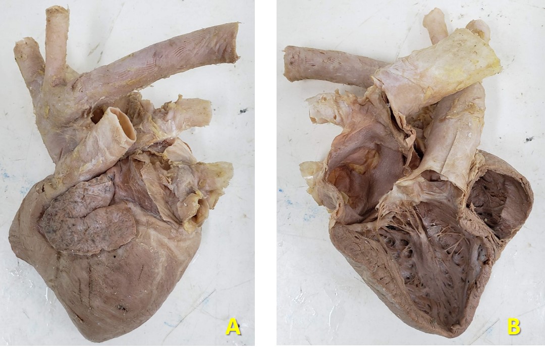 Figure 7: plastinated heart of dog. A, lateral view; B, medial view |
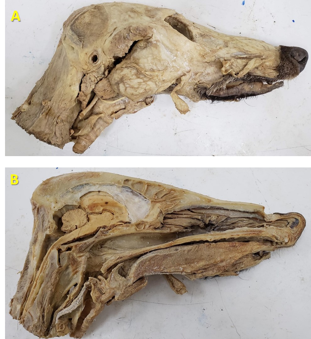 Figure 8: plastinated head of a guará wolf (Chrysocyon brachyurus). A, lateral view; B, medial view |
 Figure 9: stomachs of equines plastinated and inflated with air to maintain the anatomical position. A, lateral view; B, view of stomach with spleen |
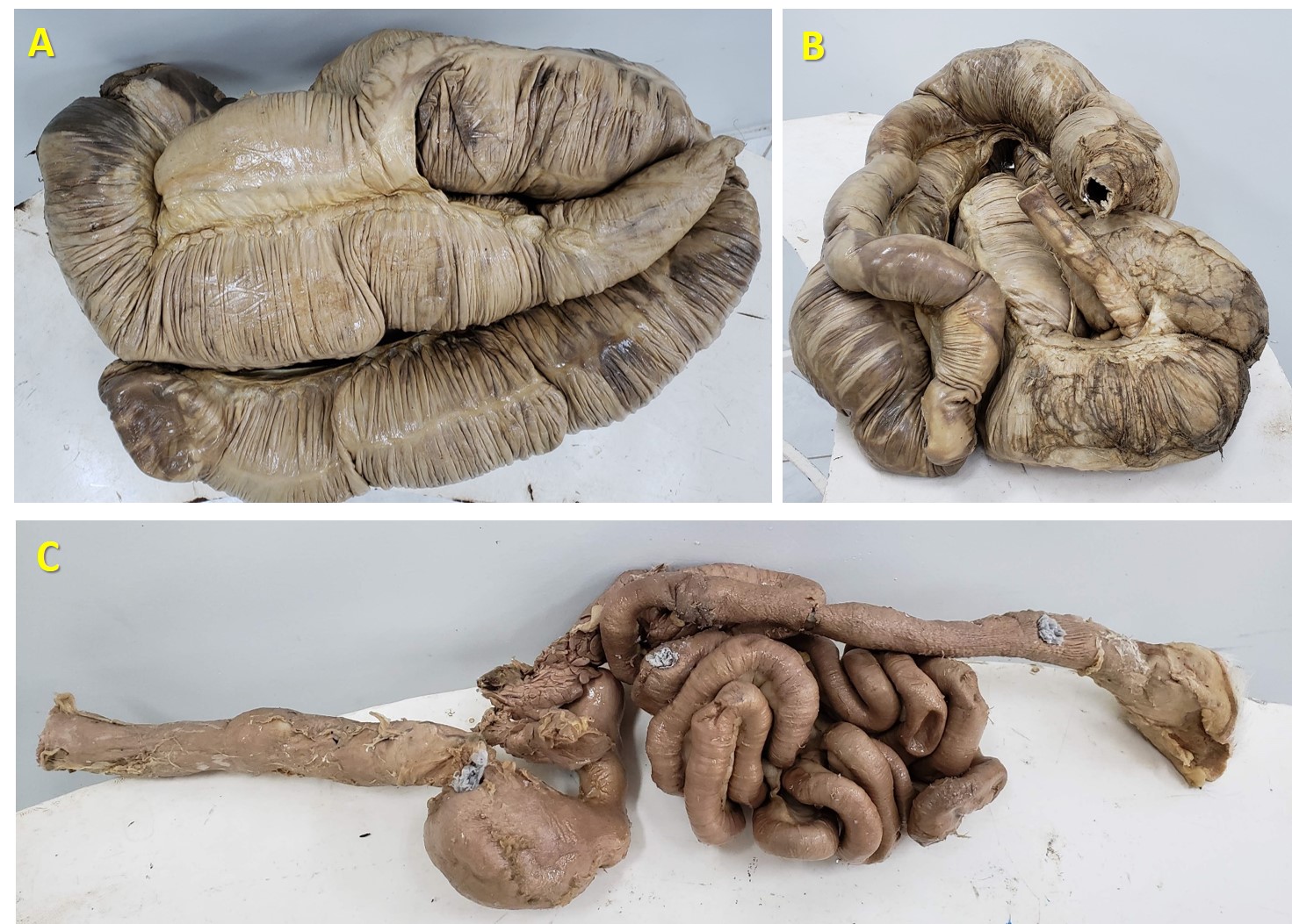 Figure 10: A, plastinated intestine of equine, dorsal view; B, plastinated intestine of equine ventral back view; C, digestive tract of dog, from the esophagus to the anus |
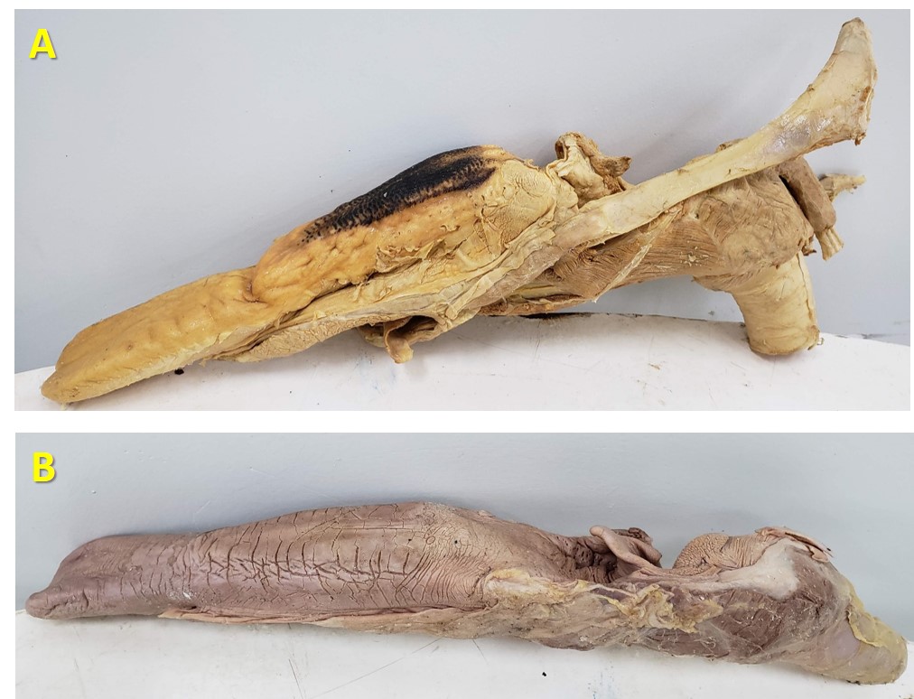 Figure 11: plastinated tongues, lateral view. A, fresh tongue of equine plastinated with hyoid bone and cartilages of the larynx; B, equine tongue previously fixed, stored in formaldehyde, and plastinated with cartilages of the larynx. |
Discussion is included in Results section.
The structural alterations implemented for the development of the dehydration and impregnation sector at the DVT/UFV plastination laboratory proved to be cost effective, including the installation of an efficient exhaust system and the reduction of the risk of fire and explosion from handling acetone. The substitution of acetone as an intermediate solvent by combustible ethyl alcohol, although not completely eliminating its use (since a degreasing bath with acetone is still recommended), considerably reduces the volume of acetone used, resulting in a safer and more economical dehydration step.
The development of a closed vacuum system with the use of a production and storage plant, a distribution line and independent impregnation chambers, has resulted in a considerable reduction of maintenance costs and equipment replacement (vacuum pump), with ease and accuracy of handling, resulting in the production of anatomical specimens with good didactic quality.
The reuse of parts from discarded and disused machinery allowed the installation of functional equipment with low investment and excellent quality. The use of anatomical specimens and old cadavers from a wet collection kept in 10% formaldehyde solution, even for a long time, is viable in the production of plastinated specimens, but those produced from fresh samples showed better quality of color and texture.
Bickley HC, von Hagens G, Townsend FM. 1981: An improved method for preserving of teaching specimens. Arch Pathol Lab Med 105:674-676.
Baptista CAC, Skie M, Yeasting RA, Ebraheim N, Jackson WT. 1989: Plastination of wrist: potential uses in education and clinical medicine. J Int Soc Plast 3:18-21.
https://doi.org/10.56507/XENF9035
Briggs CA, Robbins SG, Kaegi WH. 1997: Development of an Anatomical Technologies Laboratory. J Int Soc Plast 12(2):8-11.
https://doi.org/10.56507/YRLU4811
Graduate Catalogue UFV (2020) Graduation Catalogue, Dean of Education. Available at: http://www.catalogo.ufv.br/ementario.php?campus=vicosa&ano=2020. Accessed 02/08/2020.
de Jong K, Henry RW. 2007: Silicone plastination of biological tissue: cold-temperature technique Biodur S10/S15 technique and products. J Int Soc Plast 22: 2-14.
https://doi.org/10.56507/ZLMJ7068
Fornaziero CC,, GordanI PA, de CarvalhoI MAV, AraujoI JC, de AquinoI JCB. 2010: Integração do corpo humano e meio ambiente (Teaching of Anatomy: Integration of the Human Body and the Environment). Revista Brasileira de Educação Médica, Rio de Janeiro 34(2): 290-297.
https://doi.org/10.1590/S0100-55022010000200014
G1 GLOBO (2017) Exposição que faz 'viagem' pelo corpo humano está em Belo Horizonte. Disponível em: https://g1.globo.com/minas-gerais/noticia/exposicao-que-faz-viagem-pelo-corpo-humano-esta-em-belo-horizonte.ghtm. Accessed: 03/01/2020.
Gubbins RBG. 1990: Design of a plastination laboratory. J Int Soc Plast 4:24-27.
https://doi.org/10.56507/ZXJT8319
Henry RW, Janick L, Henry C. 1997: Specimen preparation for silicone plastination. J Int Soc Plast 12(1):13-17.
https://doi.org/10.56507/HVSK9838
Latorre RM, García-Sanz MP, Moreno M, Hernández F, Gil F, López O, Ayala MD' Ramírez G, Vázquez JM, Arencibia A, Henry RW. 2007: How useful is plastination in learning anatomy? J Vet Med Educat 34(2):172-176.
https://doi.org/10.3138/jvme.34.2.172
Oostrom K. 1987: Fixation of tissue for plastination: general principles. J Int Soc Plast 1: 3-1.
https://doi.org/10.56507/WLZH2223
Ottone NE, Cirigliano V, Bianchi HF, Medan CD, Algieri RD, Brum GB, Fuentes R. 2015: New contributions to the development of a plastination technique at room temperature with silicone. Anat Sci Int 90(2): 126-135.
https://doi.org/10.1007/s12565-014-0258-6
Ottone NE, Cirigliano V, Lewicki M, Bianchi HF, Aja‐Guardiola S, Algieri RD, Cantin M, Fuentes R. 2014: Plastination technique in laboratory rats: an alternative resource for teaching, surgical training and research development. Int J Morphol 32(4): 1430-1435.
https://doi.org/10.4067/S0717-95022014000400048
Pentea M, Hulea C, Stancu A, Butnariu M, Romeo T. 2016: Developing the plastination laboratory for the technique S10. Mater Plast 53(1): 150-152.
Ramos M L. 2018: Avaliação de diferentes fixadores na qualidade histológica de tecidos previamente plastinados. Tese - Universidade Federal de Viçosa, Viçosa, MG. 59f.
Raoof A, Henry RW, Reed RB. 2007: Silicone plastination of biological tissue: room-temperature technique. DowTM/Corcoran technique and products J Int Soc Plast 22: 21-25.
https://doi.org/10.56507/AWAC9285
Riederer BM. 2014: Plastination and its importance in teaching anatomy. Critical points for long‐term preservation of human tissue. J Anat 224(3): 309-315.
https://doi.org/10.1111/joa.12056
Santana ML. 2018: Avaliação de dados morfométricos em corações suínos submetidos a diferentes protocolos de desidratação, visando técnica de plastinação com silicone.Tese - Universidade Federal de Viçosa, Viçosa, MG. 36f.
Silva JH, Foureaux G, Sá MA, Schetino LPL, Guerra LB. 2018: The teaching and learning of human anatomy: the assessment of student performance after the use of concept maps as a pedagogical strategy. Ciênc Educ Bauru 24(1): 95-110.
https://doi.org/10.1590/1516-731320180010007
Singer C. 1996: Uma breve história da anatomia e fisiologia desde os gregos até Harvey. Campinas: Editora da Unicamp.
Sora MC, Latorre R, Baptista C, López-Albors O. 2019: Plastination-a scientific method for teaching and research. Anat Histol Embryol 48(6): 526-531.
https://doi.org/10.1111/ahe.12493
Srisuwatanasagul K, Srisuwatanasagul S, Adirekthaworn A, Darawiroj D. 2010: Comparative study between using acetone and absolute alcohol for dehydration in plastination procedure. The Thai J of Vet Med 40(4): 437-440.
https://doi.org/10.56808/2985-1130.2263
Tianzhong Z, Jingren L, Kerming Z. 1998: Plastination at room temperature. J Int Soc Plast 13(2): 21-5.
https://doi.org/10.56507/YSHV9792
UFV Graduate Catalogue 2020: Graduation Catalogue, Dean of Education. Available at: http://www.catalogo.ufv.br/ementario.php?campus=vicosa&ano=2020&disciplina=VET%20101. Accessed 02/08/2020.
UFRN https://nossaciencia.com.br/noticias/ufrn-inaugura-maior-laboratorio-de-plastinacao-da-america-latina/ Accessed 02/08/2020.
Valdés F, Vega E, Valenzuela M. 2010: Estudio comparativo de dos técnicas de plastinación. Int J Morphol 28(3): 783-786.
https://doi.org/10.4067/S0717-95022010000300020
von Hagens G. 1979: Impregnation of soft biological specimens with thermosetting resins and elastomers. Anat Rec 194(2): 247- 255.
https://doi.org/10.1002/ar.1091940206
von Hagens G. 1986: Heidelberg plastination folder: collection of technical leaflets of plastination. Biodur Products.
Weiglein A. 1996: Plastination- a teaching and research tool. Acta Anatomica 258 (1).
https://doi.org/10.1159/isbn.978-3-8055-8714-3
Zerlotini MF, Paula TAR, Silva FFR, Lopes MR. 2020: (in prep.) Minoração do uso de formol e acetona nas etapas de fixação e desidratação da técnica de plastinação na produção de peças anatômicas. Universidade Federal de Viçosa.