1Department of Pathology, Faculty of Veterinary Medicine, University National Autonomous of Mexico, Mexico DF, 04510, Mexico.
2Department of Comparative Medicine, College of Veterinary medicine, The University of Tennessee, Knoxville, TN, the USA.
Formalin-fixed pathologic specimens were impregnated via two methods to evaluate color reactivation quality. 1. The classic S10 procedure and reaction-mixture or 2. The classic S10 procedure followed by a modified S10 process enhanced by adding one part imidazole to the classic reaction-mixture. To record specimen color and size, all specimens were photographed after fixation and again after plastination. Image ProPlus 4.2 software was used to analyze the images for color change and shrinkage. Lungs and kidneys treated with the imidazole additive in the reaction-mixture preserved the characteristics of lesions and the original color. However, statistically, neither group showed a significant difference for either parameter, color or shrinkage (p>0.05). The negligible difference of shrinkage was an important finding since shrinkage is often a byproduct of plastination. Plastination is an alternative method to preserve anatomopathologic specimens, particularly with the use of imidazole which yields little shrinkage and preserves original pathological color.
plastination; S10; imidazole; pathology; specimens

![]()


Each day, the use of animals for educational purposes in the laboratory becomes more difficult in veterinary medicine. Amid somewhat routine autopsies on domestic and wild animals in daily practice, an occasional non-frequent injury or a disease that is rare to our geographic zone is observed. Conservation of such specimens that maintain diagnostic characteristics over time allows many students and professors to observe and to learn from these archived specimens. Demonstration of complete pathologic specimens or thick slices of such has worldwide acceptance and plays a very important role in medical science education and also in pathology, anatomy and zoology (Bickley et al.,
1981; Hermes, 2006; Latorre et al., 2007). Such specimens allow recognition of structures in their three- dimensional disposition. For hundreds of years, numerous attempts have lead to numerous techniques to maintain intrinsic characteristics of specimens. Plastination was a development which produced durable specimens that were easy to handle (von Hagens, 1987). Nevertheless, the traditional silicone plastination technique has three important disadvantages: loss of color, diminished consistency and shrinkage. Presently, there is no one ideal methodology that recovers the total natural appearance of plastinated organs.
The objective of the present work was to introduce the technique of reactivation of color in fixed lungs and kidneys with demonstrable pathology. Following impregnation with a traditional silicone reaction- mixture, imidazole was added to the traditional S10 plastination reaction-mixture and specimens were submerged in the mix and vacuum was lowered incrementally again. These re-impregnated specimens were evaluated for color saturation and shrinkage.
Specimen preparation
Twenty domestic swine lungs and twenty kidneys of domestic dogs were collected from necropsy.
The twenty sets of lungs consisted of eight cases of suppurative bronchopneumonia, five cases of fibrinosuppurative bronchopneumonia, two cases of fibrinosuppurative pleurobronchopneumonia and five cases showing pathologies including pulmonary congestion, hemorrhage and edema.
The dog kidneys were from a variety of breeds and ages. These organs displayed various pathological conditions including four cases of glomerulonephritis of undetermined origin, three cases of hydronephrosis and hydroureter, three cases of urolithiasis, two cases of polycystic kidney disease and one case each of cystic carcinoma, transitional cell carcinoma, metastatic mammary gland carcinoma, metastatic lymphoma, mesothelioma, renal dysplasia, acute renal infarct and kidney atrophy.
All organs were rinsed with tap water and the lungs also via the trachea until excess blood was removed. The lungs were insufflated slightly to drain excess water. The trachea was cannulated with appropriate sized tubing and later infused with 4% buffered formalin and stored in 4% buffered formalin for approximately 48 hours (von Hagens, 1986). The kidneys were submerged in 4% formalin for two days. Following 48 hour fixation, the lungs and kidneys were rinsed with tap water to dilute and remove excess formalin. The lungs were also flushed with water intra- tracheally. After flushing, the organs were stored in cold water for 12 hours in a cold room. The organs were dissected to remove most fat and excess connective tissue. After cleaning of excess tissue, the specimens were photographed with a Nikon A100 camera, at a distance of 55cm from the organ, on a dark background with two 60 watt lamps placed at 45°. A centimeter scale was included for reference. The standardized images were used for lung measurements: a) length right lobe; b) length left lobe; c) bifurcation of the trachea to the tip of the right middle lobe, and d) bifurcation of the trachea to the tip of the cranial left lobe. Kidney measurements were: a) longitudinal axis, b) length of cranial pole, and c) length of caudal pole (Fig. 1).
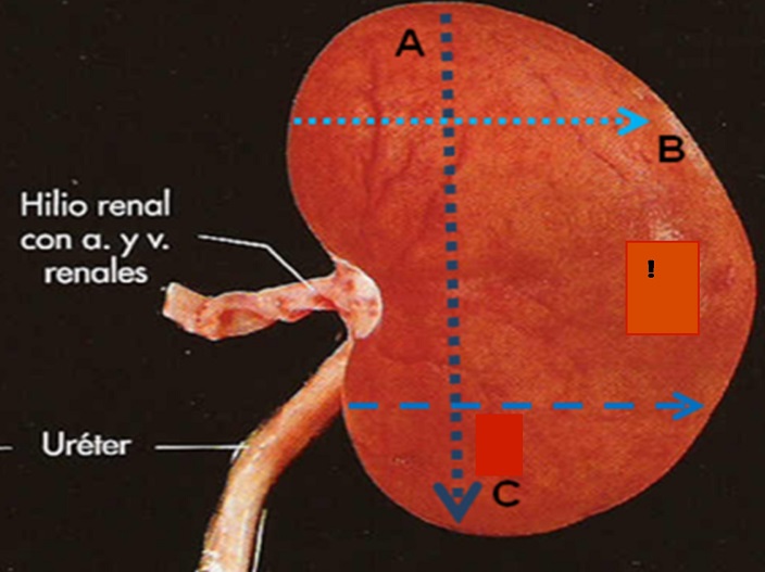
Figure 1. Kidney measurement.
Plastination
All specimens were dehydrated in cold (-20ºC) 100% acetone (freeze substitution). Each third day acetone concentration was measured with an acetonometer. When acetone percent was stable, the specimens were placed in new acetone. Four to six changes of acetone were carried out over an eight week period. The specimens were impregnated using the traditional S10 silicone (BiodurTM) and S3 catalyst (BiodurTM) in a proportion of 100:1 (deJong and Henry, 2007). Pressure decrease was regulated in the vacuum chamber by incremental closure of the valve until bubble formation ceased and pressure had been lowered nearly one atmosphere. The impregnation process took four weeks. The impregnated specimens were removed from the silicone impregnation reaction-mixture. One half of the impregnated specimens (10 lungs and 10 kidneys) were placed in disposable bags and stored for two weeks at 4º C. A color reactivation-mixture was prepared for the other one half of the impregnated specimens by preparing a saturated solution of imidazole/ethanol using a ratio of 1:3. One part of the imidazole-mix was mixed with 100 parts of the classic reaction-mixture and placed in a stainless steel container inside the cold vacuum chamber. The remaining half of impregnated specimens (10 lungs and 10 kidneys) was submerged in the silicone/catalyst/imidazole reaction-mixture in the vacuum chamber in the freezer. Initially pressure was lowered rapidly to 20mmHg (-20º C) and then incrementally lowered to the end point of 5mmHg over a four week period.
Subsequently, all the organs impregnated with silicone/catalyst or with silicone/catalyst plus imidazole were removed from the refrigerator and vacuum chamber, respectively, and allowed to drain at room temperature. The specimens were wiped of excess impregnation-mixture with paper towels and adjusted to proper anatomical position. After draining, the specimens were placed in a closed container and saturated with SH06 gas cure (BiodurTM) using a continuously running aquarium pump. The curing of specimens was complete between 1 and 4 weeks. The cured lungs and kidneys were photographed and measurements of organs were recorded. The black background of the pictures was changed to white so the image analyzer program could measure the color changes of the organ. Each one of the images of the lungs and kidneys plastinated with and without imidazole were analyzed to evaluate the color, saturation and hue using the program Image Pro Plus, version 4.2®. In order to evaluate the significance of color preservation and the degree of shrinkage of the organs, the Student t test was used from the program SPSS 10 for Windows.
It was observed that lungs and kidneys with imidazole displayed a reddish coloration and pathology was more easily differentiated (Figs. 2-7).
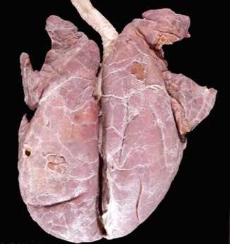 Figure 2. Plastinated porcine lung with imidazole |
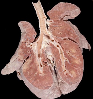 Figure 4. Plastinated porcine lung with imidazole. |
 Figure 6. Plastinated canine viscera with imidazole. |
 Figure 3. Plastinated porcine lung with imidazole. |
 Figure 5. Plastinated porcine lung with imidazole. |
 Figure 7. Plastinated canine kidney with imidazole. |
 Figure 8. Plastinated canine kidney without imidazole. |
Dysplastic kidneys showed a marked increase in red coloration (Fig. 7). Lungs with edema did not show much color differentiation. Specimens plastinated without the addition of imidizole demonstrated the classic bleaching of natural colors (Figs. 8-11). Nevertheless, no significant differences (P>0.05) were observed in the values of saturation and hue of color between plastination S10 and the plastination S10/imidazole in lungs and kidneys (Tables 1, 2). The length/width measurements from lung and kidneys, before and after, with plastination reaction-mixtures showed no significant difference in the degree of shrinkage (P> 0.05) (Tables 3, 4). The percentage of shrinkage in lungs and kidneys for both techniques was <3% (P>0.05). Specimens exposed to 4 weeks of S6 (gas cure) were firmer than those with shorter exposure.
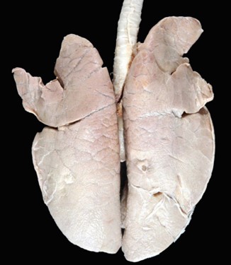 Figure 9. Plastinated porcine lung without imidazole. |
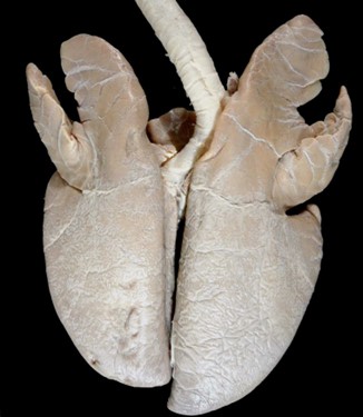 Figure 10. Plastinated porcine lung without imidazole. |
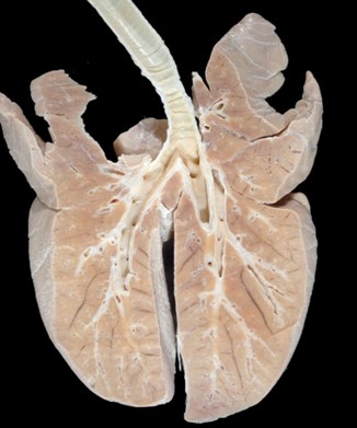 Figure 11. Plastinated porcine lung without imidazole. |
| Item | Measurements | Mean | SE | ||
| before | after | ||||
|
S10
|
Hue | 201.39 | 204.42 | 4.91 | |
| Saturation | 39.96 | 44.33 | 5.76 | ||
| Value | 123.71 | 116.94 | 8.95 | ||
| S10 plus
Imidazole
|
Hue | 199.52 | 209.56 | 5.81 | |
| Saturation | 44.33 | 44.49 | 10.24 | ||
| Value | 115.99 | 112.75 | 9.08 |
| Item | Measurements | Mean | SE | ||
| before | after | ||||
|
S10
|
Hue | 209.03 | 217.74 | 8.26 | |
| Saturation | 23.70 | 24.18 | 4.57 | ||
| Value | 134.34 | 149.42 | 11.40 | ||
|
S10 plus Imidazole
|
Hue | 207.23 | 216.55 | 8.78 | |
| Saturation | 32.49 | 37.29 | 7.42 | ||
| Value | 133.42 | 133.52 | 12.93 |
| Item | Measurements | Mean | SE | ||
| before cm |
After cm |
||||
| S10 | Length right lobe | 20.74 | 20.29 | 3.61 | |
| Length left lobe | 21.58 | 21.33 | 3.74 | ||
| Bifurcation of trachea to right middle lobe | 19.74 | 9.77 | 10.38 | ||
| Bifurcation of trachea to left cranial lobe | 9.66 | 9.73
|
1.60 | ||
| S10 plus imidazole |
Length right lobe | 20.09 | 20.31 | 2.56 | |
| Length left lobe | 22.61 | 22.92 | 2.63 | ||
| Bifurcation of trachea to right middle lobe | 10.22 | 10.33 | 1.36 | ||
|
Technique |
Mean | |||
|
S10 |
before cm |
after cm |
SE | |
| longitudinal axis | 6.81 | 6.76 | 0.42 | |
| cranial pole length | 3.69 | 3.66 | 0.34 | |
| caudal pole length | 3.84 | 3.75 | 0.32 | |
|
S10 plus imidazole |
||||
| longitudinal axis | 5.50 | 5.59 | 0.77 | |
| cranial pole length | 3.17 | 3.29 | 0.40 | |
| caudal pole length | 3.57 | 3.49 | 0.60 | |
In the present work, the lungs and kidneys plastinated using the classic S10 method preserved the characteristics of the represented pathology (Meuten, 2002; López, 2007; Newman et al., 2007). However, pathologic lesions were more evident in lungs plastinated with S10/imidazole-mix, which agrees with the work by Sakamoto et al. (2006), who used the Shin Etsu silicone polymer KE-108 with CAT-108 technique plus imidazole in one week formalin-fixed organs. Lungs with edema did not conserve the original color well, likely because the pathology (increased fluid content) was removed by dehydration. Additionally, the partial or total absence of erythrocytes in the edematous tissue block provided no hemoglobin or myoglobin for the imidazole to form complexes of hemochromogens resulting in the absence of the natural red color (Sandhyamani, 2005).
The results in both lung groups classic S10 plastination and S10 modified with imidazole showed no significant differences in shrinkage percentage which was similar to the findings of Sakamoto and co- workers (2006). Shrinkage of both lung and kidney specimens impregnated with and without imidazole was comparable to specimens treated with Shin Etsu silicone polymer KE-108 with CAT-108 plus imidazole which showed 2 to 5% shrinkage. The absence of significant shrinkage is a valued characteristic of specimens preserved by the plastination technique, especially structures as the nervous system. Impregnation is the fundamental step in the plastination process with or without substances for preservation and restoration of color. If an organ is not impregnated in its totality, it tends to shrink and acquire a dark color (Miklosová et al., 2004; Henry et al., 2006).
A natural red color was observed in kidney specimens impregnated with imidazole yet no significant difference was observed between the two different techniques. With an addition of imidazole, an organ may acquire an intense red coloration which was noted in kidneys with renal dysplasia. This intense red is not natural. It is known, that the histological composition of the organ influences the ferrohemochromogen concentration and the color (Sandhyamani, 2005).
A disadvantage associated with S10 plastinated specimens with imidazole is that over an extended period of time the surface of the organ looses color due to oxidative changes on the surface. Color changes from bright red to dark brown are likely due to surface contact with atmospheric oxygen (Sandhyamani, 2005). Another common alternative for enhancing specimen color is BiodurTM stain which is added in the last acetone bath, prior to impregnation with silicone. However, only the organ’s surface is stained with the pink color. Therefore, if the surface of the organ is damaged or removed the color is lost (Henry et al., 1997). With color reactivation, the color is restored throughout the entire specimen.
Both decreased fixation time and percentage of formalin, decrease color loss. Therefore, we chose to fix the kidneys and lungs with buffered 4% formalin for only 48 hours to aid partial preservation of natural color. Specimens dehydrated with acetone to >98% and with temperature control (-20ºC), permitted an adequate interchange of acetone with the silicone reaction- mixture under appropriate vacuum. These properly impregnated pieces did not suffer changes during the curing as shown by Miklosová et al. (2004).
Flexibility and hardness of the specimens obtained from this plastination process were influenced by the time of contact with the cure gas agent and
polymerization of the silicone chains. Longer contact with the gas cure produced harder specimens.
It will be beneficial to continue experimentation with different techniques of plastination of pathological samples because of the great variety of lesions presented in domestic animals which may significantly change its original histological composition and color.
The results of the present study suggest that the technique of plastination with addition of imidazole in organs with pathology is a good option, because it aids visualization of the pathology. Specimen shrinkage is similar to that of traditional plastination methods. Better results are obtained in compact organs like the kidney. It is recommended not to excessively wash the organs, to help retain the blood present in the organ which reacts with imidazole.
Acknowledgements
This work was supported by PAPIME-UNAM (EN212504).
Bickley HC, von Hagen G, Townsend FM. 1981: An improved method for the preservation of teaching specimens. Arch Pathol Lab Med 105:674-676.
DeJong K, Henry RW. 2007: BiodurTM S10/S15 technique and products. J Int Soc Plastination 22:2-14. Henry RW, Janick L Henry C. 1997: Specimen preparation for silicone plastination. J Int Soc Plastination 12(1):13-17.
https://doi.org/10.56507/ZLMJ7068
Henry RW. 2006: Polyester plastination technique: Specific troubles and problems. Abstract presented at The 13th International Conference on Plastination - Vienna, Austria, July 2 to 7, 2006. J Int Soc Plastination. 21:31.
Hermes B. 2006: Plastination an additional tool to teach anatomy. Int J Morphol. 24:475-480.
Latorre MR, García SM, Moreno M, Hernández F. 2007: How Use full is plastination in Learning Anatomy? JVME 34:176-180.
https://doi.org/10.3138/jvme.34.2.172
López A. Respiratory system. 2007: In: McGavin MD, Zachary JF, editors. Pathology basis of veterinary diseases. 4th ed. Mosby Elsevier: St. Louis, p 463-558. Meuten DJ. 2002: Tumors of the urinary system. In: Meuten DJ, editor. Tumors in domestic animals. 4th ed. Iowa State Press: Iowa, p 547-574.
Miklossova M, Micklos Vojtech. 2004: Plastination with silicone Method S10- Monitoring and analysis causes of failure. Biomed Papers 148:237-238.
https://doi.org/10.5507/bp.2004.048
Newman SJ, Confer AW, Panciera RJ. 2007: Urinary system. In: McGavin MD, Zachary JF, editors. Pathology basis of veterinary diseases. 4th ed. Mosby Elsevier: St. Louis, p 613-692.
Sakamoto Y, Miyake Y, Kanahara K, Kajita K, Ueki H. 2006: Chemically reactivated plastination with Shin- Etsu silicone KE-108. J Int Soc Plastination 21: 11-16.
https://doi.org/10.56507/BSRA2644
Sandhyamani S, Sindhu JK, Sriramachari S. 2005 Recolorization of museum specimens: A modification of Romahanyi's technique based on pyridine/nicotine hemochromogen reactions. Virchow's Arch 447:94-98.
https://doi.org/10.1007/s00428-005-1273-8
von Hagens G, Tiedemann K, Kriz W. 1987: The current potential of plastination. Anat Embryol 175(4):411-421.
https://doi.org/10.1007/BF00309677
von Hagens G. 1986: Heidelberg Plastination folder. Collection of all technical leaflets for plastination. Anatomish Institut I, Universitat, 2nd ed. Heildelberg, Germany.