1Department of Anatomy, School of Veterinary Medicine, University of Sarajevo, Sarajevo, Bosnia Hercegovina.
2Department of Comparative Medicine, College of Veterinary Medicine, The University of Tennessee, Knoxville, TN, USA.
Desiccated organs used for teaching purposes are susceptible to insect damage as well as damage due to improper handling. Injection of expanding foam products into the lumen of desiccated organs followed by varnishing produces anatomical specimens resistant to insect damage and more resilient to the strains of handling. This study will describe the production of such specimens.
R. W. Henry: Department of Comparative Medicine, College of Veterinary Medicine, The University of Tennessee, Knoxville, TN, USA. Telephone: 845 974 5822; Fax: 845 974 5640; E-mail: rhenry @utk.edu

![]()


For thousands of years, desiccation of biological tissue has been a useful and inexpensive means of specimen preservation (Kitchel et al., 1961; Strub and Frederick, 1 967; Church , 1 968). Plastination, though well known for its unique preservation qualities of anatomical specimens (von Hagens et al., 1987; Nicaise, 1 990; Weiglein, 1996; Latorre et al, 2001 , 2002) is still more costly than desiccation in many regions today . However, insects have a predilection for consuming organs dried in such a manner. A simple mechanism to prevent infestation of desiccated organs with insects thus preventing organ destruction would serve to greatly increase the longevity and usefulness of desiccated specimens. This study will describe a mechanism of organ preservation and protection .
Hollow organs were collected from animal cadavers for use in this study. The stomach , large colon and descending colon were harvested from a horse. Stomachs, lungs and female reproductive tracts were removed from a cow, sheep and dog. The organs were flushed with tap water until free of ingests . The adipose tissue, omenta and mesentery were removed close to the organ using caution not to damage the outer muscle layer. Once clean, the organ was prepared for classic air drying by cannulation of both ports with appropriate sized tubing and hooked to a laborato1y air source. The organ was first dilated to the desired degree of inflation . Air flow and hence organ size was either controlled by adjusting the inflow and/or the exhaust port by either occlusion or throttling by partial closure. Depending on the size of the organ, drying takes three to four days. The stomach (monogastric and ruminant) , small and large intestines, lungs and uterus and vagina have been preserved by this method. The second phase commences after drying is completed and consists of gradual injection of the plastic expanding foam, [Tekapur (Bosnia-Hercegovina) or Great Stuff ( USA)]. It was beneficial to have an exhaust in addition to the inflow. If too much loss of foam occurs via the exhaust, it can be decreased in size or closed. The next day, more foam may be injected through one of the ports to fill areas that are devoid of foam to assure complete lining and filling of the organ. The hardening time is eight hours to one or two days depending on the volume of the organ. Varnish was sprayed and brushed onto the external surface of the organs. As well, region s of the organ or the entire organ may be painted.
Cleaning and air drying the organs resulted in a desiccated specimen representative of those occurring in situ. (Fig. I ) The surfaces of the desiccated organs were dry to the touch and free of any greasy residue. The injected plastic expanded 2 to 4 times in volume in all directions and hardened gradually. Areas of the organs which par1ially collapsed following disconnection from the air source were inflated with the expanding foam . The hardening time for the injected foam is eight hours to two days depending on the volume of the organ . The resulting dried, foam filled organs were light weight and anatomically precise (Fig. 2). Application of the varnish to the outer surface of the organs was accomplished without the production of a runny, streaked appearance. Paint was applied with a brush to highlight anatomical information (Fig. 3).
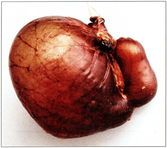 Figure I. Visceral view of canine air dried stomach . Cannula is in the esophagus. |
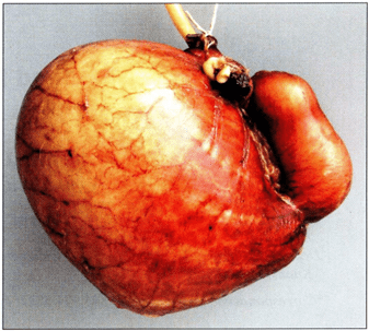 Figure 2. Visceral view of foam filled canine airdried stomach. Note fat on minor curvature that must be trimmed off |
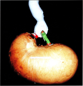 Figure 3. Parietal view of foam filled, varnished and regionally painted equine air dried stomach. |
A i r-drying of organs has been used for anatomical applications for many years (Mc Kiernan and Kneller, 1983; Henry, 1 992). However, filling air dried organs with expandable foam protects the otherwise vulnerable
inside of the organ from insect damage while the app l i cation of varnish does the same for the external
surface. The foam also protects the organ from collapsing under normal hand l i ng conditions. Various methods and products have been used to make a i r dried organs resistant to insect damage including fiberglass (Kitchel et a l. , 196 1 ), flexible plastic resin (Updike and Holladay, 1 986) and silicone (Henry and Butler, 1990). During filling it is beneficial to allow air to exhaust during inflation of organs. Continual flow of air through the specimen allows the organ to dry quicker. lf the muscular wall of the organ is cut, a herniation or blow out of the mucosa may result from inflation without an exhaust port. If too much foam is lost through the exhaust port during injection , the portal may simply be closed. It is possible this expanding foam could also be used to dilate silicone impregnated hollow organs prior to polymerization in the plastination process. It is imperative to remove all adipose tissue from the specimen prior to desiccation . The failure to do so results in greasy specimens to which the application of varnish is troublesome. The foam products tested are unstable when exposed to acetone. Acetone dissolves the foam and reduces it to a sticky substance. This would preclude the use of the foam prior to impregnation of specimens during plastination during which step the acetone is removed from the specimen. Alcohol saturates the foam but does not dissolve it which opens the possibility of its use in plastination when alcohol i s the intermediary solvent. The external surface is protected from insect damage by varnishing. Once varnished, the external surface of the organ may also be painted or labeled for demonstration of information . This method produces specimens that maintain normal anatomical form, are durable and inexpensive to produce.
Church DC. 1 968: A simple method for preserving the ruminant stomach . J Anim Sci 27: 1525-1526.
https://doi.org/10.2527/jas1968.2761525x
Henry RW , Butler J. 1 990 : Room-temperature "forced air" impregnation of dried lung with S 10/ S3-xylene mix . J Int Soc Plastination 4 (1) : 14-15.
https://doi.org/10.56507/KTGZ7837
Kitchel RL, Turnbull J , Nordine RA, Edgell SC. 1961 : Fiberglass technique of preparation of natural models of the ruminant stomach . J Am Vet Med Assoc 138:329 -331 .
Latorre R, Vaquez J M , G i l F, Ramirez G, L6pez-Albors 0, Orenes M, Martinez-Gomariz F, Arencibia A. 2001 : Teaching anatomy of the distal equine thoracic limb with plastinated slices. J Int Soc Plastination 16:23-30.
https://doi.org/10.56507/ACRF7155
Latorre R, Vaquez J M , Gil F, Ramirez G, Lopez-Albors O, Ayala M, Arencibia A. 2002 : Anatomy of the equine tarsus: A study by M RI and macroscopic plastinated section s (S 10 and P40). Abstract presented at The 11th International Conference on Plastination, San Juan, Puerto Rico, July 14-19, 2002. J Int Soc Plastination 17:6.
Mc Kiernan BC, Kneller SK . 1983: A simple method for the preparation of inflated air-dried lung specimens. Vet Rad 24(2):58 -62 .
https://doi.org/10.1111/j.1740-8261.1983.tb01539.x
Nicaise M, Simoens P, Lauwers H . 1 990: Plastination of organs: A unique technique for preparation of illustrative demonstration specimens. Vlaams Diergeneeskd Tijdschr ( Flemish Veterinary Journal) 59: 141 -146.
Strub CG, Frederick LG . 1 967 : The principles and practice of embalming. Dallas: L.G. Frederick. Updike SJ , Holladay SD. 1 986 : Preparation of flexible models of hollow organs. Anat Rec 216:207-210.
https://doi.org/10.1002/ar.1092160213
von Hagens G, Tiedemann K, Kriz W. 1987: The current potential of plastination. Anat Embryol 175(4):411 -421.
https://doi.org/10.1007/BF00309677
Weiglein AH. 1996: Preparing and using S-10 and P-35 brain slices. J int Soc Plastination 10: 22-25.
https://doi.org/10.56507/IXGV4189