Department of Anatomy, Universidade Federal do Rio de Janeiro, Rio de Janeiro, RJ, Brazil
The intent of this report is to relate the positive experience of the Department of Anatomy at Universidade Federal do Rio de Janeiro (UFRJ) in storing and making readily available large numbers of human plastinated specimens. The technique has allowed improved specimen utilization by the both professors and students representing several diverse biomedical graduate courses. A detailed description is provided of the cataloguing and storage system, which could be readily implemented by other Departments that deal with large numbers of plastinated specimens and/or whose specimens are regularly requested.
Storing, Labelling, Equipment
Rafael Augusto Dantas Prinz, Rua Gurupi, 105 Apartamento 301 - Grajau Rio de Janeiro - RJ, Brazil, CEP: 20561-100. Tel: 55 021 578 1000 / Fax: 55 021 290 0587. Email: prinz@netrio.com.br

![]()


The constantly increasing number of plastinated specimens being produced by numerous institutions worldwide has resulted in increased need for efficient methods of specimen organization (Whitten et al., 1991). Such methods must permit specific specimens within the teaching collection to be readily located and made available for teaching and research related purposes.
The Department of Anatomy at UFRJ has used specimens plastinated by the standard S10 procedure (von Hagens, 1985) since 1994, supporting a relatively large number of biomedical courses (Medicine, Dentistry, Nutrition, Nursery, Phonoaudiology, Physiotherapy, Physical Education, Psychology, Pharmacy) which together enroll over 1000 students. To effectively manage such large-scale use of these plastinated specimens, it was determined that an improved system of organization needed to be developed. This report presents that system.
Dissection and plastination of Specimens
All specimens related to the aims of this report were prepared using standard sectioning and/or dissection method. They were then dehydrated, impregnated according to the standard S 10 technique (von Hagens, 1985) and fast cured (Weiglein and Henry, 1993).
Cataloguing of Plastinated Specimens
| SYSTEM | CODE |
| Articular Cardiac Circulatory Digestive Endocrine Muscular Nervous Osseous Reproductive Respiratory Urinary | Ar Cd, Cc Dg Ed Mu Ne, Os Rp Rs Ur |
An initial-based code based on body system or body segment was developed (tables 1a and 1b). The specimen was considered as part of a system if it showed only or mainly structures of that system. Specimen which demonstrated structures representing several systems were catalogued as belonging to a body segment (table 2). The letter and number code was then applied to all specimens which had been previously produced (table 3), and new specimens were catalogued accordingly. For specimens composed of numerous parts (i.e., slices), the code was expanded to accommodate these slices (table 4). Specifically, sections were numbered from upper to lower body slices for transverse sections; from anterior to posterior for coronal sections and from right to left for sagittal sections (table 5).
Creating a Data Storage Bank
Using Microsoft Access™ software for Windows 95™ version 7.0 (trademark of Microsoft Corporation) the following data fields were created: Specimen code, description of the specimen (a brief notation of the anatomical structures present), an image of the specimen (scanned on a Hewlett Packard ScanJet 4c), date of specimen completion, specimen condition (available, in exhibit, in repair) and the person responsible for the dissection. The software also displayed the total number of specimens present in the data storage bank (figure 1). Data for any given specimen can be printed by users who are planning teaching and other specimen use. A notebook containing print-outs of all specimens contained in the data bank is maintained to further facilitate these activities
| BODY SEGMENT | CODE |
| Abdomen | Ab |
| Arm | Am |
| Elbow | Eb |
| Foot | Fo |
| Forearm | Fa |
| Hand | Ha |
| Head | He |
| Hip | Hi |
| Knee | Kn |
| Leg | Le |
| Neck | Nk |
| Pelvis | Pv |
| Shoulder | Sh |
| Thigh | Th |
| Thorax | To |
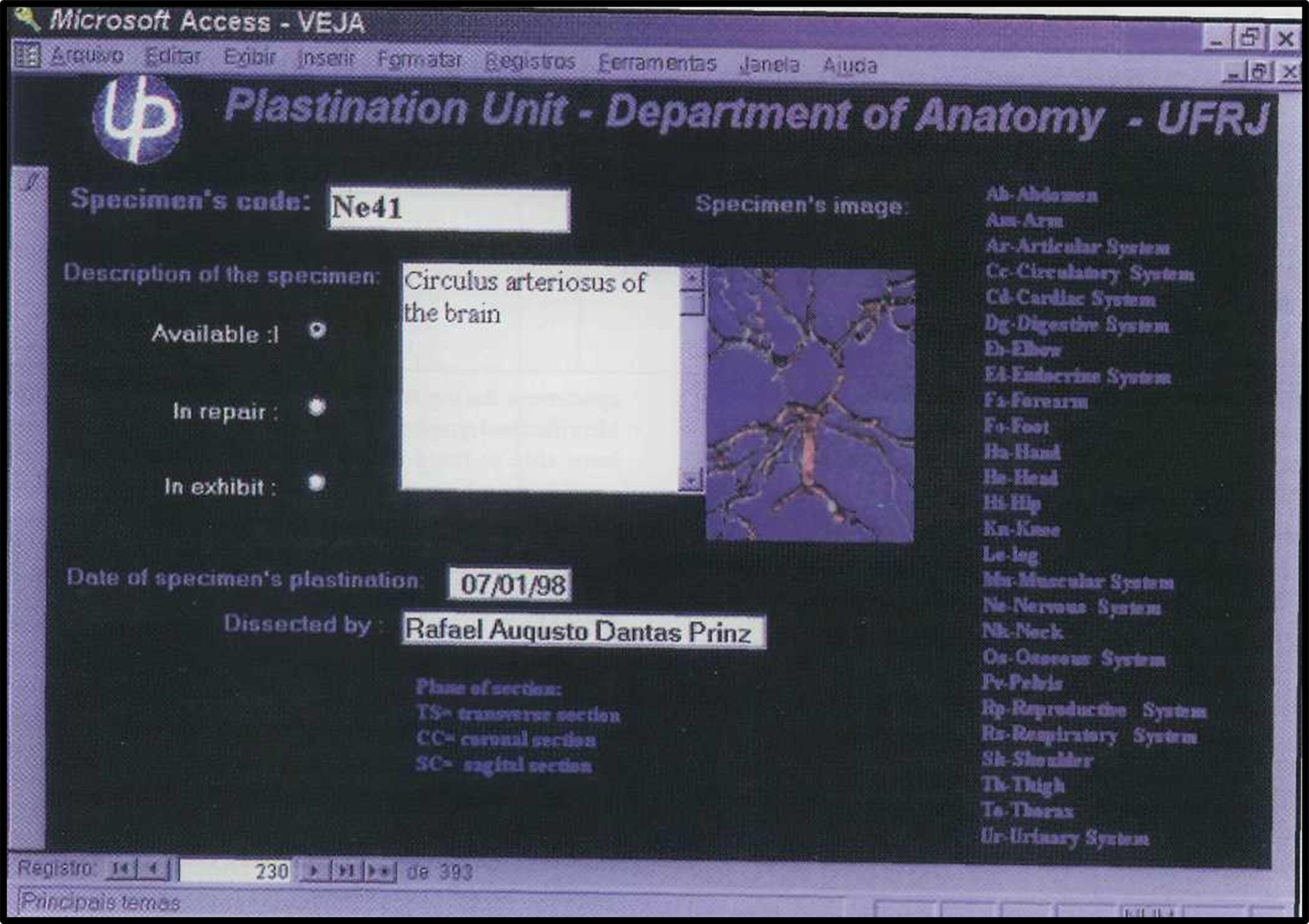
Figure 1. Computer screen showing the data of one specimen from the data storage bank.
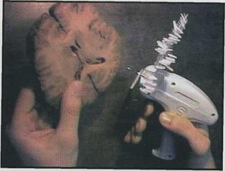
Figure 2. The tagging and labelling gun was used to
Figure 3. Example of one label. attach the labels to the specimens.
Labelling
Based on Jackson (1987), the established codes were printed on 210 x 297mm blank labels in two vertical columns, using the Microsoft Word™ software for Windows 95™ version 7.0. Width of each column was limited to 65 characters with nine codes per column (eighteen codes per sheet of paper). The page was formatted at 60 (sixty) lines per page and the labels separated by double lines. Labels were cut with scissors to 9,0 x 3,0 cm in size and both sides of the labels were manually covered with adhesive plastic. A tagging and labelling gun was used to attach the label to the plastinated specimen (figure 2). The hole was punched in the upper left side of the label and the plastic holder was then attached to a resistant part of the specimen.
Each label displays the name of the institution, department name, specimen code and name of the person responsible for the dissection of the specimen (for a professional appearance the symbol of the Plastination Unit of the Department is included on the label) (figure 3).
After a specimen is properly labeled and entered into the data storage bank, the specimen is stored within the Plastinated Specimens Facility of the Department of Anatomy. This facility consist of wooden shelves in a low- humidity environment. Shelves (56 x 45 x 2 cm) are arranged according to specimen type and have attached stickers giving the code of the specimen that is to be located in a given region on a specific shelf (figure 4).
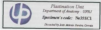 Figure 3. Example of one label. |
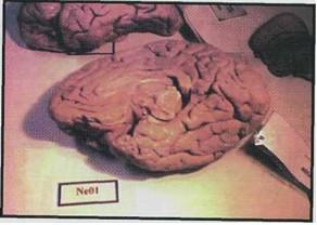 Figure 4. On each shelf we attached a sticker with the specimen's code so that we can easily find them when requested. |
Plastinated specimens have been easier to locate and manage using the described system. Professors requesting specimens typically consult the specimens notebook and then fill out a form making the formal request. Information requested on the form includes the date of the class, borrower name, requesting date (faculty are asked to make requests at least one week in advance), specimen code, and course the specimen will be used in. This provides a permanent record of specimens use. The requested plastinated specimens are taken from the storage room for use, and, after use, returned to the proper storage location by a person responsible of the plastinated specimens. Before storage, specimens are checked for damage and necessary repairs are made.
| SPECIMEN DESCRIPTION | CLASSIFICATION | CODE |
| Left cerebral hemisphere | Nervous System | Ne |
| Horizontal section of a trunk revealing the lungs, the heart, thoracic muscles, thoracic vertebrae, aorta and esophagus | Thorax | To |
| DESCRIPTION OF THE SPECIMEN | SYSTEM | BODY SEGMENT | NUMBERS OF SPECIMENS ALREADY CATALOGUED | FINAL CODE |
| Left cerebral hemisphere | Nervous | ----------- | 30 | Ne 31 |
| Horizontal section of a trunk revealing the lungs, the heart, thoracic muscles, thoracic vertebrae, aorta and esophagus | --------- | Thorax | 15 | To l6 |
| PLANE OF SECTION | CODE |
| Transverse section | TS |
| Coronal section | CS |
| Sagittal section or midsagittal section | ss |
| DESCRIPTION OF THE SPECIMEN | SEQUENCIAL NUMBER OF THE SLICE | FINAL CODE |
| The upper slice of a thorax revealing the lungs, the heart, thoracic muscles, thoracic vertebrae, aorta and esophagus | First | T0I6TSI |
| The second slice of abdomen from the anterior to the posterior region | Second | AblOCS2 |
| The fifth sagittal brain slice from right to left | Fifth | Ne31SS5 |
To date, 436 plastinated specimens have been successfully coded and stored using this system (first implemented in 1995). We have full control of all the specimens available in our institution as well as an easy way to locate and separate them for teaching. All the damaged specimens during these 3 years have also been reliably identified and repaired. Professors also indicate that they have been able to more easily find (in the data bank) those specimens which will enhance their planned teaching. Labeled specimens acquire a more professional appearance which may encourage careful use. Finally, identifying the student or preparator who performed the dissection encourages that individual to work carefully and improve his/her dissection skills.
No specimens were lost.
Acknowledgement
The authors wish to thank Mrs. Susanne Queiroz for her help with this project and everything else that is related to plastination at Rio de Janeiro.
Jackson RL: Durable labels for plastinated specimens. J Int Soc Plastination 1 (2): 9-11, 1987.
https://doi.org/10.56507/DMJV3080
von Hagens G: Heidelberg Plastination Folder. Anatomisches Institut, Universitat Heidelberg, Heidelberg, Germany, 1985.
Weiglein A, Henry RW: Curing (Hardening, Polymerization) of the polymer - Biodur S 10. J Int Soc Plastination 7(1): 32-35, 1993. https://doi.org/10.56507/ABNZ7085
Whitten D, Stamer M, Johnson J: An efficient method of storing and studying a cross sectioned, plastinated cadaver. J Int Soc Plastination 5 (1): 23-25, 1991. https://doi.org/10.56507/VRZZ9275