University of Rome "La Sapienze" Rome, Italy
The purpose of this study was to verify the structural changes of biological specimens due to plastination technique.
We plastinated some specimens with the standard technique, and then deplastinated diem. Then we processed them for L.M. technique and observed them to assess the tissue integrity.
First of all, it was necessary to isolate the best deplastinating substance. For this study we immersed eight plastinated specimens into the most frequently used substances for deplastination. We chose the best solution to remove the resin and alcohol methylbenzene. For study, we used Winstar rat organs: lung, heart, liver spleen, kidney, muscle, stomach and colon. Each organ was associated with its own control. Rat control organs were immediately processed for L.M. The other specimens, were plastinated with the standard technique of plastination, (S-10).
Finally, the plastinated specimens were immersed in the deplastination solution to remove the resin. The deplastinated organs were processed for L.M. microscopic study and showed the substantial preservation of organ structures. Nevertheless, changes in the structure of lymphatic and epithelial tissues were visible. From the results we can answer the basic question of our research: does plastination and its successive deplastination slightly modify the structure of the treated tissues.
Plastination; Deplastination; Histology
Ripani,M University of Rome "La Sapienze" Rome, Italy

![]()


Plastination is a technique that allows preservation of anatomical and surgical specimens for a long time without sur- face morphological modification. It can be effectively used for teaching Macroscopic Anatomy, and is also useful for the radiologist's understanding of the TAC-RMN images. After experience with this technique over several years, we often wondered what the results would be of histological studies of plastinated samples. We found different options in the literature. Some experts think that tissue, once plastinated, cannot be restored to its original condition. During the treatment it passed through structural changes while other experts, think the tissue remains unchanged. We therefore decided to study plastinated organs as further detailed. The purpose of our study was to ascertain whether plastination, followed by deplastination, affects structural modifications on biologically treated specimens. At the same time, we tried to locate the most suitable substance to use in deplastination, granting the alteration of the morphological structures of these organs.
In this study we used five Wistar rats weighing 250 grams each, of both male/female. Each rat was anaesthetized with ethylic ether and oxygen, and was submitted to a median sagittal thoracotomy followed by laparotomy. The rat was sub- mitted to perfusion through a special needle inserted in the left ventricle. The right atrium was opened. The perfusion was made with a blend of heparin and 10% of formalin slowly drip- ping (lasting five minutes). Then, samples of the following organs were taken: lung, heart, liver, spleen, kidney, muscle, stomach, colon. They were processed both for L. M. (light microscopy) and plastination.
Dehydration was the first step of plastination: each organ being dipped in acetone at -25°C. Because of the small size of the specimens, this step took only 48 hours. The following step was the so-called forced impregnation in a solution made up of resin and hardener (Biodur S-10 + Hardener S-3). In this phase, the escape of a small amount of bubbles from the surface of the specimens was noticed, probably due to the small dimension of the specimens. Forced impregnation at room temperature lasted 72 hours. Finally, the cure was done slowly using KOH over one week. The specimens were then submitted to the process of deplastination.
For this purpose, it was necessary to to identify the most effective substance to be used as a resin solvent. The following substances are more commonly used: alcohol, methylbenzene, methylene, bichloride acetone. Eight plastinated specimens of human ureter were used in order to carry out our research; they were submitted to transverse dissection; each specimen was 0.5 cm thick. Each sample was immersed into the various substances for the deplastination. The immersions lasted from 24 to 168 hours. From the tests, we found that the best method to obtain good results in the phase of deplastination, was first to immerse the specimens in 99% alcohol for 24 hours, and afterwards in methylbenzene for 48 hours. It is necessary to remove all the resin from the specimens before proceeding to the next phase.
The deplastinated specimens were processed for L.M. through the standard technique, i.e. using hematoxyilin and eosin stain. Then the specimens were observed with the light microscope.
Through L.M. exam of both deplastinated and control specimens, different findings were made for each organ. Deplastinated kidney showed lesions in the renal tubule with lesions of the coating epithelium of tortuous tubules of the first and second order. The cortex corticis was less seriously damaged. Lesions of the gomeruli were recognized in Bowman's capsule, as well as enlargement of the capsular space. Other glomeruli were well preserved (Figure 1a-1b).
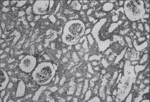 Figure 1a - Kidney: Control Specimen (L.M. 200X) |
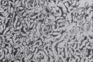 Figure 1b- Kidney: Deplastinated Specimen (L.M. 200X) |
Microscopic examinations of the liver and hepatocytes showed extensive vacuolation in the central parts. In peripheral portions, disappearance of the cytoplasm, loss of the trabecular structure in the Kieman's laminae and loss of the endothelium of the centrilobular vein was noted. The biliary epithelium was well preserved (Figure 2a-2b).
 Figure 2a - Liver: Control Specimen (L.M. 200X) |
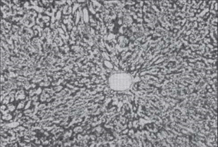 Figure 2b - Liver: Deplastinated Specimen (L.M. 200X) |
In cardiac structures the greatest lesions were found in the central areas of the specimens; in peripheral parts, it was quite well preserved. The fibre diameter became smaller, and their threadlike structure was damaged. Lesions of the cytoplasm of the common myocardial cells was also noticed.
In the spleen, a vacuolation of the parenchyma cells of the white and red pulp, (only of the central part of the pulp), was observed. The stroma connective tissue and the red blood cells were well preserved.
Skeletal muscle, parallel bundles of multinucleate fibers was well preserved; however the fibers showed loss of their original volume as well as segmentation and fragmentation, in the central portion. The cytoplasm was hyperchromatic, and the nucleus showed an accumulation of chromatin.
The lung was less severely altered than other organs. A cell coarctation and a loss of volume of the epithelium of the bronchi was observed. In addition, the chondrocytes in the cartilage of the coating plates of some bronchial branches showed vacuolation. (Figure 3-a-3b).
 Figure 3a - Lung: Control Specimen (L.M. 200X) |
 Figure 3b - Lung: Deplastinated Specimen (L.M. 200X) |
The structure of the entire stomach wall was well preserved. Volume and stain changed, in the underlying layers of the lamina propria and in the inner connective tissue of the mucous membrane. The structure of the colon was well pre- served (Figure 4a-4b).
 Figure 4a - Stomach: Control Specimen (L.M. 200X) |
 Figure 4b- Stomach: Deplastinated Specimen (L.M. 200X) |
Changes of volume (coarctation) in the cells of the loose connective tissue, of lymphoid tissue and of the cells of the epithelium infiltration were found.
From the comparisons among deplastinated and control specimens studied at the light microscope, we found out that plastinated organs maintain their separate histological and morphological characteristics. However, we have found some changes, in the structure of the fragments central zones, mainly in the epithelial and lymphoid tissues. In the specimens of deplastinated organs, these tissues presented some vacuolation and regressive changes. In other words, all the cells with changes of their small living matter during the process of plastination are those mostly damaged. We also noticed that deplastinated specimens were resistant to paraffin infiltration and we believe this is due to the induced changes on the organic structure by dehydration and by forced impregnation of biological samples, as stated in international literature. Finally, we concluded that the best substance to use in the phase of deplastination is a mixture of alcohol and methylbenzene.
From the results, we can confirm that plastination and successive deplastination modify the structure of the treated organs; and that plastination of a sample doesn't prevent other histological studies, within the above mentioned limits and interpretative caution.
Bickley, C.; Donner, R.S.; Walker, A.N.; Jackson, R.L.,1987 "Preservation of tissue by silicone rubber impregnation." J. Int. Soc. Plastination 1(1): 30-38.
https://doi.org/10.56507/XVDP9663
Bickley, C.; von Hagens,G.; Townsend, P.M., 1981. "An improved method for reservation of teaching specimens". Arch. Pathol. Lab. Med. 105: 674-676.
Gumr, ; Muller, A.; Anton, H.W.; von Hagens, G.; Bickley, H.C.. 1987. "Complete examination of mastectomy specimen using sheet plastination with epoxy resin." J. Int. Soc. Plastination 1(1): 23-29.
https://doi.org/10.56507/NXYR1705
Muller, ; Gumr,A.; Leucht,W.; von Hagens,G..1989. "Multicentricity of breast cancer. Results of a study using sheet plastination of mastectomy specimens." J. Int. Soc. Plastination 3(1): 8-14.
https://doi.org/10.56507/ZXSH4629
von Hagens, ; Tiedeman, K.; Kriz, W.. 1987. "The current potential of plastination". Anat. Embryol. 175:411-421.
https://doi.org/10.1007/BF00309677
Walker, N.; Jackson,R.L.; Powell,S., "Communicazione tecnica: Microscopia standard di tessuti deplastinati."