1Department of Animal Anatomy, Histology and Pathomorphology, National University of Life and Environmental Sciences of Ukraine, Kiev, Ukraine
2Department of Morphology, Sumy State University, Sumy, Ukraine
3Department of Anatomy, Faculty of Medicine, Masaryk University, Brno, Czech Republic
4Department of Human Anatomy National Pirogov Memorial Medical University, Vinnytsya, Ukraine
5Department of Microbiology, National Pirogov Memorial Medical University, Vinnytsya, Ukraine
6Department of Microbiology, St. Ann University Hospital in Brno, Brno, Czech Republic
The body of N.I. Pirogov has been entombed in the Church of St. Nicolas in Vinnytsia, Ukraine, since 1881, when it was embalmed by Professor Vyvodtsev. During the re-embalming of the body of N.I. Pirogov in 2018, microbiological swabs from the body and its surroundings were taken and, among other things, skin, subcutaneous tissue, and muscle and bone tissues were taken for ultramicroscopic examination. Hyphae of fungi of the genus Penicillium citrinum were found at two sites during this examination. The occurrence of fungi can be explained by the already weakened immunity before death due to cancer, and its subsequent postmortem growth due to the non-use of formaldehyde and the placement of the body in a non-sterile and relatively humid environment of the tomb without regular control.
embalming; microbiology; mildew; Penicillium citrinum; Pirogov
Jan Frišhons, Department of Anatomy, Faculty of Medicine, Masaryk University, Kamenice 126/3, 625 00, Brno, Czech Republic, telephone: +420 54949 6418, jan.frishons@fnusa.cz

![]()


The body of Prof. N. I. Pirogov was embalmed in 1881, a few days after death. Embalming was carried out by arterial injection of a solution of thymol, ethanol, glycerin, and distilled water (Vyvodstev, 1870). It was then stored without regular inspection for 27 years in the tomb of St. Nicolas Church in Vinnytsia, Ukraine. In the 1920s, the first works on the body began to be considered. In 1940, mechanical removal of mold and fungi from the face and hands of the body was performed, and the body was treated with a solution of ethanol and thymol, with the addition of water. The hands were then treated with Vyvodtseva solution: thymol, ethanol, glycerol, and water. The coffin and tomb were treated with a disinfectant solution of salts, formaldehyde, ethanol, and thymol. The relative humidity in the tomb at the time was 65-100% (Sinelnikov, 1951). Over the years, further conservation work has followed in order to stabilize the body (Matvejchuk 2013; Hunko et al., 2019). The temperature and relative humidity have only been monitored and regulated since 1989. The temperature in the tomb is between 16 and 18 °C, and the relative humidity is around 80%.
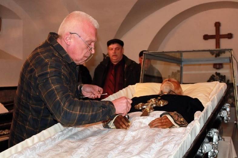 Performing microbiological smears from the body surface of Prof. N.I. Pirogov |
Before and after the 10th re-embalming of the body N.I. Pirogov in 2018, swabs were taken, and subsequent quantification of the possible occurrence of aerobic and anaerobic bacteria and fungi was carried out. Swabs were performed on the cover and on surfaces inside the sarcophagus containing the body. Immediately after opening the sarcophagus, swabs were taken from the inner surfaces of the walls of the sarcophagus, the surface of the blanket, the lapel of the uniform, the inner surface and buttons of the uniform, as well as from the scalp and beard, the skin of the right and left hands, the forehead, eyelids, nostrils, and hair from the wig, a total of 36 smears (Fig. 1). After releasing the body from the underwear, swabs were
taken from areas of skin that differed in color compared to the rest of the skin surface. These areas were located on the anterior surface of the right shoulder and right forearm, the anterior upper thigh, and the anterior surface of the left leg and foot. Abdominal swabs were taken separately from the inner surface of the anterior abdominal wall via an incision in the abdominal wall, and similarly from the anterior surface of the left tibia and anterior upper femur. A total of 48 samples were taken. The test areas were swabbed with a disposable sterile cotton swab, which had been pre-soaked in sterile isotonic sodium chloride solution. The swabs were then placed in test tubes with Amies transport medium (Sarstedt AG & Co, Germany). The collection and transport of material took place in accordance with the Decree of the Ministry of Health of Ukraine No. 234 on the organization of prevention of nosocomial infections in obstetric hospitals (10th May 2007). Quantitative determination of potential populations of each species or group of microorganisms was carried out, using Colombian agar, tryptone-soy agar (TSA), Sabouraud dextrose agar, and agar with yeast extract and glucose, according to PN ISO 7218, for selection of cultures. In addition, Schaedler-KV agar with 5% sheep blood was used to control for the presence of anaerobic microorganisms. GasPak EZ gas generating containers were used to create anaerobic conditions.
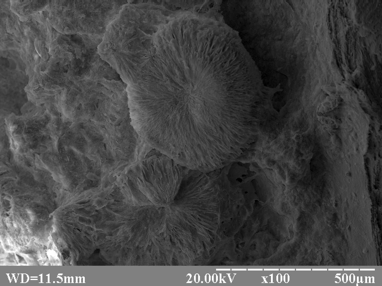 Hyphae of fungi of the genus Penicillium citrinum; REM (reflection electron microscope), magnification x 1000 |
The most microbiologically contaminated (102 colony forming units per cm2) was hair from the wig, which may be a result of the high sorption activity of hair, due to its large surface area and the laws of electrostatics. On the dense nutrient medium based on synthetic gel for microbiology cultivation, the formation of smooth colonies of a regular round shape, colored with white pigment, was observed. By combining morphological, titrimetric, biochemical, and culture characteristics in all cases, isolated bacteria were identified as coagulase-negative Staphylococcus. No microorganisms were isolated from the exposed areas of the facial skin, eyelids, hairless nostrils, and hands. The inner surfaces of the sarcophagus and clothing were not bacteriologically contaminated. Only the surface of the sarcophagus cover was contaminated with individual microbial cells of the spore-forming genus Bacillus. No bacteria were detected from the exposed surfaces in other samples. Due to the low quantitative indicators of microbial contamination, it can be assumed that their source in both cases was airborne contamination after the opening of the sarcophagus. No fungi or anaerobic bacteria were detected in any of the samples. Growth of aerobic bacteria was detected in only two samples: the anterior surface of the left tibia and the anterior upper femur. According to the combination of morphological, cultural, and biological characteristics, the isolated bacteria belonged to species that do not show proteolytic activity, from the family Micrococcaceae. Due to low quantification, these areas were likely to be accidentally contaminated during removal of clothing and other body manipulations in preparation for embalming.
Ultramicroscopically, fungal hyphae of the genus Penicillium citrinum were found without spores, around the hair follicles on the skin of the anterior abdominal wall and on the surface of the central canal of the tibial Haversian system (Fig. 2).
Because the initial embalming did not contain formaldehyde (it was first reported in 1859 by Butlerov, and not commercially available until late 19th – early 20th century), and the body was not under regular control for 45 years, the fungus had probably already formed during the disease in N.I. Pirogov’s lifetime. It is therefore probable that the body had been colonized externally and internally by the fungus Penicillium citrinum before, and at the time of, death. Subsequent gradual growth continued due to the humid, non-sterile environment of the sarcophagus and the tomb.
Penicillium citrinum is a fungus with minimal pathogenicity. It is a classic reducing organism that breaks down tissues and biological materials of plant and animal origin. It is a ubiquitous organism that occurs everywhere. Pirogov was probably in the terminal stage of oral cancer, and hence, a patient with seriously weakened immunity. It cannot be ruled out that he may have already had this fungus in him, for example in the respiratory and digestive tracts. Fragments of hyphae or spores of this fungus could have been introduced deep into the internal organs or bones in a hematogenous manner. Before his body was subjected to the embalming process and soaked in the appropriate solution, these fragments and spores could germinate in the deceased's body and grow and survive for some time. Monitoring the possible occurrence of microbial contamination of the body is important for long-term preservation of the body in good condition (Karrar Alsharif et al., 2017). Tissues preserved with formaldehyde solutions generally show no, or minimal, microbial contamination (Balta et al., 2019).
By adjusting and monitoring the relative humidity and air temperature in the tomb, hermetically sealing the sarcophagus, general cleanliness, and aseptic measures, the use of protective equipment, and clean, sterile material during re-embalming, and the appropriate composition of the embalming solution, it should be possible to prevent the growth of any microbiological organisms in and around the body. If a fungal colony occurs, its identification is required (Yaragalla & Rajput, 2017). After consultation with experts in the field of microbiology or mycology, and embalming specialists, careful mechanical or chemical removal of the fungus is possible, followed by re-embalming of the tissue.
Balta JY, Cryan JF, O'Mahony SM. 2019: The antimicrobial capacity of embalming solutions: a comparative study. J Appl Microbiol 126(3):764-770.
https://doi.org/10.1111/jam.14191
Hunko PM, Haidukov OV, Martynova ZS. 2019: Zabal'zamovane tіlo Pirogova jak unіkal'nij naukovij eksperiment [Long-term preserving of N. I. Pyrogov's embalmed body as a unique scientific experiment]. Naukovij vіsnik Nacіonalnogo muzeju іstorіi Ukraini. Zbіrnik naukovih prac. [Collection of scientific works of National Museum of Ukraine] 4: 625-639 [In Ukrainian].
Karrar Alsharif MH, Musthafa M, Elamin AY, Ibnouf EO, Taha KM, Alfaki MA, Nour YS, Aldosari KHM. 2017: A brief review on the principles of human cadaver preservation and monitoring of microbial degradation. Forensic Med Anat Res 5:19-31.
https://doi.org/10.4236/fmar.2017.53003
Matvejchuk, IV. 2013: Sovremennye dostizhenija v konservacii i dlitelnom sohranenii biologicheskih struktur i objektov N.I. Pirogov. [Modern achievements in the conservation and long-term preservation of biological structures and objects of NI Pirogov]. Voprosy biologicheskoj, medicinskoj i farmacevticheskoj chimii [Questions of biological, medical and pharmaceutical chemistry] 11: 143-147 [In Russian].
Sinelnikov RD. 1951: Vosstanovlenie ostankov N.I. Pirogova. Trudi naučnoj sessii, posvjašennoi 140-letneho so dnja roždenija N.I. Pirogova [Restoration of the remains of NI Pirogov. Proceedings of the scientific session dedicated to the 140th anniversary of the birth of N.I. Pirogov] 93-101 [In Russian].
Vyvodstev DI. 1870: Prostoj i populjarnyj metod bal'zamirovanija trupov bez vskrytija polostej [A simple and popular method of embalming corpses without opening the cavities]. Voenno-Meditsinskiy Zhurnal. Military Medical Journal (2): 71-79 [In Russian].
Yaragalla S, Rajput A. 2017: Identification of fungal growth from the internal organs of preserved human cadavers. Am J Microbiol Res 5 (1): 25-27.