University of Rome "La Sapienza", Rome, Italy
The goals of this study are: 1) to monitor and improve some phases of plastination in order to reduce any waste of reagents and time of processing of parenchyma! organs; 2) to avoid distortion of morphology; and 3) to enhance chromatic yield. This study was carried out using the standard S-10 technique.
Silicone; Biodur; S10; Plastination; Fixation; Dehydration
Mauricio Ripani University of Rome "La Sapienza", Rome, Italy

![]()


Using the protocol according to Von Hagen (1985) we have carried out the plastination technique using various Biodur polymers, (S-10, E- 12, P-35). Technical mistakes can give rise to specimen shrinkage of over 20%, changes in chromatic yield, changes in mechanical properties, and/or distortion of the whole sample. We closely examined the various steps of our plastination process in order to find an ideal protocol for processing a parenchymal organ, as well as, identify and rectify any specific problem areas of the procedures that could possibly endanger the best result of the S-10 process. In the different sections of this paper, we will outline the procedure which we found to produce the best results and will underline the changes made in the standard technique. Finally, we will suggest a new method of monitoring dehydration and forced impregnation.
Eleven parenchymal organs (liver, kidney and spleen) were used for this project. Fixation was performed in 3% and 6% formalin solutions (1,2) Dehydration was performed in cold acetone (-20°C) by freeze substitution (1-3). An alcoholometer (acetonometer) and an immersion thermometer were used to determine acetone purity.
Forced impregnation was performed in a vacuum chamber at room temperature with Biodur S-10 polymer mixed with the S-3 hardener. The basic protocol was followed according to current literature (1), however, the protocol was adapted to the different physical properties of the organs. Moreover, by observing our technical artifacts, mechanical features, and weight of the specimen, an enhanced procedure was developed.
ENHANCE PROCEDURE:
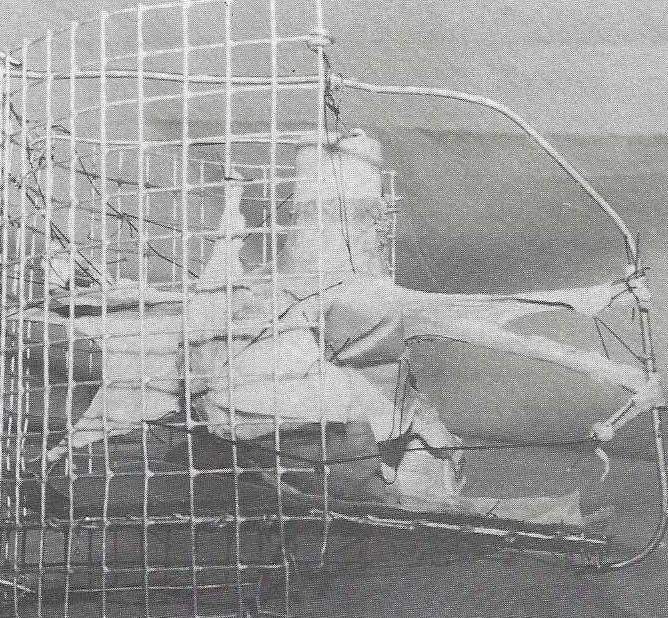
To prepare the specimen for plastination, the organ was washed in running tap water for about 24 hours. The first phase is an accurate surgical preparation of the features of the organ which are most interesting for the teaching of anatomy (e.g. hilum). Special care is taken in shape preservation. Unfortunately, most of the material we plastinate is isolated and deformable organs. To avoid any alteration of their morphological structures we cling the specimen onto a metallic grid linked to a metallic basket (1). It is necessary to make certain the specimen does not lie on the bottom of the receptacle. This enables us to move the basket into various tanks containing fixative, dehydrating, or impregnating solutions. This technique makes it easier and is harmless to the specimen. The specimen was suspended for the first three days in 3% formalin, followed by 6% formalin for 10- 15 days for organs weighing <500 g. and for 17- 22 days for organs weighing > 1000 g. Specimen weight and fixative fluid ratio was 1:5 (kg/L). The organs were fixed at +5°C to give rise to a better chromatic preservation than fixation at room temperature.
Dehydration
Dehydration was performed by freeze substitution at -20°C with specimen/acetone ratio of 1:5.
The specimen was first placed into a 97.5-98% acetone bath. From the beginning of the dehydration, between the 24th and the 48th hours the first change with pure acetone was performed. We agitated the acetone solution and controlled the acetone concentration every 2 days from the 3rd day to the 10th day for organs weighing <500 g and from the 3rd to 14th day for organs weighing >1000g. If the solution concentration was not lower than 97.75% the organs achieved the best result. Two baths are adequate only if acetone concentration in the second bath remains above 97.75%. If during the 2nd bath, the concentration falls lower than 97.75%, additional baths of 100% acetone must be used in order to keep concentration of acetone above 98%. From the 10th day to the 14th day, according to the specimen weight, should the concentration be 98% or less a new dehydration with 100% acetone should be performed for two consecutive days. When acetone concentration remains stable for a minimum of three days at least (99% - 100%), dehydration is complete. We received the best results with organs which passed through 3 or 4 baths on average. Bath duration was 11-14 days.
Forced Impregnation
Forced impregnation commenced after dehydration was complete by submerging the specimen in a mixture of Biodur S-10 and Biodur S- 3 in a vacuum chamber at room temperature. At this temperature the resin is fluid and flows in to the organ quickly. Table 1 shows the vacuum values we tried to achieve. Not increasing the vacuum for one day could be convenient to the operator and profitable for the result of plastination. From our experience it is better than interrupting the vacuum increase in an intermediate impregnating time. When absolute pressure was stabilized at 15 mm Hg for 24 hours, the specimen was considered completely impregnated. The plastinated specimen was placed in a receptacle for 28 days to let the excess polymer drip from the specimen, it was then wiped of any remaining excess polymer. Afterwards, the specimen was exposed to the action of the rapid polymerizing Biodur S-6.
| Forced Impregnation | |||||||||
| Days | 1 | 2 | 3 | 4 | 5 | 6 | 7 | 8 | 9 |
| Specimens weight <500 g | 100 | 90 | 50 | pause | 40 | 25 | 15 | ||
| Specimens weight >1000 g | 125 | 100 | 80 | 60 | pause | 50 | 30 | 20 | 15 |
| (Pressure expressed in mbar) | |||||||||
Monitoring the dehydration and forced impregnation
We carefully monitored these two phases. Because of our experience technical mistakes during dehydration and forced impregnation can give rise to artifacts which are not possible to correct, while some fixation and curing mistakes may be remedied. Dehydration time must be kept as short as possible to prevent brittleness and a decrease in the flexibility of specimens. In order to know the degree of dehydration of the specimen it is important to measure the water content of the acetone solution. However, the percentage reading of acetone purity increases with temperature. Therefore, the proper use of the alcoholometer, including temperature adjustment, is important for monitoring the acetone purity. Alcoholometers are generally calibrated to calculate the acetone concentration at a given temperature. We use the Tralle's alcoholometer which is calibrated within a
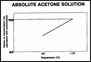
Fig.2 Linear regression by acetone concentration and temperature
range between (+10° and +30-40°C). Von Hagen (1) described the measuring of the water content of dehydrating baths by warming the acetone to the temperature for which their alcoholometer is calibrated. Warming acetone to room temperature can be time consuming. Using the following method one can quickly calculate the water content of the dehydrant while the acetone is still cold. In order to achieve this we devised a warming trough and a diagram of an average of three pure acetone solutions. Therefore, for 3 pure acetone solutions we calculated the concentration values at a temperature between -25°C and +25°C. Following this diagram the value of the pure acetone concentration (value obtained by alcoholometer) at +15°C is 100% (fig. 2). At different temperatures the calculated values of acetone concentration will be different. The difference between the real concentration and the "pure solution line" permits us to know the level of the water percentage in the dehydration solution. The impregnation can be monitored with a manometer when the pressure valve into the vacuum chamber is steady. We consider steady vacuum when release of the bubbles is very few or none. In order to allow described measurement, we calculated the pressure in the vacuum chamber, at the beginning and at end of the working day (from 8 AM to 4 PM for safety).
We suggest not exceeding the pressure valves of table N-l. Finally we can monitor the grade of an extraction from the specimen by the observation of the amount of bubbles on the impregnation solution surface. This control is easy to perform at room temperature.
According to our goals the organs we plastinated by means of this protocol achieved the good quality that we desired. The monitoring of the dehydration permitted us to know the essential minimum amount of acetone to use and reduced the impregnation time (about 10 days) (4). The use of an expensive freezer for impregnation was eliminated.
This paper may be useful for laboratories that are going to begin basic plastination or do not do a large amount of processing. Although, performing impregnation at room temperature limits the Biodur time life, on the other hand, it reduces the impregnation time. This is important if the laboratory plastinates small organs and stores the resin in a freezer at -25°C among the working intervals.
1.von Hagens, Gunther: Heidelberg Plastination Folder: Collection of all technical leaflets for April 1985.
2. Karine, Ooostrom: Fixation of tissue for Plastination: general principles. Journal of the International Society of Plastination. 1(1 ):3-9, 1987.
https://doi.org/10.56507/WLZH2223
3. Klaus, Tiedemann and Dubravka Ivic-Matiojas: Dehydration of macroscopic specimens by freeze substitution in Journal of the International Society of Plastination. 2(2):2-l4, 1987.
https://doi.org/10.56507/SCLL2742
4. von Hagens, Gunther, ; Klaus, Tiedeman; and Wilhelm Kriz: The current potential of plastination. Anatomy and Embriology. 175:411- 21, 1987.
https://doi.org/10.1007/BF00309677