1 Department of Animal Anatomy, Histology and Pathomorphology, National University of Life and Environmental Sciences of Ukraine, Kiev, 03041, Ukraine
2 Department of Morphology, Sumy State University, 40000, Ukraine
3 Department of Anatomy, Faculty of Medicine, Masaryk University, Brno, 625 00, Czech Republic
4 Department of Human Anatomy, National Pirogov Memorial Medical University, The Ministry of Healthcare of Ukraine, Vinnytsya, 21018, Ukraine
Professor Nikolay Ivanovich Pirogov (1810-1881) was an anatomist, surgeon, and scientist. He studied in Moscow and Berlin, and was one of the founders of modern surgery and aseptic procedures. He described the use of plaster for the treatment of fractures, and the use of ether as an anesthetic in combat medicine. He published a number of papers on anatomy and surgery. He died on December 5, 1881, of oral cancer. His body was embalmed by anatomist Professor David Ilyich Vyvodstev (1830-1896), and placed in a tomb in the Church of St. Nicholas in Vinnytsia, Ukraine. The first inspection of the body was performed by a commission of experts in 1927. This was followed by several re-embalmings of the body in the 1950s and 1980s by a team led by Professor Rafail Davidovich Sinelnikov (1896-1981), and several other procedures in the 1980s and 1990s by experts from Moscow's V.I. Lenina, (now VILAR: All-Russian Research Institute of Medicinal and Aromatic Plants), until 2011. In 2017, regular care of the body was taken over by Ukrainian scientists, who, in 2018, performed all tissue and fluid analyses to determine the body’s state of preservation, and subsequent re-embalmings. The results of microscopic and ultramicroscopic analysis showed some destructive changes in skin, skeletal muscle, and bone tissues. Despite these changes, however, the tissues of the body are relatively well preserved.
morphological; embalming; Pirogov; bone; skin; muscle
Jan Frišhons, Department of Anatomy, Faculty of Medicine, Masaryk University, Brno, 625 00, Czech Republic jan.frishons@fnusa.cz

![]()


Professor Nikolay Ivanovich Pirogov (1810-1881) was an anatomist, surgeon and scientist. He studied in Moscow and Berlin, and was one of the founders of modern surgery and aseptic procedures. He authored a number of papers on various anatomical and surgical topics. He described the use of a gypsum plaster cast for the treatment of fractures, and the use of ether as an anesthetic in field surgery during war. He died of oral cancer on December 5, 1881. Arterial embalming of the body of Professor Pirogov was performed by Professor David Ilyich Vyvodstev on December 9, 1881 (Vyvodstev, 1870; Hendriks, 2019). The composition of the embalming solution was most likely 5 g thymol, 45 ml ethanol, 2160 ml glycerol, and 1080 ml distilled water, although the exact composition of the embalming fluid was never published. Only a few soft tissue incisions were made to expose arteries for the purposes of perfusion. Visceral organs were not removed. His body was placed into a coffin with a glass lid in the tomb of the Church of St. Nicholas in Vinnytsia, Ukraine, without any control of temperature or relative humidity. The first inspection of the body condition was carried out by a panel of experts in 1927. In 1940, mold colonies were discovered after the coffin was opened. In 1945, the body was stabilized by the staff of the Mausoleum of V.I. Lenin; the duration of the stabilization works was 115 days. In 1956 and 1973, large-scale preservation works were carried out by the team of Professor R.D. Sinelnikov. In 1979 and 1988, the body of Professor Pirogov was transferred to the Moscow laboratory at the Mausoleum of V.I. Lenin (VILAR) for further embalming. Further re-embalming was performed by VILAR laboratory staff in 1994, 2000, 2005, and 2011. During the time when the body was in the care of the Russian experts, it was placed in a coverall with an embalming solution. Since 2017, the body has been in the care of a team of Ukrainian scientists, Professors O.P. Melnyk and Y.J. Guminskii and their colleagues (Matvejchuk, 2013; Hunko et al., 2019a, b). The composition of the re-embalming solution used in the body’s coverall is classified information owned by The National Pirogov's Estate Museum. The aim of this article is to describe the state of histological preservation of skin, muscle and bone tissues taken from the body of N.I. Pirogov 137 years after it was embalmed.
As part of the re-embalming process in 2018, skin samples were taken from the anterolateral wall of the abdomen and thigh, a sample of muscle tissue from the tibialis anterior, and a fragment of bone from the distal end of the head of the fibula. After removal, the samples were stored in the embalming solution in which the body was preserved, prior to tissue processing (Figs. 1a-c). Samples of approximately 1 mm3 were fixed in 1-2.5% glutaraldehyde solution with 0.2 M cacodylate buffer of pH 7.2 at +4 C. The samples were then washed three times (5 min/wash) in the same buffer, and additionally fixed in 1% OsO4 (osmium tetroxide) solution for 2 hours at + 2° C. Tissue samples were then dehydrated in a series of ethanol solutions of increasing concentrations (15-20 minutes each at 30, 40, 50, 70, 80, 90, 96, and 100%), and acetone. The material was placed in capsules filled with a ready-made mixture of epon (a type of epoxy) and araldite resins (araldite M: 20 ml, epon 812: 25 ml), fixed in 60 ml of DDSA sealant with DMR-30 2% catalyst (10 drops) (Sigma-Aldrich, USA). Tissue samples were placed in an oven for polymerization at 60° C for 24 hours. The ultra-microtome UMTP-6M microtome (Ukraine) was used for sectioning tissue blocks into ultrathin (40-60 nm) sections. The ultra-thin sections were then mounted on copper plates and contrasted twice. They were initially placed in a 2% uranyl acetate solution for 45 minutes and then in a lead citrate solution for 30 minutes, according to Reynolds (Reynolds, 1963). Examination of the skin samples was performed with a transmission electron microscope JEM-1230 (JEOL, Japan) with a resolution of 0.2 nm.
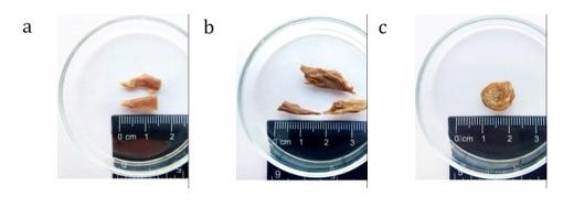 Figure 1 a) Skin samples from the anterolateral wall of the abdomen and thigh; b) a sample of muscle tissue from the tibialis anterior muscle; c) a fragment of bone tissue from the distal part of the fibula |
Samples of subcutaneous fat and muscle were fixed in 10% neutral-buffered formalin solution for two days, after which they were washed in running water for 24 h. They were then dehydrated and embedded in paraffin. An MS-2 sledge microtome was used to create 4–6 μm sections. Staining was performed using the standard hematoxylin-eosin method. The sections were observed using a light microscope Olympus BH-2 (Olympus Corporation, Japan). Histological samples were photographed with a Baumer/optronic digital camera Type: CX 05.
Before observing samples under a scanning electron microscope, they were fixed in 2.5% glutaraldehyde solution (in 0.2 M cacodylate buffer with pH = 7:2 at +4 °C for 24 hours) and postfixed in 1% OsO4 solution (for 4 hours at +4 °C). Tissue samples were then dehydrated in a series of ethanol solutions of increasing concentrations. The samples were mounted on graphite tables and air dried. Before observing the samples under a scanning electron microscope, they were sprayed with gold particles in a vacuum universal column VUP-5 and then placed in a scanning electron microscope (REM 102, Ukraine) and photographed.
Analysis of the tissues of Pirogov’s embalmed body showed destructive changes in all the analyzed samples.
Examination of the dermis revealed disruption of the spatial configuration and relationships of the fibrous elements (Fig. 2). Collagen fibers converged sharply in some places. Spaces between the fibers were narrowed or missing completely. Some solitary fibers retained their structure and showed no signs of degradation into fibrils. Deformed binding fibers were found in those inter-fibrillar bonds which were visualized well. Some of the binding fibers were strained and torn. The surface of the fibrous aggregates was rough. The surface structure did not allow determination of the inner structure of the fibrillar elements in the fibers. In the subcutaneous layer, probable fat cells were observed which were sharply deformed and compressed together.
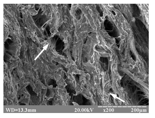 Figure 2 Dermis of the anterior abdominal wall. The spatial configuration of the fiber elements is disturbed (arrows) (scanning electron microscopy, electronic scan). |
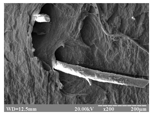 Figure 3 The surface of the skin of the anterior abdominal wall; preserved hair follicles and hair (scanning electron microscope, electronic scan) |
Preserved fragments of hair follicles were found on the skin of the anterior abdominal wall (Fig. 3). Some hair follicles were empty but had clear boundaries. The edges of the hair shaft were broken off. In general, the structure of the hair was preserved, however, some of the cuticular scales were not in close contact with each other and with the underlying layers, which led to the peeling of the cuticular scales, and a roughened appearance of the hair surface.
Examination of the skin of the anterior abdominal wall using a transmission electron microscope revealed irregularly shaped cells of the epidermal basal layer that were close together. The cell nuclei were preserved, elongated, and located eccentricaly. Individual small vesicles and fragments of mitochondria were observed in the cytoplasm of the cells. The cell membrane had uneven contours and was fragmented in places. The intercellular spaces were narrow with well-marked intercellular bridges. The basement membrane, in the form of a homogeneous band, separated the epidermis and dermis (Fig. 4).
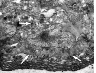 Figure 4 Basal cells of the epidermis of the anterior abdominal wall; a) basal membrane; b) intercellular connections (transmission electron microscopy) |
 Figure 5 Cells of the stratum spinosum of the epidermis of the anterior abdominal wall; |
 Figure 6 Cells of the stratum granulosum of the epidermis of the anterior abdominal wall; a) fragmented nuclei, b) tonofilament bundles and amorphous homogeneous detritus; c) widened interfibrillar spaces; d) fine-grained and homogeneous masses (transmission electron microscopy) |
The epidermal cells of the stratum corneum were of irregular shape and uneven contours. The cell membrane was not visible, only weak osmophilic fragments were observed in the areas of the desmosomes. The internal structure of the desmosomes was destroyed, and their lamellar structure was not observed. Parts of the intercellular contacts had a damaged appearance, and there were significant gaps between them. The cell nuclei were of different sizes with a polymorphic shape, without nucleoli. Residues of membranous organelles, globular inclusions, and homogenized tonofibrils were observed around the nucleus (Fig. 5).
Granular epidermal cells contained fragmented nuclei and cytoplasm containing tonofilament bundles and amorphous homogeneous detritus. Interfibrilar spaces appeared widened. Fine-grained and homogeneous masses with increased electron density were observed between the fibers (Fig. 6).
Transmission electron microscope examination of the dermis sampled from the anterior abdominal wall showed that the collagen fibers retained their internal fibrillar structure and had a clear parallel arrangement with each other. Most of the fibers appeared clearly separated. Some of the fibers did not have gaps between their individual fibrils, which gave the impression of merged fibrils. The collagen fibrils had a relatively uniform electron density in their cross sections: a round, oval, or polygonal shape with clear-cut contours (Fig. 7).
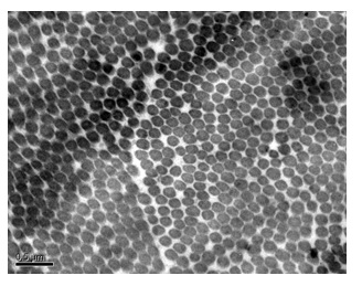 Figure 7 Collagen fibers of the dermis of the skin of the anterior abdominal wall, cross section; collagen fibrils with uniform electron density and clear contours (transmission electron microscopy) |
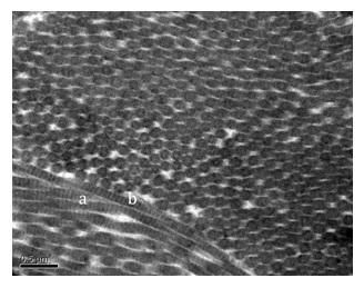 Figure 8 Dermis of the skin of the anterior abdominal wall showing longitudinal sections of the fibers (a); which appeared as dotted lines (b) (transmission electron microscopy) |
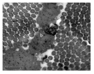 Figure 9 Dermis of the skin of the anterior abdominal wall showing a) collagen and elastic fibers; b) cellular detritus (transmission electron microscopy) |
The longitudinal sections of the fibers, which consisted of alternating light and dark parts, clearly showed the periodicity of the fibers. Elastic fibers were localized between the collagen fibers. In cross-section, the elastic fibers had an irregular shape and depth, and protrusions of the circumferential part. In the amorphous substance, there were point electron-dense fibrillin microfibrils which appeared as dotted lines. High electron-density cell detritus residues were noted between the interfiber bonds (Figs. 8 and 9).
Histological examination of the subcutaneous adipose tissue showed brown deposits in some views, suggesting possible localization of past erythrocyte clusters. Damaged vessels were also observed. The vessels were probably arteries with empty lumens, without a clearly defined wall structure (Fig. 10). Collagen and argyrophilic fibers, which form the skeleton of fat cells, appeared well defined. The intercellular membranes of adipose cells were partially destroyed, but the overall structure was preserved (Fig. 11).
Scanning electron microscope examination of the tibialis anterior muscle showed long strands of muscle fibers fitting closely alongside each other. The muscle fibers had an undulating surface which corresponded to alternating circular depressions and elevations caused by typical transverse striations of the myofibrillar apparatus (Fig. 12).
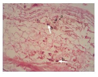 Figure 10 Subcutaneous adipose tissue; a) blood vessels with empty lumens, without a clear structure of the vessel walls; b) foci of brown staining, accumulation of erythrocytes (hematoxylin-eosin staining, magnification x 100) |
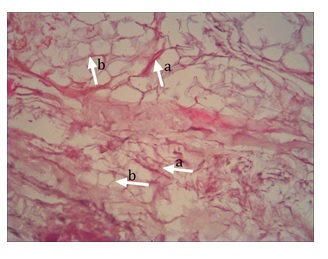 Figure 11 Subcutaneous fat; a) collagen and argyrophilic fibers; b) preserved and torn intercellular membranes of adipose cells (hematoxylin-eosin staining, magnification x 100) |
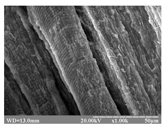 Figure 12 Muscle fibers; transverse striation of the myofibrillar apparatus is visible (scanning electron microscope, electronic scan) |
Each muscle fiber was covered with a layer of endomysial connective tissue. However, some of the muscle fibers were only partially covered by the endomysium. Small openings, similar to the exit sites of the cross-T-tubules of the sarcoplasmic reticulum, were observed on the surface of these fibers. Between the bundles of muscle fibers, there were enlarged spaces and deformed elements of the connective tissue of the perimysium, with separated and fragmented collagen fibers. The muscle fibers were of different sizes, directions, and shapes, and were unevenly colored. Wide, empty spaces were observed between the muscle fibers. Well-defined round cavities were observed at the sites of the fibers (Fig. 13). There were also small connective tissue bridges observed between the fiber bundles. Individual vessels with empty lumens, lacking a clear structure, were also identified.
Examination of the muscle tissue using a transmission electron microscope revealed that the structure of the myofibrils was largely preserved. The muscle fibrils maintained their parallel arrangement. Anisotropic "A" and isotropic "I" sarcomere discs were clearly identified in the myofibrils. Myosin and actin myofilaments as well as the "H" zone, and "M" and "Z" lines were visible. The overlapping bands of actin and myosin protofibrils were well preserved. Against the background of generally satisfactory preservation of the myofibrils, various changes were observed, from the destruction of focal discs to the fragmentary disintegration of protofibrils (Fig. 14).
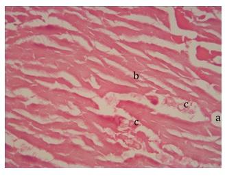 Figure 13 Muscle fibers; a) wide, optically empty slits visible in the fibers defined by round voids; b) small connective tissue bridges; c) individual vessels (hematoxylin-eosin staining, magnification x 100) |
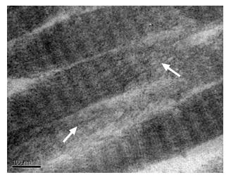 Figure 14 Fragmentary decay of myofibrils and focal destruction of discs, marked with arrows (transmission electron microscopy) |
 Figure 15 Volkmann canal with particles of loose connective tissue (arrow) (scanning electron microscopy, electronic scan) |
Examination of the fibular bone sections revealed a wide range of osteon forms with a distinct lamellar structure. The osteons differed in diameter, number of circular lamellae, and Haversian canal configurations. The surface of the osteon sections was characterized by an undulating surface, due to the alteration of bone lamellae. Interstitial bone lamellae, which are the remnants of pre-existing osteons, were preserved between the osteons. Alteration of circular bone lamellae was clearly visible; the collagen fibrils in these lamellae were directed in parallel and transverse directions to the long axis of the endosteal cavity of the fibula. The microrelief of the inner surface of the central canal was clearly visible, and the openings of the Volkmann canals were observed. In most cases, it was not possible to detect vessels and nerves in the lumen of the Haversian and Volkmann canals, only their residual elements and particles of loose connective tissue were observed (Fig. 15). Many empty spaces were observed on the surface of the central canal walls. It was possible to detect osteocytes without protrusions inside the spaces in the walls. Examination of the inner surface of the fibular shaft at the site of bone marrow conservation revealed cells that were close to the surface of the endosteum and had numerous protrusions. Bone marrow was present as an accumulation of compacted cell masses. Sinus capillaries with a wide outer diameter and numerous pores were observed in the vascular wall in the bone marrow and in the Volkmann osteon canals.
All the tissues taken from the embalmed body of Professor Pirogov showed signs of microscopic changes. The only previously published study dealing with the condition of tissues embalmed using a similar method is from 1960. The study published the histological analysis of the skin and subcutaneous and muscle tissues removed by the Soviet specialists during the last periodic re-embalming of the body of the Czechoslovakian president Klement Gottwald, seven years after the body’s initial embalming. The microstructure of the skin with the papillary and reticular layers was clearly visible. Vacuolization of cells was preserved in the germ layer of the skin, especially around the nuclei. The lobular fat cells of various sizes with the content were of the usual shapes, and easily observable. The rhobdomyocytes were clearly visible in the longitudinal section of the muscle fiber. All this proves that the tissues extracted from the embalmed remains of Klement Gottwald were in a good condition of preservation in general, and in better condition than the remains of Professor Pirogov (Frišhons, 2014). However, it is important to keep in mind the difference in the number of years after the initial embalming between the studies of Gottwald’s and Pirogov’s remains.
Analysis of the skin tissue of Pirogov’s embalmed body showed destructive changes in all its parts. The surface layers of the epidermis and its cellular elements were particularly affected. Examination of skin samples taken from the anterior abdominal wall and the anterior region of the thigh using scanning electron microscopy revealed significant surface irregularities, and detachment of the upper layers of the epidermis. Preserved fragments of hair follicles and fungal hyphae (Melnyk et al., 2021) were found on the skin of the anterior abdominal wall. Of the cellular elements of the epidermis, the basal cell layer was the best preserved. Nuclei, individual small vesicles, fragments of perforated mitochondria, and intercellular junctions were found in these cells. In the dermis, there were mainly collagen fibers with their internal fibrillar structure and transverse periodicity preserved. The elastic fibers were destructively altered. Examination of subcutaneous fat revealed brown deposits, erythrocyte accumulations, and damaged arteries with empty lumens and without a clear wall structure.
It should be mentioned that the first embalming of the body N.I. Pirogov was produced by DI Vyvodtsev in 1881 (Kuznetsov, 1999). Only in 1939-1940, in connection with the upcoming 130th anniversary of the birth of N.I. Pirogov, the NKZ of the Ukrainian SSR, and the Ukrainian branch of the Pirogov Society, was it decided to start restoring the remains of N.I. Pirogov. The special commission managed to carry out only a superficial examination of N.I. Pirogov. The fungus was partially removed from the open and easily accessible areas of the skin of the hands and face. The fungus covered the entire surface of the skin of both hands and the entire face. May 8, 1945 work on the restoration of the body of N.I. Pirogov was resumed, which ended on October 4, 1945. After rebalancing, no mold was observed on the body (Hunko, 2019a). In our work, during the microbiological study of washes from the body of N.I. Pirogov, we did not find evidence of the growth of colonies of pathogenic fungi and other microorganisms. Therefore, taking into account the historical data on the restoration of the body of N.I. Pirogov and our research, we can conclude that the hyphae of molds found on skin samples appeared during the period from 1881 to 1940. Taking into account the data of microbiological research, it can be assumed that the selected components of the embalming solution were correct, namely, their antiseptic properties.The general ultramicrostructure of the skeletal muscle was preserved, which is evidenced by the presence of the characteristic transverse banding of the muscle fibers, and connective tissue membranes of the endomysium and perimysium. At the same time, destructive changes were also seen. Histological examination of the muscle tissue revealed muscle fibers that had completely lost their structural properties; transverse banding was observed only at the edges of the sections in some views with a darkened aperture. Small areas of connective tissue were visible between the bundles of fibers, and individual vessels with empty lumens and without a clear structure, were identified. Mitochondria, sarcoplasmic reticulum elements and other muscle fiber organelles were not detected. Enlarged spaces and deformed elements of the connective tissue of the perimysium with collapsed collagen fibers were evident. Various changes were observed in myofibrils; these changes ranged from focal destruction of discs to fragmentary decay of protofibrils.
The general bone structure was also preserved. The osteons had a distinct lamellar structure. The microrelief of the inner surface of the central fibular canal was well preserved. Numerous openings of Volkman's canals were observed in the relief of the endosteal surface. Only residual elements of blood vessels, nerves, and particles of free connective tissue were observed. Cells were found in the bone marrow that was located close to the endothelial surface. Wide bone trabeculae and cells that were round, and contained wide protrusions, were also noted in the bone marrow masses. The bone marrow appeared to be formed by a cluster of compacted cell masses. Preserved sinus capillaries with a wide outer diameter and numerous pores in the vascular wall were also seen. The sinus capillaries were intertwined in the bone marrow and in the Volkmann canals of the osteons.
In conclusion, it can be stated that, despite a certain degree of destructive changes, the examined tissues taken from the embalmed body of Professor Pirogov 137 years after the initial embalming, are relatively well preserved. Continual application of the appropriate re-embalming procedures and following of other measures (such as contactless body monitoring) suggest that the body has a potential to be preserved in a good condition for many years to come.
Acknowledgments
The authors of the article thank Vinnytsia National Medical University N.I. Pirogov for providing financial support for the cost of carrying out analyses.
Funding. Analyses were funded Vinnytsia National Medical University N.I. Pirogov.
Ali Shtayeh M, Jamous R, Yaghmou R. 1998: Mycology Manual. Nablus: An-Najah National University, Palestine.
Frišhons J, Vacín L. 2014: "Aby byla zachována tvář soudruha Gottwalda": Postup a průběh balzamace těla Klementa Gottwalda [“In order to preserve the face of Comrade Gottwald": The procedure and course of embalming the body of Klement Gottwald]. Acta Musei Nationalis Pragae. Series A - Historia Praha: Národní muzeum 68(1-2):3-31.
Hendriks II, Zhuravlev DA, Bovill JG, Boer F, Gaivoronskii IV, Hogendoorn P, Deruiter MC. 2020: Nikolay Ivanovich Pirogov (1810–1881): Anatomical research to develop surgery. Clin Anat 33:714-730. https://doi.org/10.1002/ca.23493
Hunko PM, Haidukov OV, Martynova ZS. 2019a: Long-term preserving of N. I. Pyrogov’s embalmed body as a unique scientific experiment [In Ukrainian]. Naukovij vіsnik Nacіonalnogo muzeju іstorіi Ukraini. Zbіrnik naukovih prac. [Scientific Bulletin of the National Museum of History of Ukraine. Collection of scientific works]. (4): 625-639. ISSN: 2618-0235.
Hunko PM, Haidukov OV, Martynova ZS. 2019b: Body rebalancing N. I. Pyrogov: historiographic section [In Ukrainian]. Vіsnik Vіnnickogo nacіonalnogo medichnogo unіversitetu [Bulletin of Vinnytsia National Medical University] 23 (2): 330-336.
Kuznetsov LE, Khokhlov VV, Fadeev SP, Shigeev. VB 1999: Embalming and restoration of corpses. Leadership. - Moscow, 496 p.
Matvejchuk IV. 2013: Modern achievements in conservation and long-term preservation of biological structures and objects N.I. Pirogov [In Russian]. Voprosy Biologicheskoj, Medicinskoj i Farmacevticheskoj Chimii [Biological, Medical and Pharmaceutical Chemistry Issues] 11: 143-147.
Melnyk OP, Tkach GF, Frišhons J, Gumminskii YJ, Kovalchuk VP, Svoboda J, Melnyk MV, Dzetkuličová V. 2021: Microbiological aspects of the examination of the 140 year-old embalmed body of N.I. Pirogov. J Plastination (in press)
Reynolds ES. 1963: The use of lead citrate at high pH as an electron-opaque stain in electron microscopy. J Cell Biol 17(1):208-212.
https://doi.org/10.1083/jcb.17.1.208
Vyvodstev DI. 1870: A simple and popular method of embalming corpses without opening the cavities [In Russian]. Voenno-Meditsinskiy Zhurnal [Military Medical Journal] 2: 71-79.