1Cuerpo Académico en Ciencia e Inovación Tecnológica Pecuaria
2Cuerpo Académico en Medicina y Cirugía Animal
3Departamento de Anatomía FMVZ-UAEMex. Facultad de Medicina Veterinaria y Zootecnia. Universidad Autónoma del Estado de México. Toluca, México
Different pathological events occur to the male urethra, and various surgical protocols have been developed to resolve them. Despite the clinical interest involving the urethra of male dogs in the literature there are only a few reported anatomical investigations. The aim of this study was to create a three-dimensional model of the urethra by means of a corrosion cast to describe and record the structure of the pelvic and penile urethra. These casts of male dog urethras were useful to observe the pelvic and penile portions of the urethra. Morphological details of the inner surface of the urethra remained imprinted on the casts. These replicas can improve understanding of anatomical considerations needed to treat clinical and surgical pathology of the urethra.
corrosion cast; male dog; urethra
Dr. Jorge Arredondo-Ramos, Departamento de Anatomía, Facultad de Medicina Veterinaria y Zootecnia, Universidad Autónoma del Estado de México, Instituto Literario Ote. 100. Col. Centro. C.P. 50130, Toluca, México. Tel.: (+52) 7222965548, E-mail: arredondo.uaemex@gmail.com

![]()


The male urethra carries urine, semen and seminal secretions to the distal end of the penis (König y Liebich, 2009). Various pathological events affect the urethra, and to resolve them, conventional (Smeak, 2000; Burrow et al, 2017) and minimally invasive (Della Maggiore, 2013; Luckring et al, 2015) surgical protocols have been developed. Despite the clinical interest involving the urethra of male dogs in the literature there are only a few anatomical investigations recorded (Cullen et al, 1981; Creed et al, 1998; Stolzenburg et al, 2002) and there is also a significant lack of meticulous anatomic descriptions (Stolzenburg et al, 2006). The aim of this study was to create a three-dimensional model of the urethra by means of a corrosion cast to aid description of the structure and shape of the pelvic and penile urethra.
Ten male mixed breed dog cadavers were used in this study. Dogs were between 1-5 years of age with a mean weight of 24 kg (range 18-28 kg). The dogs were obtained from the Veterinary Hospital of the Autonomous University of the State of México, and were subjected to euthanasia with an overdose of sodium pentobarbital for reasons not related to reproductive or urinary disorders. All animals received humane care in compliance with the Animal Care and Bioethics Committee of the Autonomous University of the State of México.
Dissection of the caudal abdomen was made in order to clamp the vesicular trigone with a Kelly forceps to avoid filling the urinary bladder. A 16-gauge vascular catheter was introduced 3 mm into the external urethral opening, and 20 ml was injected of epoxy Biodur E20 mixture (20 ml of epoxy Biodur E20 Plus, mixed with 12 ml of epoxy Biodur E20 Hardener colored with yellow Biodur AC53 pigment). After injection of epoxy, the vascular catheter was removed, and another Kelly forceps was placed over the external urethral opening to avoid external leaking of the polymer-mix. The cadaver was then refrigerated at 4° C for 48 hours. The reproductive system was isolated from the body, maintaining the pubic bone as a reference landmark, and then corrosion was carried out using 20% solution of sodium hydroxide (NaOH) at room temperature, which was changed every 72 hours until total tissue degradation was achieved.
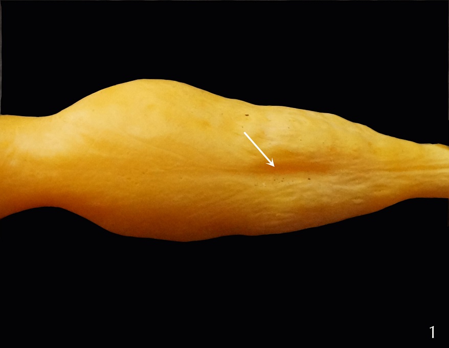 Figure 1. Corrosion cast of the male canine urethra at the level of the prostate showing considerable dilation. The arrow indicates the seminal colliculus (male uterus homologue) |
The resulting model of the male dog’s urethra had well-defined surface features. The imprinted mucosal details were visible with the naked eye. The resultant three-dimensional model was useful to observe the intra- and extra-pelvic portions of the urethra (Figs. 1, 2 AND 3). A dorsal dilation was present at the level of the prostatic gland located in between the vesicular neck and the prostatic utricle. The prostatic utricle was observed as a central longitudinal concavity located in the dorsal aspect of the cast. The prostatic ducts were represented by several elevations and depressions that were observed on and lateral to the prostatic utricle (Fig. 1).
The proper shape and size of the urethra was maintained in this corrosion cast, which made it possible to appreciate the considerable narrowing of the urethra at the level of its ischiatic curvature (Fig. 2). The position of the urethra in relation to the pelvic bones can also be achieved by enzymatic digestion of the soft tissues. The penile urethra has a dramatically reduced caliber. In our study, this portion of the urethra was seen to be laterally flattened, not only at the level of the penile bone, but also caudal and cranial to it (Fig. 3). The shape of the corrosion casts of the male canine urethra showed four different portions in the dogs evaluated: 1) a dilated prostatic portion, 2) a rounded pelvic portion, 3) a curved oval ischiatic portion, and 4) a laterally flattened penile portion.
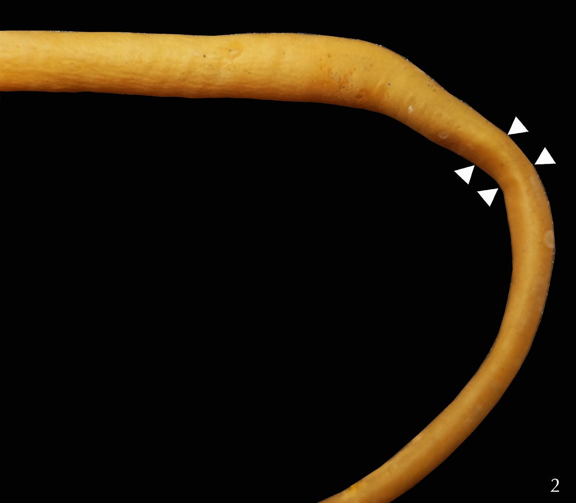 Figure 2. Corrosion cast of the male canine urethra at the level of the ischial arch (arrowheads indicate the narrowing of the urethra at the ischial arch) |
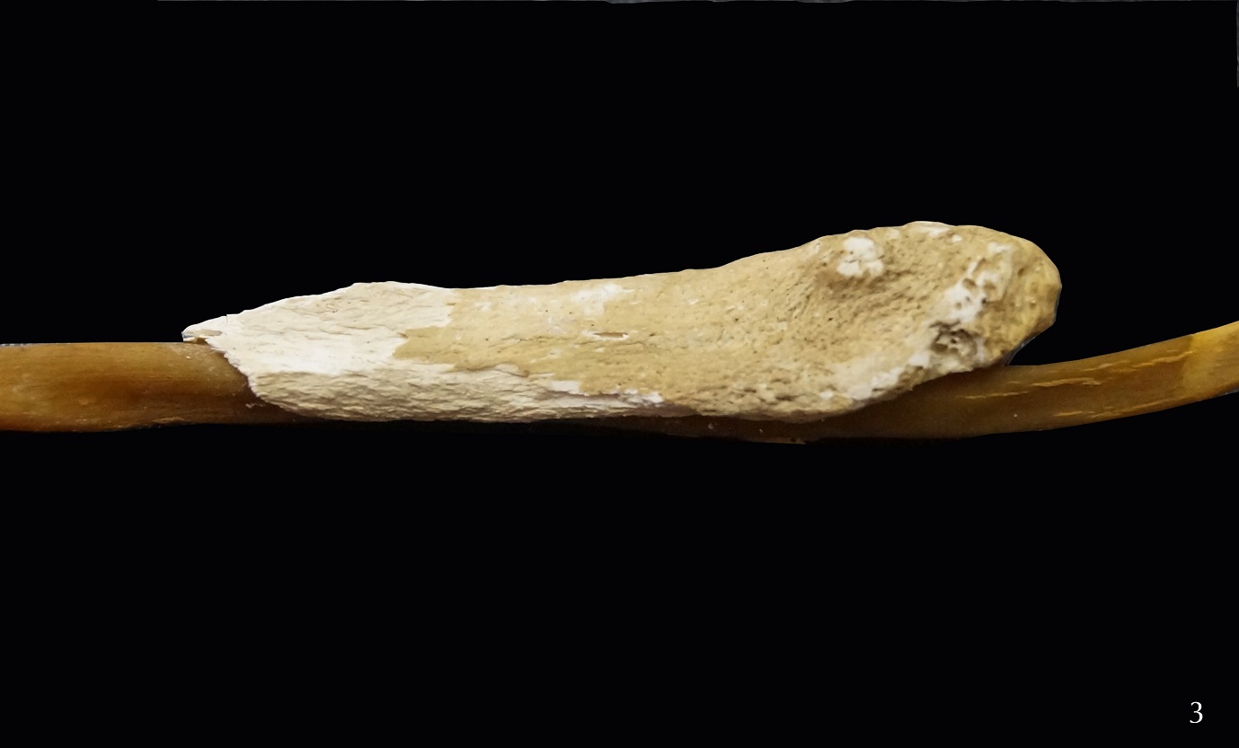 Figure 3. Corrosion cast of the male canine urethra, with the os penis in position, showing lateral compression of the urethra |
It is important to produce basic information on the anatomy of the male reproductive system, since it has considerable significance in breeding, in order to make an accurate pathological diagnosis, and to provide effective clinical treatment of diseases (AlLugami et al, 2017). The obtained urethral corrosion cast allowed visual inspection of the urethra, which led to better understanding of the anatomy needed to resolve clinical and surgical pathological conditions.
The main characteristic of the obtained three-dimensional model was maintenance of shape of the surface of all the portions of the urethra. This enabled the identification of four morphologically distinct portions of the canine male urethra, contrary to the two (Getty, 1986; Dyce et al., 2010) or three (Dellman, 1993) portions for domestic animals previously described in the scientific literature.
The total replication model obtained can also be useful to describe the total volume capability of the urethra in each portion; other studies have found a significantly variation of the urethral lumen during spontaneous urination (AlLugami et al., 2017). As mentioned previously, a dilation of the prostatic portion of the urethra was found in our study (Poogird and Wood, 1986).
The three-dimensional model of the male canine urethra by injection of epoxy E20 mix is a useful tool to perform research of the reproductive tract in the male dog, due to its capability of preserving the spaces and elevations of the lumen. This improves the understanding of anatomical considerations, which can be applied in clinical and surgical aspects of pathologies. Another major advantage of using epoxy E20 mix for urinary corrosion casts is the long-term preservation of a clean and odorless cast.
AlLugami A, von Pückler K, Wehrend A, Sickinger M. 2017: Sonography of the distal urethra in lambs. Acta Vet Scand 59:16
https://doi.org/10.1186/s13028-017-0283-2
Burrow RD, Gregory SP, Giejda AA, White RN. 2011: Penile amputation and scrotal urethrostomy in 18 dogs. Vet Rec 169(25):657
https://doi.org/10.1136/vr.100039
Creed KE, Van der Werf BA, Kaye KW. 1998: Innervation of the striated muscle of the membranous urethra of the male dog. J Urology; 159(5): 1712-1716
https://doi.org/10.1097/00005392-199805000-00099
Cullen CW, Fletcher T, Bradley WE. 1981: Histology of the canine urethra II. Morphometry of the male pelvic urethra. Anat Rec 199(2): 187-195
https://doi.org/10.1002/ar.1091990204
Della Maggiore AM, Steffey MA, Westropp JL. 2013: Treatment of traumatic penile urethral stricture in a dog with a self-expanding, covered nitinol stent. J Am Vet Med Assoc 242: 1117-1121
https://doi.org/10.2460/javma.242.8.1117
Dellmann HD. 1993: Textbook of Veterinary Histology, 4th ed. Philadelphia: Lea & Febiger, p 307-312.
Dyce KM, Sack WO, Wensing CLG. 2010: Textbook of veterinary anatomy, 4th ed. St. Louis: Saunders Elsevier, p 467-469.
Getty R, editor. 1975: Sisson and Grossman's The anatomy of the domestic animals, 5th ed. Philadelphia: WB Saunders Co Ltd, p 1736.
König E, Liebich H-G. 2009: Veterinary anatomy of domestic mammals, 4th ed. Stuttgart: Schattauer GmbH, p 127-129.
Luckring EJ, Ham K, Adin CA, McLoughlin MA, Stull JW. 2016: Laparoscopic placement and urodynamic effects of an artificial urethral sphincter in cadaveric dogs. Vet Surg 45:20-27
https://doi.org/10.1111/vsu.12496
Poogird W, Wood AKW. 1986: Radiologic study of the canine urethra. Am J Vet Res 47(12): 2491-2497.
Smeak DD. 2000: Urethrotomy and urethrostomy in the dog. Clin Tech Small Anim Pract 15(1): 25-34
https://doi.org/10.1053/svms.2000.7301
Stolzenburg JU, Neuhaus J, Evangelios L, Schwalenberg T, Ludewig E, Ganzer R. 2006: Histomorphology of canine urethral sphincter systems, including three-dimensional reconstruction and magnetic resonance imaging. Urology 67: 624-30
https://doi.org/10.1016/j.urology.2005.09.055
Stolzenburg JU, Schwalenberg T, Do M, Dorschner W, Salomon F-V, Jurina K, Neuhaus J. 2002: Is the male dog comparable to human? A histological study of the muscle systems of the lower urinary tract. Anat Histol Embryol; 31(4): 198-205
https://doi.org/10.1046/j.1439-0264.2002.00395.x