Anatomical Institute, Karl-Franzens University Graz Graz, Austria
Nerve plexuses such as the brachial plexus and the lumbosacral plexus are difficult to display by normal anatomical dissection. They are better displayed by P-35 plastination. A cadaver, 43 year old male, was prepared using Thiel's fixation (Thiel, 1992). Dissection began at the distal end of the upper extremity (membrum superius) for the brachial plexus and the distal end of the lower extremity (membrum inferius) for the lumbosacral plexus. Each nerve was marked with a colored thread to avoid any confusion. This was necessary to ensure proper identification of individual nerves after the plexus has been removed from the cadaver. The spinal column was opened from the dorsal aspect and the plexus and distal cord were removed. Plastination followed using the P-35 technique (Weber, 1994).
P35; Brain Slices; Lumbosacral plexus
Andreas H. Weiglein Anatomical Institute, Karl-Franzens University Graz Graz, Austria

![]()


Nerve plexuses such as the brachial plexus and the lumbosacral plexus are difficult to display by normal anatomical dissection. They are better displayed by P-35 plastination.
A cadaver, 43 year old male, was prepared using Thiel's fixation (Thiel, 1992). Dissection began at the distal end of the upper extremity (membrum superius) for the brachial plexus and the distal end of the lower extremity (membrum inferius) for the lumbosacral plexus. Each nerve was marked with a colored thread to avoid any confusion. This was necessary to ensure proper identification of individual nerves after the plexus has been removed from the cadaver. The spinal column was opened from the dorsal aspect and the plexus and distal cord were removed. Plastination followed using the P-35 technique (Weber, 1994).
DEHYDRATION
Dehydration was done by freeze substitution. Each of the specimens were immersed in 15 litres of 99% Acetone at - 25°C for 72 hours. There was no need to change the acetone after the initial dehydration took place.
IMMERSION
Immersion of the specimens was carried out in two baths of P-35/A9 mixture for 24 hours each. During both of the immersions the specimens were held below the surface of the mixture using a grid.
FORCED IMPREGNATION
During forced impregnation the specimens were placed in a fresh P-35/A9 mixture for 24 hours. After impregnation they were placed on a glass plate for orientation and trimmed. Proper orientation of the plexuses was very difficult because the nerves were tangled and the specimens became less flexible as time passed. After correct positioning, which was facilitated by the identifying colored threads on the individual nerves, the specimens were embedded.
EMBEDDING
The double glass chamber system of embedding was used.
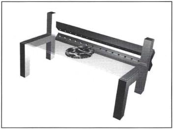
Fig. 1: Compressed air cooling system.
CURING
Curing was started using UVA-light on both sides of the specimen for 3 hours. Ventilation is very important during light curing because the resin temperature in the embedding chamber must never exceed 35°C. To prevent this, a special cooling system (Fig. 1) was developed for the specimens. The cooling system consisted of two metal tubes with many small holes in them. One tube was placed on each side of the double glass chamber housing the specimen to be cured, and compressed air was forced through them. The air passed through the tubes and out through the holes facing the embedding chamber. As the forced air flowed over the surface of the specimen chamber the heat produced by the exothermic reaction of the curing polymer was dissipated, thus maintaining the curing temperature within safe limits. Using this type of system for curing was much better than using desk-top ventilators because the air flow could be easily adjusted. Curing was completed using a well ventilated oven at 42°C for a period of 3 days.
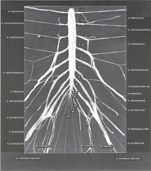 Fig. 2: Lumbosacral plexus plastinated using the P-35 technique and placed on a plexiglass plate which has the names of the nerves engraved in it. |
Subsequently, the glass plates were removed and the plastic trimmed. For demonstration purposes the P-35 plexus plate was placed on a larger plexiglass plate, on which the names of all the nerves shown in the specimen were engraved (Fig. 2 ). For examination purposes the P35 plexus plate could be easily removed from the plexiglass plate.
| STEP | TIME |
| Dissection | 16 h |
| Dehydration | 72 h |
| 1. Immersion | 24 h |
| 2. Immersion | 24 h |
| Forced Impregnation | 24 h |
| Light curing | 3h |
| Heat Curing | 96 h |
| TOTAL | approx. 2 weeks |
The P-35 technique for plastination of brain slices has been used as a teaching tool for several years (Weiglein, 1993). By using our method we have developed a very suitable procedure for displaying nerve plexuses and keeping them for long periods of time in perfect condition.
ACKNOWLEDGMENT
We gratefully appreciate the assistance of our laboratory technician, Johann Eder, who assisted us with the plastination procedure and development of the compressed air cooling system.
Thiel, W. 1992- Die Konservierung ganzer Leichen in naturlichen Farben. Ann Anat 174:185-195.
https://doi.org/10.1016/S0940-9602(11)80346-8
Thiel, W. 1992. Eine Arterienmasse zur Nachinjektion bel der Konservierung ganzer Leichen. Ann Anat 174:197-200. https://doi.org/10.1016/S0940-9602(11)80347-X
Weber, W. 1994. Sheet plastination of brain slices. J Int Soc Plastination 8 (1):23.
https://doi.org/10.56507/OWYV2878
Weiglein, A. 1993. Plastinated brain slices in the anatomical curriculum at Graz University. J Int Soc Plastination 7 (1):3-7. https://doi.org/10.56507/EHRX7749