Department of Oral Pathology & Microbiology, Kothiwal Dental College and Research Center, Mora Mustaqueem, Kanth Road, Moradabad, U.P.-244001, India
Objective: The study was conducted to overcome specific limitations of formalin-preserved specimens at negligible cost.
Materials and Methods: The study was conducted on museum specimens collected from the Department of General Anatomy, Kothiwal Dental College. All the collected specimens were plastinated employing previously utilized laboratory consumables, such as disposable culture plates, and xylene, mixed together in a fixed proportion to form a homogeneous paste. After complete dehydration in alcohol, clearing was done in acetone followed by impregnation with reactive polymer under vacuum which was created with a modified suction apparatus. Curing of the specimen was done under direct sunlight.
Results: This technique has proved its advantages over formalin-preserved specimens as it has produced dry, life-like specimens. The only limitation of our technique was that it has showed marked shrinkage after curing.
Conclusion: We have devised a new and cost-effective method of plastination that involves no specialized equipment and prepares plastinated specimens that retain much of their natural features in a life-like manner.
formalin; heart; larynx; museum; specimen handling
Dr. Ambika Chaudhary, M.D.S. Student, Department of Oral Pathology & Microbiology, Kothiwal Dental College and Research Center, Mora Mustaqueem, Kanth Road, Moradabad, U.P.-244001, India, Phone No: 09897433239, Email address: a.ambika14@gmail.com

![]()


Handling and managing gross specimens is an important part of the training in general and in maxillofacial anatomy and pathology. Unfortunately handling formalin-fixed specimens carries several disadvantages such as repulsive odor, loss of color vibrancy due to long-term fixation and difficulty in maintaining the 3D orientation of luminal and branching patterns of the specimens (Jain et al. 2014). The potential carcinogenicity of long-term exposure to formalin is also a considerable concern. Novel solutions have been explored over the years to overcome and eliminate these drawbacks.
A technique which carries inventive solutions for most of these difficulties is plastination. Dr. von Hagens in 1978 invented the technique of plastination for museum specimen preservation at the University of Heidelberg (Jain et al., 2014). Plastination is a word of Greek origin, taken from Plassein which has a literal meaning to shape or to form (Dudanakar et al., 2014). Due to its many advantages plastination has gained wide recognition all over the world, and the first paper describing this original method was published by Dr. von Hagens in 1979. Plastination is basically a combination of science, technological phenomenon and artistry along with cultural aspects of life and death. Plastinated specimens have several advantages over formalin-conserved ones in that the former are clean, dry, odorless, durable, non-toxic, non-infectious, do not exude damaging vapors, have finer esthetics, can act as educative tool in patient counselling, can be handled with bare hands and do not require any special storage conditions or care (Jain et al. 2014). According to the available literature, very few have tried this technique for oral specimens (Ravi &Bhat, 2011).
The technique of plastination, as described by von Hagens, and followed across the world, is quite effective and durable. However, it requires the use of specialized materials and equipment that make it unsuitably expensive. The challenge for us is to develop and adapt the technique in a way that is economically viable and suited for use on a routine basis. Here we describe a novel way to implement a plastination method that was developed keeping these requirements in mind. We have tried to use locally-sourced, cheap and easily available materials, and, using only the routine lab armamentarium along with laboratory waste reagents, we have developed a practice for plastination that may be adopted easily in museums and labs across the country
The anatomical specimens taken for plastination were those preserved in the anatomy museum of Kothiwal Dental College and Research Centre. These were being stored in 10% formaldehyde as study models including specimens of larynx, kidney, fetal stomach and fetal heart.
The materials to perform plastination in our laboratory were drawn from previously used xylene, disposable sterilized culture plates, acetone, alcohol, suction machine, eosin stain and hydrogen peroxide. The detailed list of materials is discussed in table 2.
Routine plastination methods were implemented utilizing the following procedures.
Fixation
Specimens utilized for plastination were prefixed in 10% formalin solution for several years for the purpose of teaching of anatomy. In the case of fresh specimens, fixation time depended on the size and nature of specimen. However, a standard 48-hour fixation time was followed for small specimens.
Staining & Bleaching
Stored specimens were discolored and brownish in appearance due to the long duration of fixation. A staining and bleaching step was introduced into the methodology to remove this pigmentation and achieve a more life-like appearance for the finished plastinated specimen.
The fixed specimens were first immersed in routinely used eosin-Y dye (10% in 80% ethanol) and 0.5% glacial acetic acid (1gm) in to it to sharpen the stain. Thereafter, the specimen was dipped twice into hydrogen peroxide (50%) for bleaching and removing the excess stain. This procedure improved color differentiation of the specimen.
Processing
Processing of specimens for plastination was performed under two steps that included dehydration and clearing. All the procedures were performed at room temperature (37°c). The specimens were immersed in 70% alcohol for 24 hours and 100% alcohol for next 48 hours with one change in alcohol (24 hours). Anhydrous copper sulfate was used as an indicator to ensure complete dehydration that was considered complete when colorless. For clearing, specimens were immersed in acetone (4-5 hours) depending on the size of the specimen.
Formation of reactive polymer
Reactive polymer for impregnation was formed using disposable culture plates and used filtered xylene. Autoclaved used culture plates (200gms) were dissolved into Xylene (1000ml) under adequate ventilation. Once the plates were fully dissolved, it formed a homogenous viscous fluid that was used as the polymer solution for impregnation.
Impregnation
Forced impregnation of polymer into the specimen was performed under vacuum (25-30mmHg). A clinical suction apparatus was modified to create the vacuum. The vacuum was created in a single jar of the suction apparatus by blocking its second jar, which generally helps to neutralize the excessive vacuum in the container. A safety valve incorporated in the design ensured that any pressure in excess of 30mmHg was released, thereby reducing the risk of any explosion. During the impregnation period, pressure in the vacuum chamber was consistently monitored using the meter indicator of the suction apparatus. Specimens were submerged in the polymer bath for 3-5 days depending on the size of the specimen. The end-point of impregnation was considered as the stage at which air bubbles were no longer seen escaping from the surface of the specimen. This indicated that the specimen was now saturated with the polymer solution.
Curing
Once specimen were completely impregnated with the polymer, they were taken out of the polymer bath, leaving the residual polymer in the jar, and allowed to stand for few hours at room temperature so that excess polymer drained from the surface of specimen. The curing was performed under direct sunlight by placing the specimen in a transparent glass jar.
In previous trials with the polymer, we had found that it could be cured by exposure to direct sunlight, presumably by the UV present, and this method was used. The end point of curing was determined as the stage at which the specimen hardened and no residual xylene was detected in the specimen, typically after 2-3 days.
Plastination produced odorless, durable, non-hazardous, easy-to-handle, formalin-free and life-like specimens that could be handled without personal protective equipment (Figs. 1-4). Eosin dye imparted a crisp and well-differentiated color to the different structures in the specimens. The structural integrity and anatomical details of the specimens were well preserved. On the other hand, a small amount of shrinkage was also observed in all the specimens.
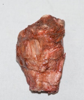 Figure 1 -Plastinated specimen showing the anterior view of the larynx |
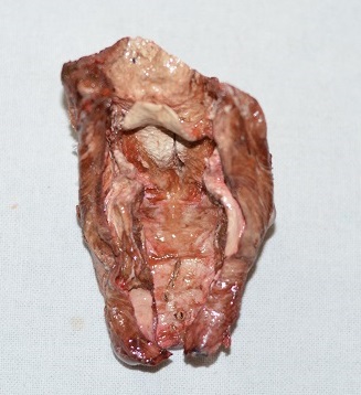 Figure 2 -Plastinated specimen showing the posterior view of the larynx |
 Figure 3 - Plastinated specimen showing the external aspect of kidney. |
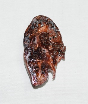 Figure 4 - Plastinated specimen showing the internal aspect of kidney. |
Plastinations are permanent, non-hazardous, durable, dry museum specimens, which are excellent tools for teaching the anatomy and morphological characteristics of various specimens. The use and interest in plastination has steadily increased since it was first developed. However, the degree to which it may impact the developing countries appears to depend on cost-effectiveness and practicability of execution as well as training of human resources. In the past, it cost approximately $15,152 to setup a laboratory for plastination (Torre et al., 2004).
The general belief is that production of plastinations including the cost of equipment is an expensive proposition and hence is not an economically viable option, particularly for small to medium scale labs and institutions. However, our method of plastination warrants a thorough rethinking of this mindset. We have innovated a method using regularly-used laboratory equipment that includes a suction machine with suitable modifications, and laboratory wastes. With minimum expenditure in terms of materials and armamentarium, we have been able to create plastinations of the laryngeal complex, adult human kidney, fetal heart and fetal stomach. Our method is particularly useful and effective in the case of small to medium-sized specimens, particularly those routinely encountered in an oral pathology department. Our method of plastination has opened an era of great opportunities for oral pathologists to develop specimen libraries. Although this technique is not new to the field of oral pathology, the reduced cost of polymer, insignificant cost of laboratory setup and impregnation unit gives exclusivity to our study.
However, our study and the method has limitations. For one, only specimens small enough to fit into the jar of the suction machine can be plastinated this way. It was also difficult to maintain adequate vacuum for extended periods in the modified suction machine. There was also the risk of explosion of the suction unit if the pressure inside exceeded beyond the safe level (30mmHg). Shrinkage of specimens noticed during impregnation and curing was also a drawback. In spite of these limitations, this technique was very advantageous in many ways including its cost-effectiveness and the fact that additional lab setup and impregnation unit were not required.
Utilizing long-term fixed specimens for plastination, resulted in good preservation of specimens for observation-based teaching. The structural integrity of the specimens remained sound and aesthetically agreeable. The preservation of anatomical specimens that retain much of their natural features has been a long-standing goal of pathologists in India. With this technique we can reduce the cost of plastination by 90%. The expenditure in our method was limited to the cost of acetone, eosin dye and hydrogen peroxide that is significantly less than the usual cost. We recommend further studies on larger number of specimens to explore other possible innovations in reactive polymer and impregnation units at lower temperature to reduce shrinkage.
Cannas M, Fuda P. 1991: Plastination of old formalin-fixed specimens. J Int Soc Plastination 5:11-15
https://doi.org/10.56507/IYFR4714
Dudanakar M, Joshi PS, Kempwade RS, et al. 2014: Plastination revisited: a teaching aid in oral pathology. Int J Contemp Dent 5:1-4
Jain M, Kasetty S, Sudheendra U S. 2014: Plastination: an intricate and real display of oral hard and soft tissue specimens. J Res Prac Dent: 1-6
https://doi.org/10.5171/2014.639870
Marks DL, Chaney EJ, Boppart SA. 2008: Plastinated tissue samples as three-dimensional models for optical instrument characterization. J Opt Soc Am A 25:1156-1164
https://doi.org/10.1364/OE.16.016272
Patil S, Rao RS, Ganavi BS. 2013: The museum maze in oral pathology demystified: part ii. J Contemp Dent Pract 14:987-992
https://doi.org/10.5005/jp-journals-10024-1438
Ravi SB, Bhat VM. 2011: Plastination: a novel, innovative teaching adjunct in oral pathology. J Oral Maxillofac Pathol 15:133-137.
https://doi.org/10.4103/0973-029X.84475
Ravikumar C. 2014: Plastination. J Pharm Sci Res 6:271-273
Rumph PF, Garrett PD, Marshall AE. 1987: A versatile vacuum control system for plastination. J Int Soc Plastination 1:4-8
https://doi.org/10.56507/IZLI7746
Suganthy J, Francis DV. 2012: Plastination using standard S10 technique- our experience in Christian Medical College, Vellore. J Anat Soc India 61:44-47.
https://doi.org/10.1016/S0003-2778(12)80012-8
Torre FR, Rodriguez-Baeza A, Domenech-Mateu JM. 2004: Setting up a plastination laboratory at the faculty of medicine of the autonomous University of Barcelona. Eur J Anat 8:1-6