1- Department of Anatomy 2, Anatomical Institute, Vienna University, Austria
2-Department of Anatomy 1 , Anatomical Institute, Vienna University, Austria
One human brain was used for this study. The brain was fixed in 5% formalin for two months, rinsed and cut in two halves on the sagittal plane. Both brain halves were sagittal sliced at a thickness of 4 mm. From each brain half we selected 8 slices and plastinated them with P40 using different immersion and impregnation conditions. Two points were marked on each slice and subsequently an imprint of the slices was drawn on transparency film. After dehydration in -25°C acetone, the slices of the left-brain half were immersed at -25°C, for two days in P40 and then impregnated for 24 hours. The slices of the right brain half were immersed at +5°C for two days and impregnated at room temperature at+15°C for 24 hours. All impregnated slices were cured with UV light. The imprints of the fixed brain slices were scanned into a computer, as well as the plastinated slices. By using a Kontron KSA 400 v. 2.0 (ZEISS) software we calculated the area of the plastinated brain slices as well as the area of the scanned imprints. By comparing the obtained data, we were able to determine the shrinkage rate of the slices. The slices processed at -25°C showed a shrinkage rate of 4.41%. In comparison the slices immersed at +5°C and impregnated at +15°C showed a shrinkage rate of 6.96%.
Presented in part at the 9th International Conference on Plastination, Trois-Rivieres, Canada, July 5-10, 1998.
Brain, Polymer P40, Impregnation, Temperature
M.-C. Sora, MD, Anatomical Institute, Vienna University, Währingerstrasse 13/3, A-1090 Vienna, Austria. Telephone: 43 1 4277 611 50 / Fax: 43 1 4277 611 42. Email: mircea-constantin.sora@univie.ac.at

![]()


Brain slices plastinated with the P40 technique (von Hagens, 1994) are shrinking and sometime this shrinkage is very annoying. In order to find out the shrinkage degree we used two different immersion and impregnation conditions. One set of slices was immersed and impregnated at -25°C and an other set of slices was immersed at +5°C and impregnated at room temperature (+15°C). These temperature conditions being both described (von Hagens, 1994; Barnett, 1997), the purpose of this study was to find out the most suitable plastination conditions for brain slices.
Fixation and Sectioning
A human brain was obtain at post-mortem from a cadaver. The brain was fixed in 5% formalin for two months. Before being serially sectioned the brain was washed in running tap water for two days.
The brain was first cut in two halves. By using a meat slicer each half was sliced in 4 mm thick sagittal slices. Sixteen slices were selected for this study: 8 slices from the left brain half and 8 slices from the right brain half. As we intended to find out the shrinkage degree of the slices we placed in each slice two plastic markers, one anterior and one posterior (figure 1). The distance between the marker was measured and subsequently an imprint of each slice was drawn on transparency film. The imprints drawn from the fresh slices (figure 2) were scanned into the computer and the area was calculated by using a Kontron KSA 400 v.2.0 (ZEISS) software. In a next step the plastinated brain slices were scanned into the computer and the area of the plastinated slices was determined by using the same Kontron KSA 400 v.2.0 (ZEISS) software. The distance between "A" and "B" was necessary in order to calibrate the scanned images. From those brain slices where the cerebellum was not attached to the slice, imprints were made only from the cerebrum. The marker placed anterior was named "A" and the marker placed posterior was named "B". The obtained slices were placed into two grid baskets and into distilled water at +5°C overnight (von Hagens, 1994).
Dehydration
Both sets of slices were placed into an acetone bath at -25°C (Barnett, 1997). After two days the grid baskets containing the slices were moved into an other acetone bath, also at -25°C. Dehydration was completed after one week.
Immersion
The slices of the left brain half were placed with the grid basket into the immersion bath of P40 at -25°C for 2 days. The slices of the right brain half were placed with the grid basket into the immersion bath of P40, at +5°C, for 2 days.
Impregnation
The grid basket containing the left brain slices was taken from the immersion bath (-25°C) and placed in cold P40 at -25°C in the vacuum chamber. The vacuum chamber was then placed in a freezer at -25°C. Impregnation was undertaken for 12 hours and completed at a pressure of 2mm Hg (von Hagens, 1994).
The grid basket containing the right brain slices was taken from the immersion bath (+5°C) and placed in an impregnation bath at +5°C. The impregnation bath was placed into a vacuum chamber at room temperature (Cook and Barnett, 1996). Impregnation was undertaken for 12 hours and completed at a pressure of 10mm Hg (von Hagens, 1994).
Casting and Curing
The casting and curing conditions were the same for both brain halves. All slices were casted in flat chambers and the chambers cured with UV lights for 3 hours.
| RIGHT HALF | LEFT HALF | |
| DEHYDRATION | -25°C, one week | -25°C, one week |
| IMMERSION | +5°C, 48 hours | -25°C, 48 hours |
| IMPREGNATION | +15°C, 24 hours | -25°C, 24 hours |
| LINEAR SHRINKAGE
(mm) |
2.60% | 1.92% |
| SURFACE SHRINKAGE
(mm2) |
6.96% | 4.41% |
In order to calculate shrinkage, the distance between the marker "A" and "B" was measured on all slices before and after plastination. One dimensional shrinkage could be calculated by comparing the distance between "A" and "B" on fresh and plastinated slices. At -25°C the average shrinkage is 1.92% and at room temperature it is 2.60%. By comparing the area of the fresh and of the plastinated slices a two dimensional shrinkage was calculated. On slices impregnated at -25°C we found a shrinkage of 4.41% and on slices impregnated at room temperature the shrinkage was of 6.96%.
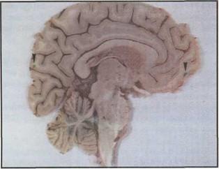 Figure 1. Fixed brain slice illustrating the markers (arrows) that were placed to measure the linear shrinking during the process. |
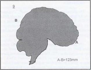 Figure 2. Scanned imprint from a brain slice used to calculate the surface of each slice before and after plastination in order to measure the shrinkage of the whole surface. |
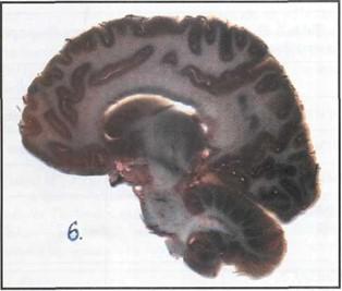 Figure 3. P40 plastinated brain slice after immersion at -25°C and impregnation at -25°C. |
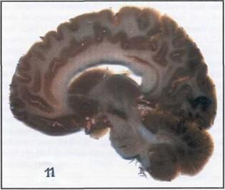 Figure 4. P40 plastinated brain slice after immersion at |
The aim of the present study was to compare 2 conditions of immersion and impregnation. Although we all know that dehydration leads to shrinkage (Schwab and von Hagens, 1981; Holladay, 1988; Tiedemann and Ivic-Matijas, 1988) we did not measure the shrinkage following dehydration for 2 reasons. First all 16 slices were dehydrated in the same way and second we considered that taking the slices out of the acetone to measure them could have produce drying of the slices, increasing of the shrinkage and could have damaged them.
The study suggests that immersion and impregnation of brain slices should occur at -25°C. Shrinkage can not be appreciated only by comparing linear distances. Precise appreciation of the degree of shrinkage can be obtained by area comparison or better by volumetric comparison. The shrinkage values determined in the present study represent only a two dimensional shrinkage. We all know that the total shrinkage of a specimen after plastination is three dimensional so the values for the real shrinkage will be greater than our findings.
The finding that the degree of shrinkage is about 4.41 % after immersion and impregnation at -25°C is excellent. P40 plastination is in our opinion the best method to preserve brain slices. Furthermore we think that the P40 technique is much better than the S10 standard technique for plastination of brain slices, where shrinkage can reach up to 10% (Suriyaprapadilok and Withyachumnarkul, 1997).
Even if the degree of shrinkage is 6.96% at room temperature the method could be used if a -25°C impregnation is not possible. Both shrinkage values are acceptable as we know that unsaturated polyester resins can usually shrink 5 - 8% during the transition from the liquid to the solid state (Arpe et al., 1992; Kirk-Othmer, 1996).
Arpe H-J, Schulz G, Elvers B ed.: Ullmann's Encyclopedia of Industrial Chemistry. 5th Ed., Weinheim, VCH publishers, vol 21, pp 217-225, 1992.
Barnett RJ: Plastination of Coronal and Horizontal Brain Slices using the P40 technique. J Int Soc Plastination 12 (1): 33-36, 1997.
https://doi.org/10.56507/YJVS5787
Cook P, Barnett R: Practical applications in plastination. Instructional videotape, Departments of Anatomy, University of Auckland and University of Otago, New Zealand, 1996.
Holladay SD: Experiments in dehydration technique. J Int Soc Plastination 2 (2): 17-20, 1988.
https://doi.org/10.56507/RSDZ6598
Kirk-Othmer: Encyclopedia of chemical technology. 4th Ed., New York, Wiley, vol 19, pp 654-677, 1996.
Schwab K, von Hagens G: Freeze substitution of macroscopic specimens for plastination. Acta Anat 111: 139-140, 1981.
Suriyaprapadilok L., Withyachumnarnkul B: Plastination of stained section of the human brain: Comparison between different staining methods. J Int Soc Plastination 12 (1): 27-32, 1997.
https://doi.org/10.56507/YISQ6047
Tiedemann K, Ivic-Matijas D: Dehydration of macroscopic specimens by freeze substitution in acetone. J Int Soc Plastination 2 (2): 2-12, 1988.
https://doi.org/10.56507/SCLL2742
von Hagens G: Plastination of brain slices according to the P40 procedure. A step-by-step description, pp. 1-23, 1994.
https://doi.org/10.56507/OWYV2878