1 School of Biological Sciences, University of Bristol, Bristol, UK.
2 School of Chemistry, University of Bristol, Bristol, UK.
The lifelike preservation of three-dimensional plant material poses particular challenges, and there is still no established method for it. The aim of the present study was to develop a method to preserve the trapping leaf of a carnivorous pitcher plant in its natural shape and coloration for long-term display in a public exhibition. Fresh pitchers were subjected to one of the following preservation methods: freeze-drying, coating in PDMS, and plastination. The resulting specimens were then compared against fresh and air-dried material. Plastination was found to be superior to the other preservation methods in yielding lifelike specimens for display. In particular, plastinates retained their shape better and exhibited no obvious shrinkage. However, the process altered the coloration significantly due to the loss of chlorophyll and mobilisation of anthocyanins (red–blue pigments) during the dehydration and impregnation stages. Exposure of the finished plastinated specimen to bright light also caused it to turn brown over a period of several weeks. Further work is needed to refine the procedures for plastination of botanical material. In particular, a method should be sought for fixing chlorophyll and other plant pigments. These issues notwithstanding, plastination shows promise as a 3D preservation method to supplement herbarium material and educational displays.
S10 method; room temperature plastination; freeze drying; PDMS coating; color retention
Michal R. Golos (michal.golos@bristol.ac.uk) and Anne-Kristin Lenz (anne-kristin.lenz@bristol.ac.uk), School of Biological Sciences, University of Bristol, 24 Tyndall Avenue, Bristol, BS8 1TQ, UK.

![]()


The importance of lifelike biological specimens as teaching tools has long been recognized. Preservation methods vary depending on the type of specimen. While vertebrates are typically stuffed (Péquignot, 2006) and invertebrates are either air dried and pinned, preserved in ethanol, or critical-point dried (Huber, 1998; Quicke et al., 1999), plant material is generally pressed and dried (Miller and Nyberg, 1955; MacFarlane, 1985). While this method yields durable and easy-to-store specimens, it is not well suited to highly three-dimensional organs such as pitcher plant traps (e.g. genus Nepenthes and Sarracenia) or complex flowers such as those of orchids or pipe vines (genus Aristolochia). Nepenthes pitchers are hollow, cup-shaped leaves (Cheek and Jebb, 2001; Clarke, 2001) specialized to capture and digest predominantly invertebrate prey (Moran and Clarke, 2010). Each pitcher is connected to the main leaf blade via a thin tendril (Fig. 1A). Other distinctive features are a pair of ventral ‘wings’, the collar-shaped pitcher rim (peristome), and a lid shielding the opening from rain (Clarke, 2001). Pressing dramatically alters these distinctive geometries, thereby obscuring taxonomically relevant morphological information, and potentially producing herbarium specimens of limited use for the identification of fresh plant material (Shivas, 1983; Lamb, 1989; Clarke and Moran, 2011).
Historically, these limitations spurred the production of botanical models and replicas, as perhaps best exemplified by the Blaschka “glass flowers” at Harvard, which number more than 4000 and are known for their craftsmanship and general scientific accuracy (Parke, 1983; McNally and Buschini, 1993). But even so, certain botanical inaccuracies have been noted (Rossi-Wilcox, 2008, 2015). Wax was also used to create lifelike replicas – a giant Nepenthes rajah pitcher was ‘preserved’ in this manner at the Royal Botanic Gardens, Kew (Nelson, 1991). While these techniques have their merits, it is desirable to preserve real botanical material wherever possible.
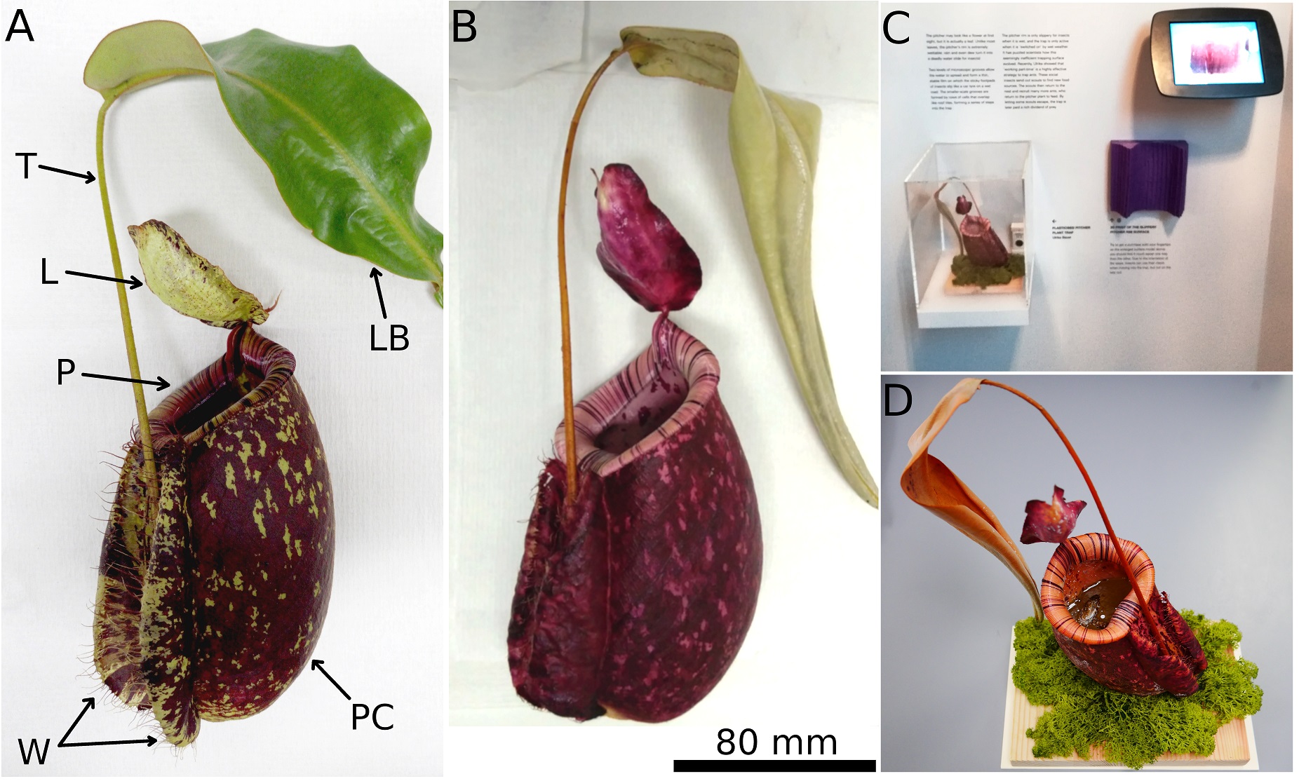 Figure 1. Pitchers of Nepenthes × hookeriana before and after preservation. A) Fresh pitcher. Labelled parts: Tendril, Lid, Peristome (pitcher rim), Wings, Pitcher Cup, Leaf Blade. B) Pitcher plastinated with the refined room temperature method. C) Final exhibit on display at Cambridge University Library, March 2019. D) Closeup of the same exhibit in September 2019. Note the color change of the pitcher and leaf which is likely due to bright light exposure, and the simulated pool of pitcher fluid with cockroach prey. Photographs A–C by MR Golos and A-K Lenz; D by Rachel Sawicki (Conservator, Cambridge University Library). |
Previous efforts to preserve pitcher plant traps in their three-dimensional form varied in outcome. Shivas (1983) freeze-dried pitchers at −50°C under vacuum, achieving good shape but only partial color retention. Stewart (2008) employed a method which, though described as such, was not true freeze-drying, as it involved placing the pitcher in an ordinary freezer at standard pressure. The method resulted in complete loss of coloration as well as lid and wing curling, as the specimens thawed and then dried following removal from the freezer. It was also a lengthy process, taking four to eight weeks (Stewart, 2008). Other methods include preservation with glycerin and encasing in blocks of acrylic. However, the former gives an unnaturally dark, oily appearance, while latter is a challenging material to work with (Stewart, 2008) and has the obvious disadvantage of precluding close examination.
Shanos (1985) and Stewart (2008) described a method of desiccating pitcher plants and other carnivorous plants by covering them in silica gel in a sealed container. The pressure exerted by the silica beads on all sides maintained the shape and orientation of delicate parts such as the lid and wings and prevented shriveling in thin-walled species. But the dehydrated specimens were very fragile (Shanos, 1985; Stewart, 2008) and sensitive to moisture, and required carefully monitored storage conditions. Protective coatings, while increasing durability, will alter both texture and optical appearance.
The aim of the present study was to develop a preservation method that could yield a lifelike, three-dimensional pitcher plant specimen for display at an exhibition of the Cambridge Philosophical Society. In particular, we explored the potential of plastination, a method that is unrivalled in the preservation of three-dimensional vertebrate specimens, from organs to whole bodies (von Hagens et al. 1987). Recently, this method was successfully applied to mushrooms (Diz et al., 2004a, 2004b; Looney and Henry, 2014; Henry et al., 2016). Plant plastination, however, remains considerably more obscure: while brief mentions are scattered across various publications (Shama Sundar, 2010; Henry et al., 2016) and even appear in the earliest patents related to the technique (von Hagens, 1978, 1980), to our knowledge it has not previously been detailed in the academic literature.
Plant material
Pitcher plants (Nepenthes × hookeriana) were grown in a climate-controlled chamber on a 12-hour photoperiod with an average daytime temperature of 30°C and 60% humidity, and a night-time temperature of 24°C and 80% humidity. Initial trials to compare different preservation methods were performed on eight pitchers, each approximately 10 cm tall. For the final display, we plastinated a pitcher of 24 cm height. Pitchers were cut from plants, and subjected to one of the below-described preservation methods within 30 minutes. The pitchers were handled by their tendrils whenever possible as this was deemed the part most resistant to mechanical damage. One pitcher was left to air-dry at room temperature for comparison.
Freeze-drying
A total of four pitchers were freeze-dried for 48 hours using a FreeZone 1L benchtop freeze-drying system (Labconco Corp.). Prior to freeze-drying, each pitcher was subjected to one of the following treatments: 1) submersion in liquid nitrogen (−196°C) until bubble formation ceased; 2) submersion in 20% ethylene glycol solution for several days followed by submersion in liquid nitrogen; 3) freezing at −80°C; and 4) freezing at −20°C. These temperatures were chosen as they are readily achievable in common labs. The rationale behind rapid cooling with liquid nitrogen was to produce amorphous ice, thereby minimizing crystal formation and associated tissue damage.
PDMS coating
A single freshly cut pitcher was submerged in absolute ethanol and kept at −20°C to dehydrate the specimen. After a week it was removed from the freezer, submerged in polydimethylsiloxane (PDMS; Sigma Aldrich) and, using a nylon string, hung upside down in an oven at 40°C for three days to cure the coating.
Plastination
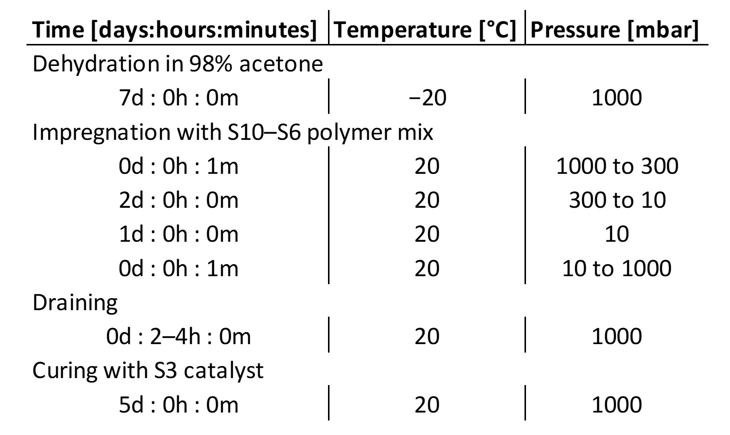 Table 1. Time, temperature and pressure parameters for the refined plastination method for pitcher plants. |
In total, four pitchers were plastinated. For the first, we followed the room temperature method for plastination as described by Henry (2007b) and Looney and Henry (2014). This method comprises three main steps: dehydration, impregnation, and curing. In order to improve the result, we slightly modified this method for the remaining three specimens by omitting the initial acclimation step during the impregnation phase (Table 1). Both acetone (used in the standard method) and ethanol were trialed as dehydrating fluids. While both were found to mobilize green (chlorophyll) and red pigments (anthocyanins) to some degree, acetone appeared to have a less severe effect and was therefore used for all further plastination work.
The first pitcher was dehydrated in 98% acetone at −20°C for seven days, then transferred to a polymer mix of ten parts S10 polymer and one part S6 cross-linker (Biodur), and allowed to equilibrate overnight. It was then placed in a vacuum chamber (Vacuum Oven Digital; Fistreem International) with an attached diaphragm vacuum pump (Rotavac Vario Pumping Unit; Heidolph Instruments) and the pressure rapidly reduced to 300 mbar. Over the following 48 hours the pressure was incrementally decreased to 10 mbar, where it was held for an additional 24 hours to ensure complete replacement of the acetone by the polymer mix. After that, the pressure was increased back to atmospheric pressure within a minute before the pitcher was removed from the polymer bath, hung upside down, and left to drain for several hours.
The specimen was then sprayed with S3 catalyst (Biodur) on its inner and outer surfaces and wrapped in cling film to create a saturated environment in which the vaporized catalyst could more efficiently diffuse into the specimen. The catalyst treatment was repeated three times over the course of five days. Paper towels were used to wipe away excess catalyst from the outer surface of the specimen.
When plastinating subsequent pitchers, the acclimation step between dehydration and impregnation was omitted, to improve the result for the thin plant material. Further refinements to the curing step were made when plastinating the large pitcher for the exhibition. The catalyst was sprayed only on the inside of the pitcher as well as on the undersides of the lid and leaf. Spraying only one side of each surface in this way was sufficient to initiate the desired chain reaction while minimising the exposed areas that might retain a liquid layer of excess catalyst following curing. When wrapping in cling film, particular care was taken not to deform the fragile lid and wings; a loose roll of cling film was placed between the wings to help maintain their shape. Finally, the large pitcher was partially filled with polymer mix to simulate a pool of pitcher fluid, complete with insect ‘prey’ (Fig. 1D). This also helped maintain the structural integrity of the exhibit upon curing.
Evaluation
The shape and color of the specimens resulting from different preservation methods were monitored and recorded over a total of four months (one month for the exhibit pitcher). A qualitative assessment of the changes was made by visual comparison against fresh pitchers. At the end of this period, images of all specimens were taken. All specimens were kept at 20°C in a room with low-intensity artificial lights. Additionally, the exhibit pitcher was monitored for the first few weeks of the exhibition, during which time it was kept in a Perspex box and exposed to natural light (Fig. 1C), and examined after the conclusion of the six-month exhibition (Fig. 1D).
None of the tested methods was able to deliver a fully lifelike result, but plastination with the aforementioned refinements yielded the best specimen for display (Fig. 2). While some methods excelled at preserving color, others retained the original shape better. In comparison to a fresh sample (Fig. 2A), all drying approaches (air drying and freeze drying with and without prior treatment) resulted in some degree of shrinkage, and specimens became brittle. Particularly obvious deformations were curled-up wings, shrunken pitcher rims and tendrils, and downward folding of the lid (Fig. 3).
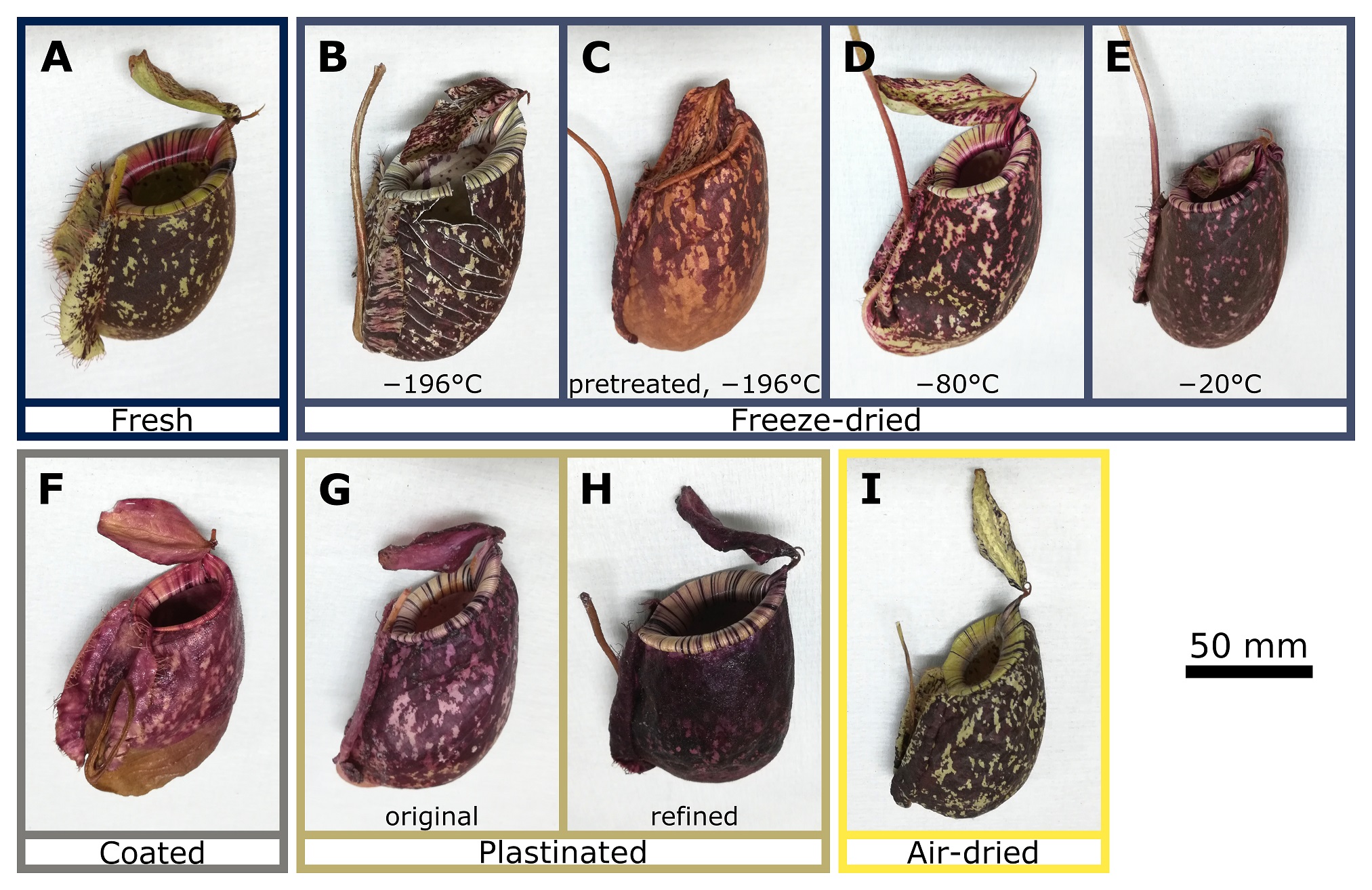 Figure 2. Comparison of Nepenthes × hookeriana pitchers subjected to different preservation methods. A) Fresh pitcher. B–E) Freeze dried after (B) plunge-freezing in liquid nitrogen (−196°C), (C) submerging in ethylene glycol for several days and plunge-freezing in liquid nitrogen (−196°C), (D) freezing at −80°C, and (E) freezing at −20°C. F) Dehydrated in ethanol, then PDMS coated. G) Plastinated at room temperature following the original method. H) Plastinated at room temperature using our refined method (omitting the acclimation step after dehydration). I) Air dried without further treatment. Photographs A–I by M. Golos and AK. Lenz |
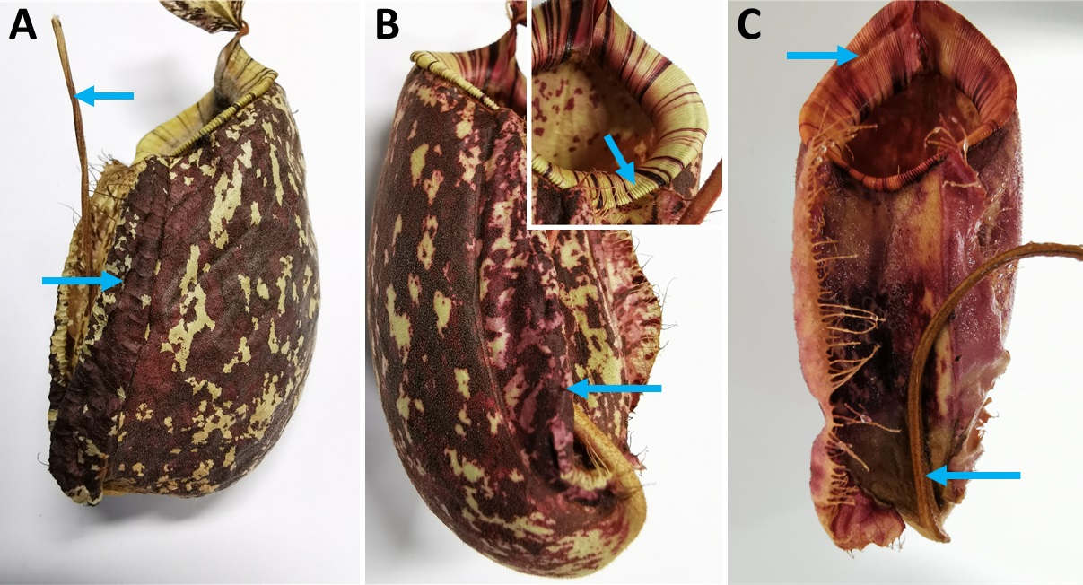 Figure 3. A) Air-dried pitcher, showing shriveled tendril (top arrow) and wings (bottom arrow). Note the uneven surface of the pitcher body resulting from shrinkage, which nonetheless showed excellent color retention. B) Pitcher freeze dried at −80°C, showing buckling of peristome surface (top arrow; inset) and pitcher wing deformation (bottom arrow). While the pitcher body did not contract significantly, ‘bleeding’ of red pigments was obvious. C) PDMS-coated pitcher, showing severe drying artefacts affecting the peristome (top arrow) and tendril (bottom arrow). Color retention was very poor, though the wings were surprisingly well preserved. |
Shrinkage was most severe in the air-dried pitcher (Figs. 2I, 3A), but color retention was better than for any other method. The liquid nitrogen-treated, freeze-dried pitcher (Fig. 2B) shrank less but developed multiple long cracks across the pitcher cup. Color retention was good. Ethylene glycol treatment (Fig. 2C) reduced the cracking, but the resulting specimen gradually lost its natural color and turned brown over the course of several weeks. Pitchers frozen at −80°C (Fig. 2D) and −20°C (Fig. 2E) showed significant shrinking of the pitcher rim (Fig. 3B) but otherwise good shape retention, particularly those treated at −80°C. Cracks did not occur with this method; however, color retention was poorer with increasing temperature, and red pigments leached out into adjacent tissues.
PDMS coating (Fig. 2F) did not cause cracking and achieved generally good shape retention. The delicate wings were preserved well in their natural position; however, the pitcher rim shrank more than in all other preservation methods apart from air drying (Fig. 3C). Coating with PDMS preserved the red pigments well, but led to a complete loss of green color. In addition, the lower half of the pitcher turned brown during the coating process. In contrast to all other methods, the PDMS coating also resulted in an unnaturally glossy appearance.
Plastination using the standard room temperature method (Fig. 2G) led to slight shrinkage, with the rim and wings affected most severely. Omitting the overnight acclimation phase after the transition from acetone to polymer mix (Fig. 2H) eliminated this shrinkage almost entirely. However, both plastination methods caused severe discoloration. The green chlorophyll was lost during dehydration in acetone, and the red pigments moved within the tissue, which led to a red-colored pitcher with increased saturation. In comparison to the dried pitcher, the plastinated samples were less brittle, but the wings in particular remained fragile as they consist of very thin tissue. Stuffing cling film between the wings prevented them from rolling inwards and improved the result in the final exhibit (Fig. 1B).
Apart from the bleaching of green to near-white, the final plastinated exhibit (Fig. 1B) appeared lifelike. However, in contrast to previous plastinated pitchers that were kept under low-light conditions at 20°C, the final exhibit gradually turned brown over the six-month course of the exhibition (Fig. 1D). This might be a result of the exposure to stronger lighting or higher temperatures in comparison to our storage conditions. To disentangle the relative contributions of light and temperature, we kept three plastinated pitchers in different environmental conditions for a period of two weeks and recorded the temperature continuously. The results suggest that high light intensity rather than increased temperature contributed to the discoloration observed in the exhibit.
We successfully adapted and applied the plastination method for tissue preservation to a three-dimensional plant organ for the first time. Because the tissue of our specimen was not more than a few millimeters thick, the procedure was much faster than that for e.g. zoological specimens or mushrooms (the room-temperature process for the latter taking around three weeks; Looney and Henry, 2014). The entire plastination process took only two weeks. Our refined method yielded a reasonably robust specimen with good shape retention and intense red pigmentation. In terms of 3D shape preservation, plastination proved superior to air drying, freeze drying and PDMS coating. However, freeze drying at both −196°C and −80°C achieved better color preservation, especially of the green pigments. Both plastination and freeze-drying therefore excelled in different aspects of preservation. Since Nepenthes pitcher coloration is highly variable in nature (McPherson, 2009), exact color preservation was deemed less important than natural shape retention in an educational context, and the refined plastination method was used for the final exhibit.
Several additional modifications could and should be trialed to further improve the results of both methods. For freeze drying, the use of a dry ice-acetone cooling bath could help to maintain the temperature of the sample stable at −78°C. This might help to eliminate shrinkage. For plastination, fixation of the plant tissue in glutaraldehyde or FAA (formaldehyde, acetic acid, and ethanol) could be trialed. In addition, an acetone dilution series could be tested as a milder alternative to direct transfer into 98% acetone. Cold temperature plastination (de Jong and Henry, 2007; Henry, 2007a; Looney and Henry, 2014) could also lead to improved results.
For use in a public exhibition environment, light-induced discoloration was a significant problem. The color change only became apparent after two to three weeks. Therefore, the procedure is not currently suitable for the preparation of long-term exhibits with exposure to strong natural or artificial lighting, and further research is necessary to understand the effects of light and temperature more thoroughly. Nevertheless, plastination can be useful for educational purposes whenever it is not possible to have a live plant, particularly for structures whose three-dimensional shape is essential for explaining their biological function, as for carnivorous plant traps, kettle trap flowers, or flowers with moving parts that play an essential role in their pollination biology.
Plastinated pitchers could also potentially serve a scientific function and be deposited alongside type material in herbaria. Under adequate cool and dark storage conditions, browning should be reduced or prevented entirely. Plastinates could supplement pressed specimens in the same way as “wet”, alcohol-preserved specimens. By providing vital information about 3D structure, plastinated specimens could facilitate taxonomic identification and enhance functional understanding. Future work should investigate the applicability of this method to other plant species and tissues.
Room-temperature plastination was successfully adapted and used to preserve the three-dimensional pitcher trap of a carnivorous plant (Nepenthes × hookeriana). The resulting specimen (Fig. 1B–D) was displayed at the Cambridge Philosophical Society exhibition (University Library, Cambridge) from March to September 2019 (Dean, 2019). Color changes were observed during both the dehydration and impregnation steps of the plastination process, and significant browning occurred post-curing, most likely as a result of light exposure. Further refinements are needed to better preserve the natural pigmentation. Other methods such as drying, freeze-drying and PDMS coating obtained inferior results with regard to shape preservation, but freeze-drying at low temperatures (−196° C and −80° C) yielded better color retention.
Acknowledgements
We thank Christopher Burgess, Rachel Sawicki and the rest of the team at Cambridge University Library for their tremendous work in putting together the pitcher plant display, which was seen by approximately 56,000 visitors during the six months of the exhibition.
Cheek MR, Jebb MHP. 2001: Flora Malesiana. Series I - Seed plants. Volume 15: Nepenthaceae. Leiden, Netherlands: Nationaal Herbarium Nederland. iv + 164 p.
Clarke CM. 2001: Nepenthes of Sumatra and Peninsular Malaysia. Kota Kinabalu, Malaysia: Natural History Publications (Borneo). x + 32 p.
Clarke CM, Moran JA. 2011: Incorporating ecological context: a revised protocol for the preservation of Nepenthes pitcher plant specimens (Nepenthaceae). Blumea 56(3): 225-228.
https://doi.org/10.3767/000651911X605781
Dean K. 2019: Discovery: 200 Years of the Cambridge Philosophical Society. Cambridge University Library Special Collections. Available from: https://specialcollections-blog.lib.cam.ac.uk/?p=17330
de Jong K, Henry RW. 2007: Silicone plastination of biological tissue: cold-temperature technique - Biodur™ S10/S15 technique and products. J Int Soc Plastination 22: 2-14.
https://doi.org/10.56507/ZLMJ7068
Diz A, Martinez-Galisteo A, Berlango J, Conde-Pérez A. 2004a: Some aspects on fungi plastination. Murcia, Spain: 12th Int Conf Plastination. (Abstract in J Int Soc Plastination 19: 55-56.)
Diz A, Martinez-Galisteo A, Sanchez-Rodriguez M, Conde-Pérez A. 2004b: Plastination of fungi as an aid in teaching botanic classification. Murcia, Spain: 12th Int Conf Plastination. (Abstract in J Int Soc Plastination 19: 55.)
Henry RW. 2007a: Silicone plastination of biological tissue: cold temperature technique - North Carolina technique and products. J Int Soc Plastination 22: 15-19.
https://doi.org/10.56507/DGZJ6845
Henry RW. 2007b: Silicone plastination of biological tissue: room-temperature technique - North Carolina technique and products. J Int Soc Plastination 22: 26-30.
https://doi.org/10.56507/FSNZ3092
Henry RW, Wilton J, Iliff S. 2016: Plastination of fungi and fragile biological specimens. Toledo, US: 18th Int Conf Plastination. (Abstract in J Plastination 28: 22.)
Huber JT. 1998: The importance of voucher specimens, with practical guidelines for preserving specimens of the major invertebrate phyla for identification. J Nat Hist 32(3): 367-85.
https://doi.org/10.1080/00222939800770191
Lamb R. 1989: Herbarium samples and preserving CP specimens. Carniv Plant Newsl 18(3): 83, 85-86.
Looney B, Henry RW. 2014: Fruitbody Worlds, plastination of mushrooms. Fungi 7(1): 45-49.
MacFarlane RBA. 1985: Collecting and Preserving Plants for Science and Pleasure. New York, US: Arco Publishing. viii + 184 p.
McNally RS, Buschini N. 1993: The Harvard Glass Flowers: materials and techniques. J Am Inst Conservat 32(3): 231-240.
https://doi.org/10.1179/019713693806124893
McPherson SR. 2009: Pitcher Plants of the Old World: Volume One. Poole, UK: Redfern Natural History Productions. xvi + 631 p.
Miller AG, Nyberg JA. 1995: Chapter 27 Collecting herbarium vouchers. In Collecting Plant Diversity
Technical Guidelines, Guarino L, Ramanatha Rao V, Reid R (eds.). CAB International [for updated version see Davis 2011] p 561-573.
http://cropgenebank.sgrp.cgiar.org/images/file/procedures/collecting1995/Chapter27.pdf
Miller AG, Nyberg JA. 1995: Collecting herbarium vouchers. In: Guarino L, Ramanatha Rao V, Reid R, editors. Collecting Plant Diversity: Technical Guidelines. Wallingford, UK: CAB International, p 561-573.
Moran JA, Clarke CM. 2010: The carnivorous syndrome in Nepenthes pitcher plants. Plant Signal Behav 5(6): 644-648.
https://doi.org/10.4161/psb.5.6.11238
Nelson EC. 1991: The waxing of a glorious rajah. Curtis's Bot Mag 8(2): 81-89.
https://doi.org/10.1111/j.1467-8748.1991.tb00361.x
Parke M. 1983: The glass flowers of Harvard's Botanical Museum. Endeavour 7(3): 116-122.
https://doi.org/10.1016/0160-9327(83)90003-0
Péquignot A. 2006: The history of taxidermy: clues for preservation. Collections 2(3): 245-255.
https://doi.org/10.1177/155019060600200306
Quicke DL, Lopez‐Vaamonde C, Belshaw R. 1999: Preservation of hymenopteran specimens for subsequent molecular and morphological study. Zool Scr 28(1-2): 261-267.
https://doi.org/10.1046/j.1463-6409.1999.00004.x
Rossi-Wilcox SM. 2008: From reference specimen to verisimilitude: the Blaschkas' penchant for botanical accuracy. J Historical Biol 20(1): 11-18.
https://doi.org/10.1080/08912960701677432
Rossi-Wilcox SM. 2015: A brief history of Harvard's Glass Flowers Collection and its development. J Glass Stud 57: 197-211.
Shama Sundar NM. 2010: "Plastination" - by Indigenous Method. Plastination.in. Available from: http://plastination.in/plastination_article.html
Shanos GT. 1985: A simple technique for the preservation of CP. Carniv Plant Newsl 14(3): 66-67.
Shivas RG. 1983: Preservation of Nepenthes pitchers by freeze drying. Carniv Plant Newsl 12(3): 62-63.
Stewart SE. 2008: Freeze-drying carnivorous plant pitchers at home. Carniv Plant Newsl 37(2): 47-51.
von Hagens G. 1978: DE patent 2710147: Preserving human, animal or plant specimens - by impregnation with a polymerisable plastic material without affecting outline. European Patent Office. Available from: https://worldwide.espacenet.com/publicationDetails/biblio?CC=DE&NR=2710147
von Hagens G. 1980: US patent 4205059: Animal and vegetal tissues permanently preserved by synthetic resin impregnation. Google Patents. Available from: https://patents.google.com/patent/US4205059A/en
von Hagens G, Tiedemann K, Kriz W. 1987: The current potential of plastination. Anat Embryol (Berl) 175(4): 411-421.
https://doi.org/10.1007/BF00309677