Anatomisches Institut, Karl-Franzens-Universitat Graz, Harrachgasse 21, A - 8010 Graz, AUSTRIA
The study of sectional anatomy of the human body goes back to the earliest days of systematic topographic anatomy. The beautiful drawings of sagittal sections of the pregnant uterus and of horizontal sections of the lower limb by Leonardo da Vinci (1452 - 1519) are well known (Fig. 1). These are the first known examples of cross-sectional anatomy. Due to the softness and the fast process of decomposition of the nervous tissue and for lack of hardening reagents or freezing method, da Vinci was not able to do brain sections. That is why Leonardo da Vinci only described a method for injection of the ventricles, but the structures of the brain itself stayed unexplored. The main handicap to detailed sectional studies was, of course, the problem of fixation.
P35; Da Vinci: Brain;
Andreas H. Weiglein Anatomisches Institut, Karl-Franzens-Universitat Graz, Harrachgasse 21, A - 8010 Graz, AUSTRIA

![]()


The study of sectional anatomy of the human body goes back to the earliest days of systematic topographic anatomy. The beautiful drawings of sagittal sections of the pregnant uterus and of horizontal sections of the lower limb by Leonardo da Vinci (1452 - 1519) are well known (Fig. 1). These are the first known examples of cross-sectional anatomy. Due to the softness and the fast process of decomposition of the nervous tissue and for lack of hardening reagents or freezing method, da Vinci was not able to do brain sections. That is why Leonardo da Vinci only described a method for injection of the ventricles, but the structures of the brain itself stayed unexplored. The main handicap to detailed sectional studies was, of course, the problem of fixation.
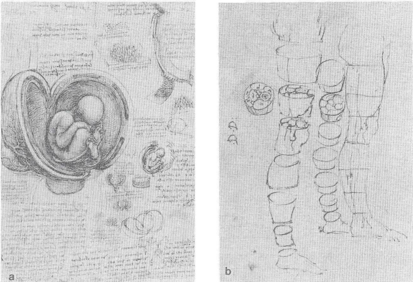 Figure 1. First known examples of cross-section anatomy by Leonardo da Vinci (1452- 1519) of the pregnant uterus (a) and of the lower limb (b). |
The use of formaldehyde as a hardening and preserving fluid was introduced by Gerota in 1895 (Kaiserling, 1895), but formaldehyde is not harmless. At the least, it causes burning of the nasal mucosa, reddening of the conjunctiva, and toxic edema of the lung; at the worst, it may cause cancer (Pabst, 1985). Furthermore, formaldehyde-fixed brains have poor distinction between grey and white matter and need constant care because they gradually deteriorate. At the turn of the century thermoplastics, including paraffin, were used as
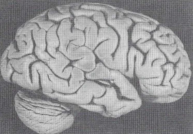
Figure 2. Photograph of a S 10 plastinated brain.
preservatives. However, biological substances shrink in baths of hot paraffin, grow opaque and loose their fine detail, including distinction of grey and white matter. Paraffin-embedded slices are unwieldy and, because of their thinness, not appropriate to obtain a general view. Several slices were needed to get a three- dimensional impression and, hence, were cumbersome for students usage.
In 1977, when Gunther von Hagens invented plastination, it was a special year for all anatomists, as well as, for all students of medicine (von Hagens, 1979a, b; Bickley, 1981 ). Almost all of the problems were solved at once. The S 10 standard technique allowed the production of dry, clean, odorless, durable and, above all, nontoxic and noncarcinogenic samples of all organs including the brain (Fig. 2). The one remaining problem - the distinction between grey and white matter - was
solved only a few years later. A slightly changed method made it possible to replace the water in the cells of the nervous tissue with a polyester resin called P 35 (von Hagens, 1987, 1990). This resin has an index of refraction which causes a unique grey-white distinction in direct and transmitted light as well (Fig. 3).
Plastination procedures (Table 1)

Figure 3. Photograph of a P 35 plastinated horizontal brain slice. Note the well defined Lentiform nucleus: ar ro w - Gl ob us pall id us a n d p - Putamen, and Thalamic nuclei:
* - medial nuclei, v - ventrolateral nuclei and arrowhead - pulvinar nuclei.
The principles of the P 35 technique are almost the same as of standard plastination: fixation, dehydration, forced impregnation and curing (von Hagens, 1987). To avoid shrinkage, the brain slices are immersed twice in the P 35 resin before forced impregnation. Only the curing procedure is different from that of the S 10 technique, because styrol would evaporate during polymerization and the polymerization temperature would destroy the specimen. For these reasons, brain slices are placed into flat glass chambers for curing. Furthermore, instead of gas, light and temperature are used for curing. As I pointed out during the 5th conference in Heidelberg, it is possible to use only temperature, because light curing needs a lot of time (Weiglein, 1990), To gain a better distinction between grey and white matter in S10 brain slices, staining procedures with Astra blue or aldehyde fuchsin are also recommended (Ulfig, 1989, 1990).
| S 10 - STANDARD TECHNIQUE | P 35 - TECHNIQUE |
| FIXATION DEHYDRATION FORCED IMPREGNATION CURING (gas) |
FIXATION DEHYDRATION IMMERSION in P35 IMMERSION in P35 FORCED IMPREGNATION CURING (light & heat) |
The use of plastinated brain specimens (Table 2)
In Graz, we use S10 standard plastinated whole and half brains, as well as, 4 mm thick P 35 brain slices. The samples are used in teaching students and postgraduate radiologists and neurosurgeons; S 10 brains are very helpful in topographic anatomy, because you can put them into the skull and realize the topographic relations directly (Resch, 1990). S 10 and P 35 specimens are applied, of course, in the lectures on neuroanatomy and in the neuroanatomic courses. Plastinated specimens are well accepted by students of medicine as well as by physiotherapists, medical laboratory and x- ray technicians. To show the principles of brain circulation and . the special anatomy of the blood vessels of the brain, P 35 slices are helpful, when the vessels are not rinsed before fixation, because the red blood cells become black during plastination and so no extra injection is needed.
| (L) Anderhuber F: (L) Anderhuber F: (L) Weiglein A: (p) Anderhuber F, Reimann R, Tesch P' Weiglein A: (L) Reimann R: (L) Tesch P: (L) Weiglein A: (L) Weiglein A: |
Neuroanatomy Special Anatomy of blood vessels of the brain Fundamentals of radiology anatomy and CT Neuroanatomy course Neuroanatomy for Medical Laboratory Technicians Neuroanatomy for Hospital Nurses Neuroanatomy for Medical X-Ray Technicians Neuroanatomy for Physiotherapists |
| (L) Lecture, (P) Practical course | |
In my lectures on "Fundamentals of Radiographic Anatomy and Computed Tomography", P 35 slices are best to use to compare anatomic and CT-slices (Cooper, 1990).
Students of medicine, as well as, physiotherapists, medical laboratory and x- ray technicians can compare plastinated specimens using drawings without staining their books, their clothes or their hands. It has been my experience that students have gained a better knowledge of neuroanatomy since we began using plastinated specimens. This is further evidenced, by the cleanliness and convenience of the samples, which induces a closer look, allowing the student a more detailed study of the specimen (Lischka, 1981; Purinton, 1990; Scott, 1990).
| Plastinated specimens are: | ||
| Dry Clean Odorless |
Durable Material- Saving No special care needed |
Non-Toxic Nor-Carcinogenic |
Upon questioning students about comparing plastinated brains to formalin- fixed brains, you get one evaluation value for plastinated specimens: 100%, and one value for formaldehyde specimens: 0%. A summary of the main advantages of plastinated specimens in comparison with formaldehyde-fixed specimens, is presented in Table 3. Plastinated specimens are dry, clean and odorless; therefore, comfortable to use. Because of their durability, these plastinated specimens help save material. Also, because these specimens need no care, as well as being easy to store, help to save time. Moreover, since P 35 slices have an excellent grey-white distinction, they have the clarity of macroscopic slices and are almost as detailed as histologic slices. Last but not least, they are nontoxic and noncarcinogenic.
Bickley, HC, G von Hagens, FM Townsend: An improved method for preserving of teaching specimens. Arch Pathol Lab Med 105:674-676, 1981.
Cooper, M: The technique and use of plastinated specimens in teaching and research: Gross anatomical sections of the head and neck. Presented at The 5th International Conference on Plastination, Faculty of Medicine, University of Heidelberg, Germany July 1990 J Int Soc Plastination 4(1 ):4, 1990.
von Hagens, Gunther: Emulsifying resins for plastination. Der Praparator 25/H.2:43-50, 1979.
von Hagens, Gunther, K Tiedemann, W Kriz: The current potential of plastination. Anat Embryol 175:411-421, 1987.
https://doi.org/10.1007/BF00309677
von Hagens, Gunther: Principles of the P 35-technique. 5th International Conference on Plastination , Heidelberg, 1990.
Kaiserling, C: Uber die Konservierung von Sammlungspraparaten mit Erhaltung der natiirlichen Farben. Fortschr Anat Entw Gesch 2(2):221, 1895.
Lischka, MF, R Mayr, W Mayerhoffer: Erfahrungen mit "plastinierten" Praparaten im Anatomieunterricht. Acta Anat 111:92, 1981.
Pabst, R: Welche Gefahren bestehen beim Umgang mit Formaldehyd? Anat Anz 161:154, 1986.
Purinton, FT: The use of plastinated specimens in teaching veterinary gross anatomy: substitution or supplementation. Presented at The 5th International Conference on Plastination, Faculty of Medicine, University of Heidelberg, Germany July 1990 J Int Soc Plastination 4(1): 11, 1990.
Resch, KDM, A Pernecsky: The use of plastinated head specimens in planning microsurgical approaches to the skull and brain base. Presented at The 5th International Conference on Plastination, Faculty of Medicine, University of Heidelberg, Germany July 1990 J Int Soc Plastination 4(1): 12, 1990.
Scott, TM: Plastinated specimens in medical anatomical teaching. Presented at The 5th International Conference on Plastination, Faculty of Medicine, University of Heidelberg, Germany July 1990 J Int Soc Plastination 4(1 ):13, 1990.
Ulfig, N, M Wuttke: Plastination of stained sections of the human brain. Anat Anz 170:309-312, 1990.
Ulfig, N: Staining of human fetal and adult brain slices combined with subsequent Plastination. J Int Soc Plastination 4(1):33-38, 1990.
https://doi.org/10.56507/THKE9781
Weiglein, A: P 35-technique without light curing. 5th Int Conf on Plastination, Heidelberg, 1990.