Department of Anatomy and Radiology, College of Veterinary Medicine, University of Georgia
A combination of computer assisted learning modules with a series of plastinated dissections of sheep and dog brains has been developed at the University of Georgia, College of Veterinary Medicine for use in teaching the laboratory portion of veterinary neuroanatomy.
The objectives of this project were to improve efficiency and reduce frustration of students while they attempt to identify structures and pathways on transverse sections of the brain and correlate them three-dimensionally with the whole brain.
Computer assisted; Plastination; Brain; Neuroanatomy
P. T. Purinton Department of Anatomy and Radiology, College of Veterinary Medicine, University of Georgia

![]()


A combination of computer assisted learning modules with a series of plastinated dissections of sheep and dog brains has been developed at the University of Georgia, College of Veterinary Medicine for use in teaching the laboratory portion of veterinary neuroanatomy.
The objectives of this project were to improve efficiency and reduce frustration of students while they attempt to identify structures and pathways on transverse sections of the brain and correlate them three-dimensionally with the whole brain.
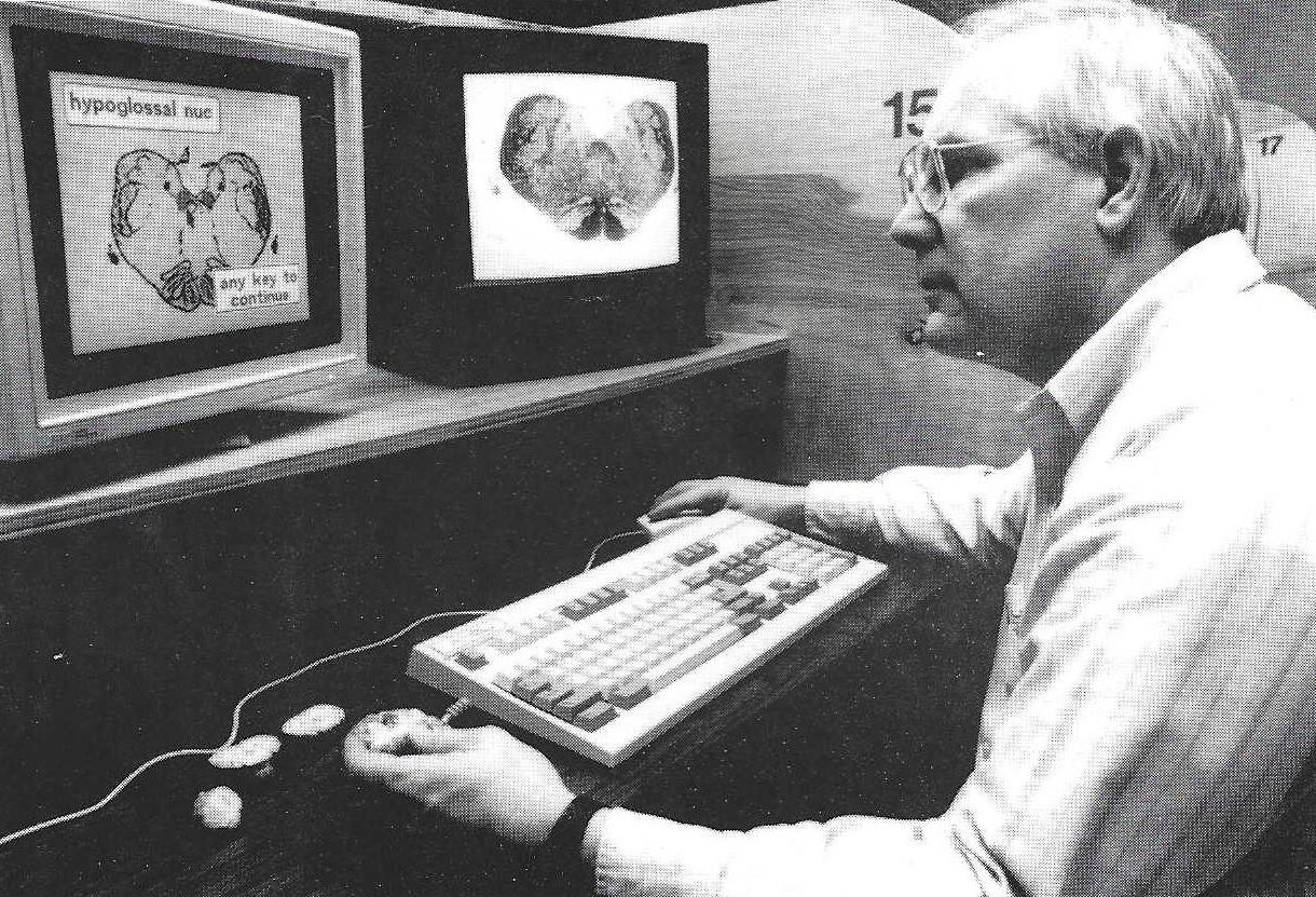
Figure 1. Work station with 286 PC computer, laser video disk player, and dual monitors.
A series of 13 modules, covering all of the structures and pathways studied, has been generated. Using minimal text, each module will lead the student through a pathway via a series of video disk images of transverse sections through a dog brain. The video disk images were in turn integrated with labeled graphic illustrations which identified structures on transverse sections or whole brains. Additional instructions and graphics direct students to identify structures on plastinated tissues available at each work station. The Computer Assisted Learning Center (CALC) at the University of Georgia, College of Veterinary Medicine has 16 work stations available. Each has a 286 PC compatible computer with 20 MB hard disk and VGA graphics display, Pioneer LD- V4200 video disk player, and Sony color Video monitor (Fig. 1).
Brains of sheep and dogs were prepared using the standard S10 silicone rubber technique (von Hagens, 1985). A set of plastinated specimens for a work station included (Fig. 2): a sheep brain and dog brain, each had one cerebral hemisphere and one-half of the cerebellum removed to expose the brain stem; a dissected sheep brain illustrating the hippocampus, caudate nucleus, and lateral ventricle; a sheep brain with cerebral cortex removed to demonstrate the corona radiata and corpus callosum; and (Fig. 3) two sheep brains cut into transverse sections. For the transverse sections, each brain was cut at three levels such that the set of two brains illustrates sections at six different levels.
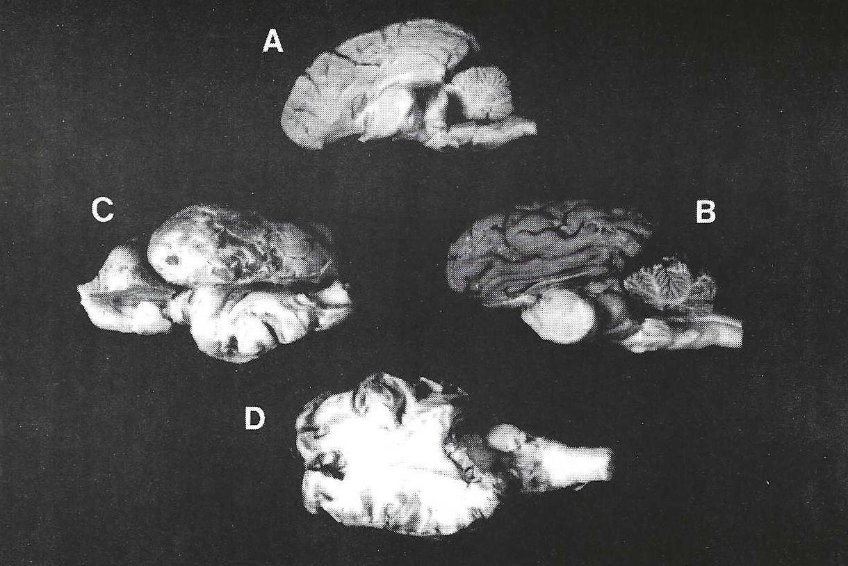 Figure 2. A. Dog brain; B. Sheep brain; C. Deep gray matter of sheep brain; D. Corpus callosum and corona radiata of sheep brain. |
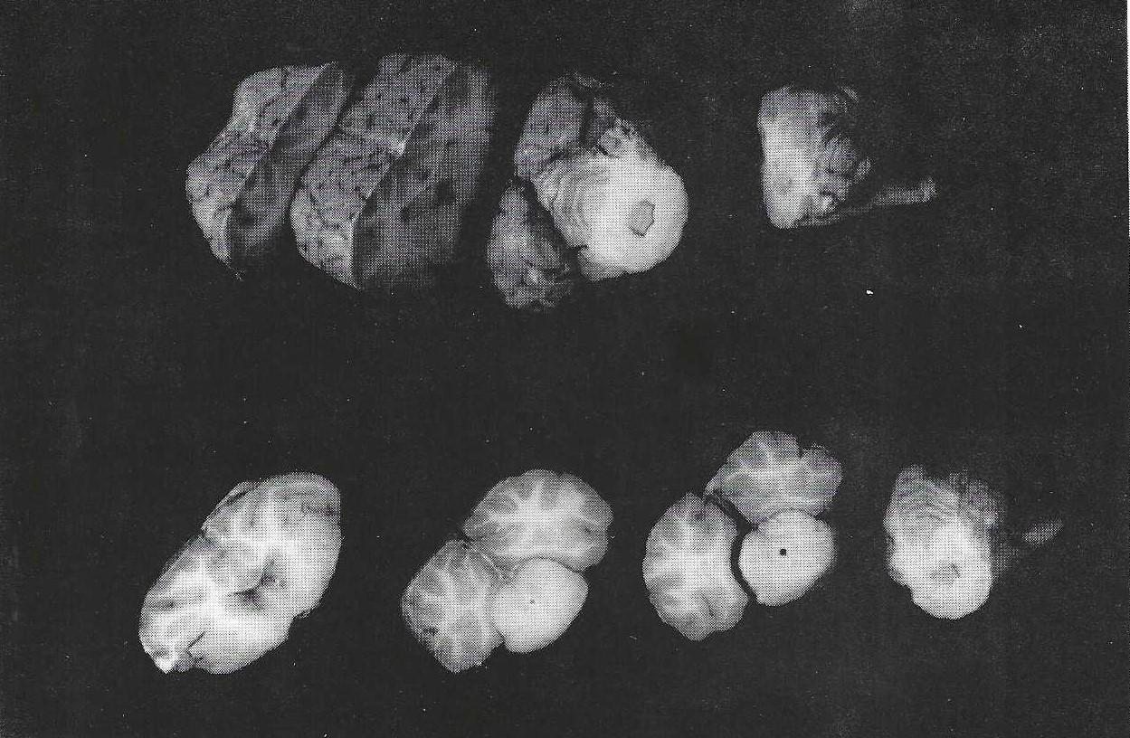 Figure 3. Sheep brains, transverse sections. |
The modules have been used two years with revision prior to the second year.
Plastinated tissues are ideal for use in the computer assisted learning center. They are dry, therefore, there is no risk of spilling formalin or other liquids on the delicate electronic equipment. Plastinated specimens are aesthetically acceptable to the students which encourages hands-on usage. Absence of specialized storage methods insures ready access. For the transverse sections, each brain was cut at only three levels so that each slice of brain was two centimeters or more in thickness. Previous usage suggested that thinner sections were too fragile for student use.
Prior to this project, students worked in the neuroanatomy laboratory in groups of four with formalin fixed sheep brains and a series of 2 X 2 slides of stained transverse sections of the brain. Even though students had a laboratory guide, textbooks, and line drawings of the transverse sections, it was observed that with only two faculty members for eighty students, a great deal of time was wasted waiting for assistance to answer questions or confirm identification. Frustration and irritation were expressed by the students because of long periods of waiting for an instructor and of questionable identification of structures which were later found to have been incorrectly identified.
The use of computer assisted learning modules with plastinated tissues has several advantages for both the students and instructors. For the students, there is little wasted time in working through the modules. Errors in identification are reduced by having a labeled drawing on the computer screen linked with an unlabeled slide on the video disk monitor. The combining of plastinated tissues as an integral part of each computer lesson is important, because historically students have tended to separate the study of the gross brain from the study of the histologic sections of the brain. This often resulted in failure to synthesize the two parts into an integrated whole. By using onscreen instructions and labeled graphics to direct students to plastinated tissues, integration is facilitated.
Students generally work in groups of two or three which encourages discussion and interaction. Students have textbooks, lecture notes, illustrations of pathways, and unlabeled line drawings to use in conjunction with the computer and plastinated materials thus allowing multiple modes of learning.
Instructors also have noted that the number of questions has been dramatically reduced. Two instructors were unable to keep up with student requests for help in the old system, but in the computer lab, questions are easily handled by the two instructors and often one faculty member would be sufficient. Questions that are asked tend to be more substantive rather than just asking for confirmation of structures which are being identified. Although the volume of information has not been reduced, fewer students have expressed a feeling of inability to master the subject.
In addition to subjective evaluation by the instructors, students were asked to score specific statements evaluating the system on a six point scale, with a six being in agreement with the statement and a one being disagreement. Two of the statements and the percentile of responses for each score are given in Table 1.
| Score: | 6 | 5 | 4 | 3 | 2 | 1 |
| 1. Modules were over all helpful: | ||||||
| 1990 | 52.8% | 30.5% | 5.5% | 8.4% | 0 | 2.8% |
| 1991 | 90.9% | 9.1% | 0 | 0 | 0 | 0 |
| 2. Plastinated brains were helpful in correlating three dimensional structure with transverse sections | ||||||
| 1990 | 16.7% | 30.6% | 25.0% | 19.4% | 2.8% | 5.5% |
| 1991 | 69.6% | 17.4% | 8.7% | 4.3% | 0 | 0 |
In general, students found the computer programs and plastinated brains very helpful in learning neuroanatomy. The improvement in scores on both statements (especially the second statement) for the second year are a reflection of the modifications implemented. Improvements consisted of increasing the number of plastinated specimens available at each work station and adding on-screen instructions and labeled illustrations specifically for the plastinated tissues. Additional dissections and more labeled illustrations should further improve student evaluation.
The modules are installed on the hard drive of the CALC computers and the plastinated brains are on library reserve, thus students have access to these learning materials outside of assigned class time, whenever the reading room is open and the computer center is not otherwise occupied. This also allows access by upper class students wishing to review details of neuroanatomy for courses such as clinical neurology.
The next major addition to this system is to develop a series of interactive self-evaluation and quiz modules. This will allow students to monitor their progress in mastering the subject matter.
von Hagens, G: Heidelberg Plastination Folder: Collection of all technical leaflets for plastination. Anatomisches Institute 1, Universitat Heidelberg, 1985.