1- Department of Pathology, Faculty Of Medicine, Khon Kaen University, Khon Kaen, Thailand.
2- Department of Anatomy, School Of Medicine, University Of Auckland, Auckland, New Zealand.
3- Anatomisches Institute I, University Heidelberg, Heidelberg, Federal Republic of Germany
The modified standard silicone technique (S-10) has been used to plastinate our pathology specimen s for teaching and museum exhibition proposes. All steps have been carried out at room temperature in Thailand where the average temperature is about +25°C to +30°C. The specimens have been collected, fixed in 10% buffered formalin for at least 1 week and dehydrated in increasing grades of ethanol (50%, 60%. 70%. 80%, 90% and 100%), with one week in each concentration, and then 2 baths of acetone for complete dehydration and degreasing. The specimens remain in each acetone bath for about 2 weeks until water content is below 1%. Forced impregnation and gas curing have also been done at room temperature. It takes about 2 weeks for forced impregnations, 1 week for pre-cure and 2 weeks for gas curing. The total time required is about 16 weeks.
The resulting plastinated specimens are dry to the touch, odorless, durable, life-like and non-toxic. They do not deteriorate with time, are maintenance free, and resistant to damage from mechanical strain caused by handling. The finished specimens maintain their original shape and are close in color and consistency. They can be stored at room temperature indefinitely so they are suitable for exhibition proposes. They are useful in the teaching of pathology especially when used in small groups and self directed learning. This technique provides satisfactory results with a minimum of equipment.
Plastination, standard silicone technique , room temperature, preservation of biological specimens.
Churairat Kularbkaew Department Of Pathology, Faculty of Medicine, Khon Kaen University, Khon Kaen, Thailand

![]()


Plastination is a relatively new process (1982) that is now widely used to preserve perishable biological specimens with high water content. In this technique, tissue water and lipid are replaced with curable polymers. The completely impregnated specimen is cured by a gaseous vapor.
Dr. Gunther von Hagens of the University of Heidelberg, Germany, developed the suitable polymers and four variations of plastination techniques based on the same fundamental process providing for difference of specimens, (von Hagens, 1979a, b, 1985/1986, 1987). Silicone impregnated specimens (S-10) are resilient and flexible and are mainly used in teaching purposes. Whole organs, limbs, student prosections and even whole bodies may be plastinated with the silicone impregnation method. Specimens produced with polymerized emulsions (P.E.M.) are as opaque as the silicone specimens but are rigid and to some extent breakable. The use of this technique is in production of thick body slices exhibiting a sharp contrast be- tween fat tissue, which shows up white, and all other more intensively stained parenchyma. Transparent body or organ slices are produced with epoxy resins. This process is known as E-12. For research purposes, these 2.5 mm. to 4.5 mm. thin slices which allow macroscopic study of the topography of all body structures with the naked eye in an uncollapsed and non-dislocated state. In addition, the specimens are useful in advanced training programs in sectional topography (resident training in CT and NMR). As with specimens produced with polymerized emulsions, the epoxy imbedded specimens are cured with heat. Opaque brain slices (P-35 process) of 4 mm. thickness, are impregnated with polyester resin, and allow a unique discrimination between fiber and nuclear areas as a direct result of contrast properties within the resin activated by curing the specimen by U.V.A. light sources.
The general procedure of Standard silicone technique S-10 (von Hagens, 1985/1986) has 4 steps.
In Thailand, because of the limited availability of some equipment, especially the large deep freezer (-25°C), we try to modify the standard silicone technique to allow all steps to be carried out at room temperature (average room temperature is about +25 to +30°C).
We use surgical specimens that are sent to the department of pathology for pathological diagnosis. The specimens are fixed in 10% buffered formalin at room temperature. Polymer (S-10), hardener (S-3) and gas cure (S-6) are required . They are only available from Biodur (Heidelberg, F.R.G.) Ethanol and acetone are also required.
FIXATION
The pathology specimens have been collected and fixed in 10% buffered formalin at room temperature for at least 1 week. In this step, care is taken that the specimens maintain their natural shape, or the shape they will exhibit when finished.
DEHYDRATION AND DEGREASING
We use stepwise dehydration in increasing graded ethanol (50%, 60%, 70%, 80%, 90% and 100%, one week for each concentration), and then 2 baths of acetone for complete dehydration and degreasing. The specimens remain in each acetone bath for about 2 weeks at room temperature or until water con tent is below 1%.
FORCED IMPREGNATION
This is the most important step in plastination, in which the intermediate solvent (acetone) is replaced by curable polymers by means of a vacuum during forced impregnation. The curable polymer is composed of a silicone base material (S-10) and hardener (S-3). We use 10 Kg. of S-10 mixed with 0.1 L. of S-3. Many kinds of tissue with varying thickness from 0.5-10 cm can be done at the same time. It takes about 2 weeks at room temperature.
GAS CURING
The specimen s are removed from the mixture of cur able polymers and then wiped of excess polymers. The specimens remain in pre-cure at room temperature for about 1 week to allow the excess polymer to drain off. After that, the specimens are cured by a silicate containing gas, evaporating from a fluid (S-6) for about 2 weeks. This step ends when the specimens become solid and dry to the touch.
The total time required is approximately 16 weeks.
This technique provides satisfactory results with mini mum equipment, because all steps can be done at room temperature. This technique can be used in all laboratories without a large deep freezer (-25°C).
The resulting plastinated specimens are dry to touch, odorless, durable, lifelike and non-toxic. They do not deterio rate with time, are maintainance free and resistant to damage caused by handling. They maintain their original shape and are close in color and consistency (figure 1 and 2). They can be stored at room temperature indefinitely. They are useful in the teaching of pathology, especially when used in small groups and are suitable for exhibition purposes as museum specimens.
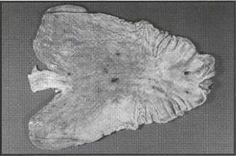 Figure1- Plastinated specimen of stomach showing stress ulcer. |
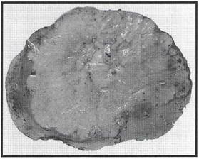 Figure 2- Plastinated specimen of liver showing cholangiocarcinoma mass. |
The specimens preserved by plastination techniques are superior to those preserved in formalin. In this method we modify the standard silicone technique so that all steps can be done at room temperature. In the step of fixation we use surgical specimens that are sent to the department of pathology for pathological diagnosis. As the specimens can be used in the plastination process only after complete diagnosis, fixation in 10% buffered formalin at room temperature for at least 1 week is necessary. This makes the tissue firm, and reduces susceptibility to shrink age in the subsequent plastination process. However, little change of color can be observed. This problem can be solved by using the Kayserling-Fixation method or staining with BIODUR STAIN C (von Hagens 1985/1986).
Dehydration in graded ethanol causes considerable shrinkage and is time consuming (Schwab and von Hagens, 1981; von Hagens, 1987) This problem is reduced by using the specimens fixed in 10% buffered formalin for a long time and not over dehydrated. The specimens which have high fat content, such as breast tissue, still shows shrinkage.
Following dehydration and degreasing, the cellular spaces within the specimens originally filled with water and lip ids are now occupied by a volatile intermediary solvent (acetone). In forced impregnation the intermediary solvent is replaced by curable polymers by means of a vacuum. The intermediary solvent has a high vapor pressure and a low boiling point (acetone: +56°C), while the polymer solution has a low vapor pressure and a high boiling point. Therefore, when a vacuum is applied, only the intermediary solvent is continuously extracted gradually out of the specimens and through the surrounding polymer solution in the form of gaseous bubbles. (von Hagens, 1987). It takes about 2 weeks to complete forced impregnation. Various types of tissue with varying thicknesses (0.5-10 cm) can be done at the same time. The residual polymer in the force impregnation bath can be reused. In this experiment the step of forced impregnation is carried out at room temperature so the viscosity of the polymer increases. This problem is solved by mixing the curable polymers with acetone before reuse for a new batch of specimens.
Following forced impregnation, the specimens are cured by a silicate containing gas, evaporated from a fluid S-6. The hardener S-3 contained in the impregnation bath initiates the curing of the silicone molecules by end to end polymerization. Due to crosslinking during the final gas curing, the silicone rubber within the specimen will become solid and dry (von Hagens, 1987). This step ends when the specimens become dry to the touch. The degree of flexibility of the finished specimen is de pendent on the ratio of S-3 hardener to the number of days of the S-6 gas curing.
This technique provides both a satisfactory result with minimum equipment and can be done in all laboratories without a large deep freezer (-25YC).
Bickley HC, von Hagens G, Townsend FM: An improved method for the preservation of teaching specimen Arch Pathol Lab Med 105:674-676, 1981.
Bickley HC, Walker AN, Jackson RL, et al: Preservation of pathology specimens by silicone plastination . An innovative adjunct to pathology Am J Clin Pathol 88:220-223, 1987.
https://doi.org/10.1093/ajcp/88.2.220
Dawson TP, James RS, Williams GT: Silicone plastinated pathology specimens and their teaching potential. J. Pathol 162:265-272, 1990.
https://doi.org/10.1002/path.1711620314
von Hagens G: Impregnation of soft biological specimens with thermosetting resins and Anat Rec 194: 247-256, 1979a.
https://doi.org/10.1002/ar.1091940206
von Hagens G: Emulsifying resins for plastination . Der Praparat 25:43-50,
von Hagen s G: Heidelberg Palstination Anatomisched Institut I, Universitat Heideberg, D-6900 Heidelberg, FRG, 1985/1986.
von Hagens G, Tiedemann K, Kriz W: The current potential of plastination . Anat Embryol 175: 411-421, 1
https://doi.org/10.1007/BF00309677
Hawley DA, Marlin DC, Cook DC, et al: Specimens for teaching forensic pathology , odontology and I. Soft tissue. Am J Forensic Med Pathol. 12:164-169, 1991.
https://doi.org/10.1097/00000433-199106000-00015
Pond KR, Holladay SD, Luginbuhl JM: Technical note: preservation of tissues and gastrointestinal tract portions by plastic coating or plastination J. Anim Sci 70:1011-1014, 1992.
https://doi.org/10.2527/1992.7041011x
Schwab KH, von Hagens G: Freeze substitution of macroscopic specimens for plastination. Abstract: Sixth European Anatomical Congress, Acta Anat 111, 12:339, 1981.