Clinic of Neurosurgery, University of Heidelberg, West Germany (before: Anatomical Institute I)
The evolution of scientifically prepared anatomical specimens for teaching has been beset with conceptual problems since its beginning. The principle difficulty has always been, "to see or not to see". Leonardo da Vinci demonstrated that "to see is to understand". In 1487, he proposed two principles for anatomical study of the cranium: 1-"There is an art to rendering invisible structures "2- "Thought must be used as a method of communicating anatomical concepts and the anatomical aspect must show a concept" .
It seems that sketches and photographs, even accurate models, often are too far removed from reality to understand anatomy for practical, clinical purposes. During the study of medicine at the University of Heidelberg, an acquaintance with Dr. Gunther von Hagens and the novel art of plastination (von Hagens, 1979a, 79b, 82, 85, 86, 87) was begun. Plastination was recognized as a means of combining both accuracy and beauty for depicting and preserving anatomical principles with unprecedented clarity. At this juncture, we began to prepare plastinated specimens of the cranium and use them in clinical experiences. This paper is essentially a chronicle of these experiences.
art; S10; Plastination; Biodur; Silicone
D. M. Resch Clinic of Neurosurgery, University of Heidelberg, West Germany (before: Anatomical Institute I)

![]()


The evolution of scientifically prepared anatomical specimens for teaching has been beset with conceptual problems since its beginning. The principle difficulty has always been, "to see or not to see". Leonardo da Vinci demonstrated that "to see is to understand". In 1487, he proposed two principles for anatomical study of the cranium: 1-"There is an art to rendering invisible structures "2- "Thought must be used as a method of communicating anatomical concepts and the anatomical aspect must show a concept".(Fig. 1). A third de facto principle is based on religion. Since prehistoric times, efforts have been made to preserve perishable materials, no doubt reflecting a universal desire for eternal life. But, what is life without beauty? And thus, a compromise, a durable life form with lasting beauty emerged as Italian wax models (Fig.2). The example in Figure 2 was produced by Paolo Mascagni during the 13th century, in Siena, Italy. It can be seen today in the Josephinum in Vienna.
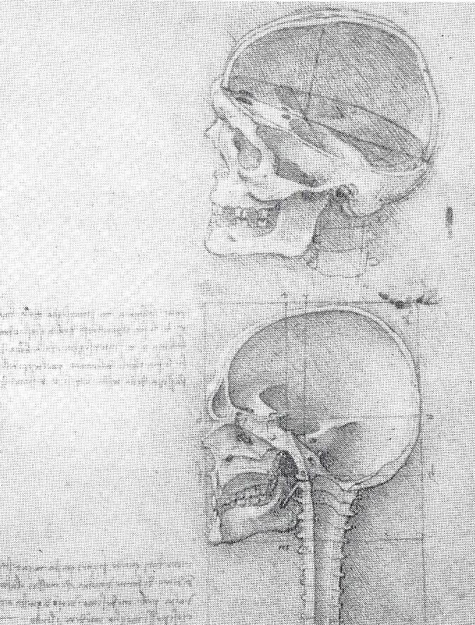 Figure 1. 1487 anatomical graphs by Leonardo da Vinci. This type of preparation shows normally invisible structures, not only the anatomical aspect but also a concept of a topographical system that helps to understand the gestalt of the skull (Hoffman 1979, Royal bibliothek of Windsor castle). |
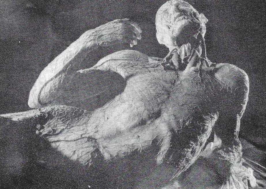 Figure 2. 1457 anatomical wax model by Paolo Mascagni which shows the lymphatic vessels of man. Using his 3-D method, anatomical detail and beauty of life was preserved by the Italian renaissance (Josephinum - Medical Historical Institute, University of Vienna). |
It seems that sketches and photographs, even accurate models, often are too far removed from reality to understand anatomy for practical, clinical purposes. During the study of medicine at the University of Heidelberg, an acquaintance with Dr. Gunther von Hagens and the novel art of plastination (von Hagens, 1979a, 79b, 82, 85, 86, 87) was begun. Plastination was recognized as a means of combining both accuracy and beauty for depicting and preserving anatomical principles with unprecedented clarity. At this juncture, we began to prepare plastinated specimens of the cranium and use them in clinical experiences. This paper is essentially a chronicle of these experiences.
The amputated, formal in-fixed cranium is supported on the dissection table and the operator views it through a dissection microscope equipped with a photo adapter to take pictures of various levels of dissection. Instruments are selected to carry out the dissection through a keyhole that may be as deep as 15 cm.
The dissection microscope is indispensable for such an intricate procedure. The magnification and the illumination provided, enable the operator to prepare the finest possible dissection while working in an ambience of microanatomic objects. This by itself is an excellent learning experience. Even with the use of this microscope, considerable skill and patience must be developed, not only in dissecting but in holding the same posture for many hours.
Four specimens have been prepared, all of which were plastinated with S10 and are now on exhibition.
Cranium I: This specimen was perfused with formalin, dissected, and plastinated. The veins, arteries and nerves were highlighted after plastination with lacquer color. The dissected specimen was prepared to demonstrate general syntopy and topography (Figs. 3, 4).
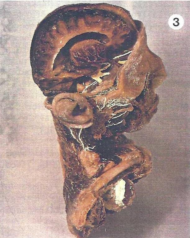 Figure 3 . Right view of plastinated cranium #1, showing deep structures of the brain and a superficial preparation of the face and collum. |
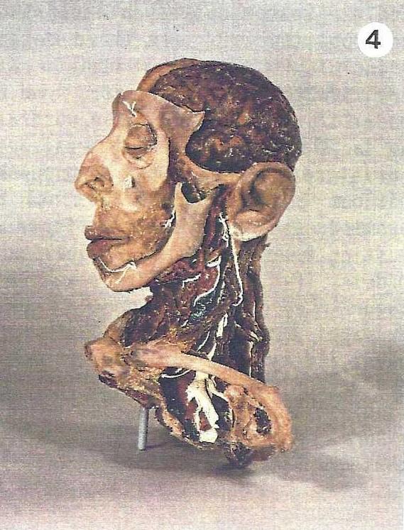 Figure 4. Left view of cranium #1, showing a superficial preparation of the brain and the deep structures of the face and collum. |
Cranium 2: The vessels were injected with colored PEM prior to amputation. The specimen was fixed by immersion in formalin and perfusion of the ventricles and subarachnoid space, dissected, and plastinated. The clinical anatomy of anterior approaches to the brain stem are demonstrated (Figs. 5, 6). One approach to the eye was too small to observe without magnification and was enlarged after plastination.
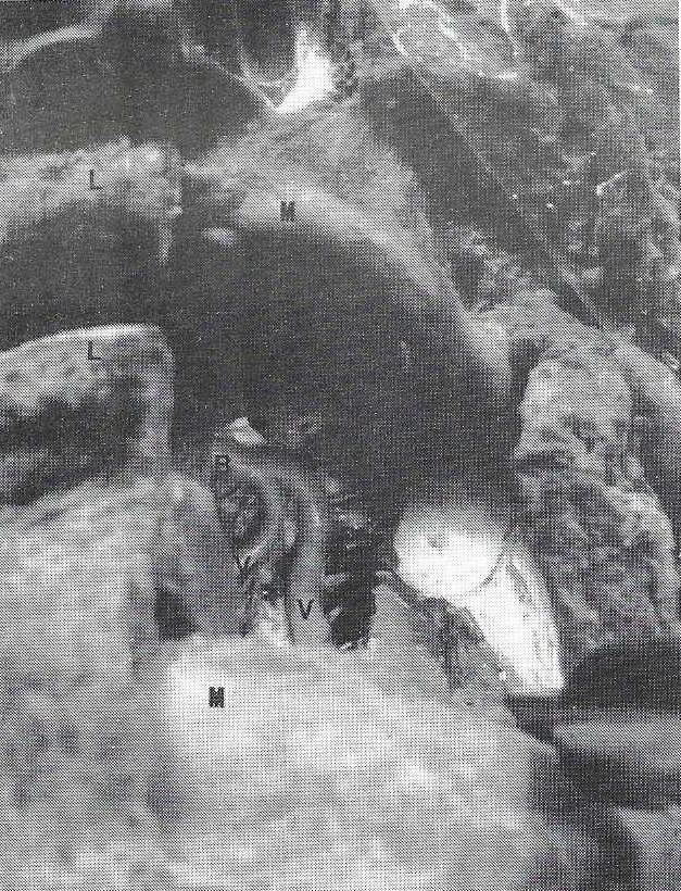 Figure 5. Close up PEM injected, S10 plastinated cranium #2. Demonstrating a transoral approach to the base of the brain and the associated vessels. |
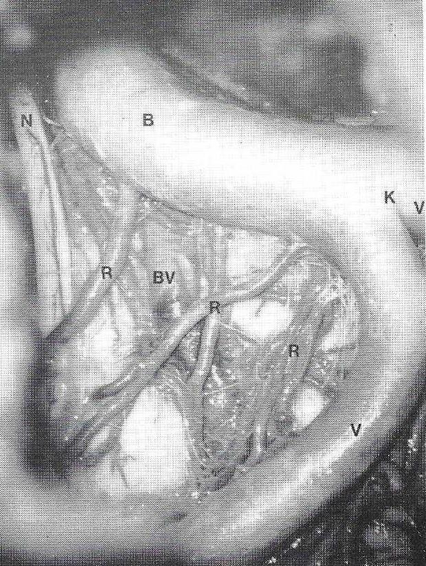 Figure 6. High magnification of figure 5. The vertebral arteries (K) forming the basilar artery |
Cranium 3: Contrast media was injected via the carotid and vertebral arteries and angiographic, radiographic and computer tomography analysis were done to gain anatomical information for the microdissections and for later comparison with the plastinated specimen. Later the vessels were injected with colored PEM. The specimen was fixed via immersion and perfusion of the ventricles and, subarachnoid space, dissected and plastinated. The clinical anatomy: of the base of the cranium and the brain are demonstrated.
Cranium 4: After amputation, the neck was stabilized with a cuff. To ensure fixation and to stabilize the larger vessels, 150 ml of 4% formalin was injected. The vessels were injected with colored silicone [S 10 + S3 (1%) + S6 (2%) + color (1%)] and not with PEM. Polymerization time was 20 hours. After dissection of the lateral approaches to the base of the cranium, the specimen was plastinated.
Four anatomical specimens which demonstrate accurate, three- dimensional structure and are not fragile were produced. What can one do with these products?
Each of the four plastinated specimens has been used to demonstrate the clinical anatomy of microsurgical approaches to the cranium and the brain on numerous occasions. They have been displayed in the "anatomical exhibition center" of the Institute of Anatomy at the University of Heidelberg. The specimens have been used for anatomical study by medical and nursing students and anatomical examination of medical students.
Neurosurgeons at the Clinic for Cranial Surgery of Heidelberg, as well as, surgeons in other departments used the specimens for their clinical training and review of the regional clinical anatomy. The specimens were used for similar reasons by the neurosurgical clinic of the University of Vienna and by the departments of ENT and Neurosurgery at the University of Zurich.
Because of having introduced plastination to the area of clinical anatomy of the cranium, the author was invited to participate and demonstrate the use of plastinated craniums in two international workshops "Microanatomy applied to neurosurgery" and "Micro- surgery applied to neurosurgery". The craniums have been shown and used for demonstrations during three conferences: the "Third International Conference on Plastination", San Antonio, Texas, 1986 and the "Sixth International Symposium: Neurological Surgery of the Ear and Skull Base", Zurich, 1988. The fourth plastinated cranium was prepared especially for the Zurich conference and to complete the set of microsurgical approaches to the cranium. At the 40th conference of the German Society for Neuro- surgery, Wurzburg, 1989, Cranium 2 was used in conjunction with a poster on "Transoral approach to the brain stem".
For review of anatomy and training as a neurosurgeon, the plastinated craniums have great value to the author. He would prefer not to use wet specimens in formalin or alcohol. With the plastinated models, his spatial imagination can be stimulated every day without added expense.
Since the introduction of the microscope to surgery in 1886 and the first application of constructive microsurgery to the human nervous system in 1957 (Donaghy, 1979), the approaches to the brain and the cranium have become more and more sophisticated. Complex approaches are demonstrated much better using the plastinated craniums than with graphs or slides, since the craniums show the approach and associated structures in three-dimension. To date, this is the best method to demonstrate the spatial problems associated with microsurgical approaches to the head. With each usage of the plastinated heads, overwhelming positive responses are obtained and their originality and accuracy applauded. For teaching nurses and medical students anatomy, no better method or specimens have been found. Experienced surgeons have confirmed the unprecedented clarity of the plastinated heads.
In the future, to solve spatial problems inherent in sophisticated approaches to the cranium, production of more prosected plastinated specimens which highlight the clinical anatomy of the cranium will be indispensable. At the clinic of head surgery at the University of Heidelberg, plastinated craniums will soon be used to plan operations and to develop new approaches to the brain and cranial base. "To see" the plastinated craniums "is to understand" what the author is talking about.
Donaghy RMP: The History of micro- surgery. Clin Neurosurg 26:619- 625, 1979.
https://doi.org/10.1093/neurosurgery/26.CN_suppl_1.619
Hoffman W, E Schaar, C Pedretti, K Keele: Leonardo da Vinci, Anatomische Zeichnungen asu der Koniglichen Bibliothek auf SchloB, Windsor. Prisma 1979.
von Hagens G: Impregnation of soft biological specimens with thermosetting resins and elastomers. Anat Rec 194:247-256, 1979a. https://doi.org/10.1002/ar.1091940206
von Hagens G: Emulsifying resins for plastination. Der Praparator 25:43-50, 1979b.
von Hagens G: Verfahren zur verbes- serten Ausnutzung von Kunststoffen bei der Konservierung biologischer Praparate. Offenlegungsschrift DE3232761, Deutsches Patentamt, Munchen, 1982.
von Hagens G: Heidelberg Plastination Folder: Collection of all technical leaflets for plastination. Anato- misches Institut 1, Universitat Heidelberg, 1985.
von Hagens G: The current potential of plastination. Anat Embryol 175:411-421, 1987.
https://doi.org/10.1007/BF00309677