School of Medicine University of Auckland Auckland, New Zealand
The development and perfecting of plastination techniques in the Department of Anatomy, at the Auckland School of Medicine, has provided new avenues for the production of specimens for teaching of modern clinical anatomy. Particularly, the preparation of cross sections of the human trunk and brain, which when accurately correlated with modern diagnostic imaging techniques, provides an invaluable teaching aid.
Plastinated specimens have also proven to be of great value providing a means of producing teaching material which can be kept for longer periods of time, with little or no maintenance, than can traditional prosected specimens. This helps to assure us that we will be able to provide specimens for teaching in the future even though we may experience budget restraints or depleted bequests from donors.
Plastination; P35
Peter Cook School of Medicine University of Auckland Auckland, New Zealand

![]()


The acquisition, use and disposal of bequeathed human material in New Zealand is governed by strict regulations specified in the Human Tissue Act of 1964 (Dept. of Health, New Zealand, 1965). The University of Auckland is fortunate to have an adequate supply of body donors and because of this, a valuable teaching resource has been established in its medical school.
The use of teaching specimens prepared using plastination techniques has become an essential component of this resource. The use of plastinated specimens has greatly assisted students in their understanding of anatomy and their being able to correlate these specimens with radiographical images of the human body. Using these plastinated specimens has resulted in the establishment of self directed learning stations which the students may use for their study of anatomy.
PLASTINATION METHODS
In 1983, in the Department of Anatomy at the University of Auckland, School of Medicine, experiments in plastination were carried out on cadaveric specimens using the PEM 27 and PEM 30 methods (von Hagens, 1985), however, these methods gave varying results. Initial trials using the S-10 technique (von Hagens, 1985) and ethanol for dehydration also produced mixed results.
After acquiring an explosion proof deep freezer the S-10, ethanol dehydration procedure was discontinued in favor of the standard S-10, freeze substitution method of plastination (von Hagens, 1985). All subsequent S-10 plastination has been done in this manner.
The majority of specimens acquired for plastination were obtained from embalmed, dissected cadavers. A smaller number of specimens were retrieved from fresh cadavers during autopsies and were used for plastination with good results.
Experiments were carried out using various types of fixatives (i.e. Wentworths' Fixative, Jores' Fixative). We found that an arterial embalming fluid formulated as outlined in a publication entitled "An Improved Method of Embalming Suited to Plastination Techniques" by Cook and Dawson (1995) was the most suitable for our purposes.
Until 1993 all forced impregnation of specimens to be plastinated was carried out at room temperature, at 26°C, in a converted laboratory centrifuge, sealed, vacuum tight, and with a 3cm thick clear perspex lid Plastination at room temperature allowed quicker impregnation of specimens than that carried out in a deep freezer at -25eC. However, because S-10 resin polymerizes more readily at room temperature, it became necessary to find an alternate method to cut down on the expense of buying new polymer.
The acquisition of funding and the design and construction of a new vacuum infiltration chamber gave us the capability of doing all forms of plastination. The chamber was constructed of stainless steel and measured 92.0 cm 42.0 cm. It is covered with a toughened glass plate lid which is hinged to the foam rubber gasketed rim of the chamber. A Javac 60 L/minute double stage vacuum pump is connected to the chamber and the vacuum is controlled by a Whitey needle valve situated between the chamber and the pump. Pressure readings are obtained using a low vacuum gauge during the initial stages of infiltration and nonmeter gauge in the latter stages of infiltration when readings of 1.0 mm of Hg or lower are required. Pump oil is changed after each S-10 impregnation run or after 3-4 runs of E-12 or P-35. Changing of the oil is very important because it removes all traces of acetone or methylene chloride which may have been deposited in it during the infiltration process. These deposits can be harmful to the pump seals. This type of forced impregnation system has proven extremely effective.
We have found that slow seepage, of greasy residues (probably a combination of bone marrow and phenol-glycerine components of the embalming solution used) from some S-10 plastinated specimens has been a problem in the past. To combat this, specimens are immersed in a mixture of 50-90% ethanol containing 5-20% Hydrogen Peroxide. This aids in extracting of embalming fluid ingredients prior to dehydration. Specimens are processed using the standard S-10 technique (von Hagens, 1985).
At present many old and valuable dissected prosections are being processed using the standard S-10 silicone method.
At the Auckland School of Medicine strong emphasis has also been placed on the use of serial sectioned cadavers and organs for teaching. These specimens have been used in correlation with computerized tomography (CT) scans, magnetic resonance imaging (MRI), ultrasound scans and traditional x-rays for teaching and instruction. Serial horizontal sections of head and neck, trunk and pelvis are being presented to the students in large display units for demonstration purposes (Figs. 3&4). Recently, 25 coronal specimens of the head and neck have been added. These specimens are displayed in removable mounts with corresponding annotated photographs and radiographs.
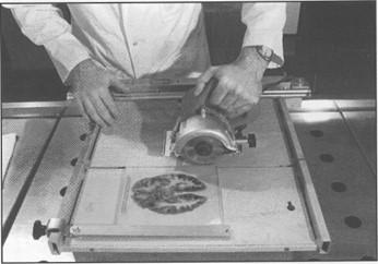
Cutting and Finishing
Head specimens, serial sectioned in the coronal plane and plastinated using the E-12 method (Weber, 1993), have been particularly well received for study purposes by both the students and staff. Certain aspects of the intricate anatomical structure of the human head, not clearly identifiable in traditional prosected cadaver material, have been emphasized using these specimens.
Brain sections, plastinated using the P-35 technique (Weber, 1994) have also been produced. These specimens not only show superb white and gray matter differentiation (Fig. 5) but they are free of formaldehyde odor and have a clean, smooth finish which is aesthetically pleasing to the eye.
Department of Health, New, Zealand. 1965. Memorandum on the Human Tissue Act 1964. Government Printer 1896- 65G, Wellington.
Weber, W 1994. Sheet plastination of brain slices. J Int Soc Plastination 8(1):23
Weber, W. 1993. Sheet plastination. Workshop proceedings, 3rd. Interim Conference on Plastination, Mobile, Alabama.
von Hagens, G. 1985. Heidelberg Plastination Folder: Collection of all technical leaflets for plastination. Universitat Heidelberg.
von Hagens, G. 1970a. Impregnation of soft biological specimens with thermosetting resins and elastomers. Anat Rec 194:256-257.
von Hagens, G., Tiedermannm, K., Kriz, W. 1987. The current potential of plastination. Anat Embryol 175:411-421.