University of Durban Westville, South Africa
In collaboration with the clinical Department of Orthopaedics at the University of Natal we have developed a method to simultaneously show the ossification of bone and the elaborate arterial network in the fetus during its development. We specifically wanted to elaborate on the arterial supply of the odontoid peg (dens) of the axis vertebra. This technique uses a combination of procedures involving fetus clearing, bone staining, arterial opacification (Thompsett, 1970), and Plastination (von Hagens, 1985).
Bone; Clearing; KOH
M.R. Haffajee University of Durban Westville, South Africa

![]()


In collaboration with the clinical Department of Orthopaedics at the University of Natal we have developed a method to simultaneously show the ossification of bone and the elaborate arterial network in the fetus during its development. We specifically wanted to elaborate on the arterial supply of the odontoid peg (dens) of the axis vertebra. This technique uses a combination of procedures involving fetus clearing, bone staining, arterial opacification (Thompsett, 1970), and Plastination (von Hagens, 1985).
The process developed can be broken down into the following stages:
Stage 1: Radiological Opacification of the Arterial Tree
An injection media was prepared by combining radio- opaque barium sulphate (Micropaque) with commercial latex (1:1). This injection media is used because of its low viscosity. The amount required for a 900g fetus is approximately 10-20 ml.
To begin the dissection and exposure of the arterial system, using a fresh warmed fetus, enter the thoracic cavity using an inverted 'V shaped incision in the region of the thoracic inlet. Remove the thymus gland, which is quite large, by blunt dissection. Take care not to cut any small vessels as this will prevent leakage when injecting them.
Locate and ligate the internal mammary artery.
Open the pericardial sac surrounding the heart and great vessels and locate the aorta and pulmonary trunk. Tie off the route of both vessels to prevent the venous circulation from filling during injection.
Pierce the aorta using a 20 gauge flexible IV cannula and secure in place.
Using a soft material, position the fetus with the neck extended.
With a 2 ml syringe, using very little pressure, inject the arterial tree with the prepared injection media until it opacities.
Using a magnifying glass check the fine vessels to be sure they have filled.
Ligate the aorta above the cannula when injection is complete.
Take radiographs of the fetus at this time.
Stage 2: Clearing of the Fetus and Alizarin Red Staining
Once radiological examination of the fetus is complete place the fetus into a transparent plexiglass container and immerse in 1-2% KOH (Potassium Hydroxide) solution. Within the next 24-72+ hours the fetus will become semitransparent and the skin and subcutaneous tissue will be partially macerated (check periodically).
If possible, using the fingertips of surgical gloves as mitts and socks, tie them loosely over the hands and feet of the fetus for protection during handling.
When the fetus is cleared enough to see the bony structure (i.e. 3-4 days for most fetuses) place the fetus into a bath of 0.1% Alizarin Red (O.lg Alizarin Red Stain/100ml dist H2O) and leave until the bones take on a reddish color. Remove and re-immerse in 1-2% KOH until clearing is complete.
Stage 3: Excision of the Skin and Subcutaneous Fat
Gently remove the fetus from the KOH solution and place on a dry cloth.
Strip off the skin by gently tugging it with blunt nosed forceps and excise using a small scalpel (#11) blade.
Scrape off the subcutaneous fat using the blunt side of a scalpel blade. The subcutaneous fat is minimal in premature still-births so this is a fairly easy task.
Following these procedures the fetus should look completely semi-translucent and the arteries should be clearly visible in the remaining soft tissues and bone.
At this stage it is possible to further opacify one half of the fetus by placing it in a weak solution of KOH (1.0%) for a short length of time. This will make the muscles and tendons of that side more prominent in appearance.
Stage 4: Plastination
After treatment with hydrogen peroxide the specimen is washed briefly in gently running tap water and transferred to 5.0% formalin solution.
The specimen is left overnight in 5.0% formalin to firm up flimsy tissues and give the fetus a rubbery consistency.
Following fixation it is useful to excise the anterior abdominal wall to further clarify the injection of the internal abdominal vessels.
After excision of the abdominal wall the fetus is dehydrated in increasing concentrations of acetone at -25°C and vacuum infiltrated with S-10 resin at -25°C (von Hagens, 1985; von Hagens et al., 1987).
After infiltration the fetus was cured using S-6 (von Hagens, 1985 ). Following infiltration, very little curing was necessary because the tissues of the specimen are very thin.
The ossifying bone of the dens was stained red by the Alizarin and the arteries and soft tissues surrounding it were clearly visible. Under the dissecting microscope it was possible to see nutrient arteries entering the bone. The remaining arterial supply of the fetus was also clearly visible and the soft tissues and developing bone and cartilage could be seen without difficulty. The specimen could then be compared to the radiographic images made earlier.
It was found that one of the problems associated with this procedure was that the limb tissues showed marked contraction. We concluded that this was probably due to the fact that our freezer would not go as low as -20°C during the infiltration stage of plastination.
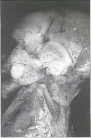 Fig. 1: Photograph of the left side of the head showing the arteries and bones of the scalp, mandible, face and neck. |
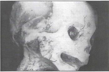 Fig. 2: Photograph of the right side of the head showing an intact right globe of the eye and arteries of the palate. |
It was found that this procedure does produce specimens which are useful for the study of developing bone. As well, the simultaneous staining of cartilage may help to assist researchers in gathering information regarding fetal development.
This procedure also produced specimens that showed the relationship between vasculature, and soft and hard tissues in the fetus. Clarity was lacking in specimens which had been prepared in the past using resin cast modeling (Thompsett, 1970).
Because the plastination procedure was used, the specimens could be handled and used by students, clinicians and researchers without fear of damaging them.
Thompsett, D,H. 1970. Cleared anatomical specimens. Anatomical Techniques. 2nd. Edition, Chap. 35:248-258.
von Hagens, G. 1985. Heidelberg Plastination Folder: Collection of technical leaflets for plastination.
von Hagens, G., Tiederman, K., Kriz, W. 1987. The current potential of plastination. Anat. and Embry. 175: 411-421.
https://doi.org/10.1007/BF00309677