Department of Anatomy and Structural Biology, University of Otago, Dunedin, New Zealand.
Stringent requirements for obtaining consent for acquisition of human tissues in New Zealand have led to difficulty in obtaining fresh brain tissue from post-mortem. In contrast, the Medical School's body donor program is well supported, and body donation often includes permission to retain organs, including the brain. However, it has been difficult to obtain brains from bodies before embalming due to higher priorities necessitating that most cadavers to be embalmed. Therefore, we have recently trialled plastination with the P35 technique on thin slices from two cadaveric brains, one embalmed with an alcohol-based mix and the other with a I 0% formalin formulation. The brains were sliced into 3mm slices and processed separately. Each basket of slices was flushed in running water, immersed in distilled water at 5°C, dehydrated in -25°C acetone, immersed with P35 at 5°C for 48 hours then placed under vacuum at room temperature for 24 hours. Impregnated slices were cast individually in float glass chambers and initially cured under UVA lights for 3 hours followed by heat-curing in an oven at 45°C for 5 days. Slices were finally removed from the glass chambers and the edges trimmed, sanded and polished. The brains from the two cadavers have yielded P35 slices of similar quality to those obtained from formalin fixed, post mortem brains.
plastination; P35; polymer; embalmed; brain; neuroanatomy; education
R Barnett: Telephone: 64 3 479 7360; E-mail: russell.barnett@stonebow.otago.ac.nz

![]()


P40 plastinated brain slices have been used extensively in our neuroanatomy courses (Jones and Barnett, 1998). These P40 slices were produced from fresh brains removed from postmortem cadavers for the intent of slice production. Then the slices were fixed in 5% neutral buffered formalin (NBF) at 5°C with four changes of fixative over a period of 9 weeks. This procedure yielded high quality P40 slices with high contrast between white and grey matter (von Hagens, 1994; Barnett, 1997). Brains fixed in I 0% formalin have also been processed successfully with the P35 technique. (Weber, 1994; Weiglein, 1996).
The New Zealand human tissue Act of 1964 allowed use of human tissue, from postmortems at government-controlled facilities, for the purpose of teaching and research. This act does not require consent for human tissue to be used in this manner. Until 1996 it was common practice for the department to receive brains from government controlled post-mortem facilities (public hospitals) without consent. Since 1996, due to the change of the ethical climate, the practice of using tissue without consent is considered to be inappropriate. (Jones and Galvin, 2002). Thus, due to this conflict concerning acquisition of fresh tissue from post-mortem sources, fresh brain material for plastination is rarely available. For this reason, we have turned to cadaveric material obtained through the University of Otago 's body donor program in order to obtain brain specimens for dissection and plastination. Our university program can provide the possibility for fresh tissue use.
However, due to the demand for cadaveric material in various areas of teaching and research 111 our department, fresh cadaveric specimens including brains are rare, as all of the cadavers are embalmed. Therefore, brains are only available from embalmed cadavers and not in the fresh state. Most polyester protocols preferably use fresh formalin fixed brains. Results of slice-plastination trials with the P35 technique on brains from two cadavers embalmed by two conventional means are reported. Slice plastination of embalmed cadaveric brains with the P40 technique was also tried, but was unsuccessful when compared with the P35 technique, and so is not further reported.
Fixation was carried out by embalming with a Portaboy 95 and a closed-circuit perfusion system. Each body was perfused with 20L of one of the two embalming fluids. The first cadaver was embalmed with Crosado mix which consisted of 60% alcohol, 15% glycerine, 15% water, 7.5% phenoxytol and 2% formalin. The second solution used was Dodge anatomical arterial mix (supplied by Regal Manufacturers, Wellington, New Zealand). For commercial reasons, we have been unable to find out the chemical composition of this fixative other than it contains 4% para formaldehyde ( I 0% formalin solution). The brains were removed from the cadavers after a minimum time of six months post-embalming.
The cadaveric brains were removed from the bodies, sliced into 3mm coronal slices on a conventional bacon slicer and stacked in a grid basket. The brain slices fixed with the Crosado mix were well-preserved, so they were washed for 24 hours in running tap water. The centre of the brain slices fixed with the Dodge mix were pink in colour and soft, indicating they were not thoroughly fixed. Therefore, they were immersed in I 0% NBF for three clays until the pink colouration was gone. They were washed for 24hours. Each basket of brain slices was immersed in distilled water at 5°C overnight.
Each basket of slices was dehydrated by the freeze substitution method using I 00% acetone at -25°C for 24 hours and changed into fresh acetone for another 24 hours.
Immersion and impregnation were carried out according to the standard procedure for the P35 technique (von Hagens, 1990; Weber and Henry, 1992; Weiglein, 1996). This consisted of two immersion baths of P35 A9 ( I 00:2) for 24 hours each at 5°C. Vacuum was applied for 24 hours at room temperature to the slices. The second 1mmersron bath was used for the impregnation mixture. Vacuum was increased until a pressure of 10mmHg was attained.
The basket of slices was removed from the vacuum chamber. Each slice was cast in an individual glass chamber consisting of two float glass plates 250 x 220 x 2mm. Silicone tubing (8mm) was placed between the two glass plates to provide a gasket and the chamber was clamped on three sides. The glass chambers were placed upright, and the slices slipped into the top of the chamber with a spatula. Each chamber was filled with a fresh mixture of P35/A9 ( I 00:2). Air bubbles were removed with the aid of a wire.
The chambers were placed between UV A lights at an angle of approximately 15 degrees with the open side of each chamber raised so the resin would not leak out. The brain slices were then positioned in the centre of the chamber with a wire before the lights were switched on. Light curing was undertaken for three hours and fans were used to cool the surfaces of the chambers.
Following light curing the glass chambers were placed in an oven at 45°C for five days. The oven was then switched off and allowed to cool before the chambers were removed.
After curing was completed the glass chambers were dismantled and the edges of sections were trimmed on an electric planer, sanded, and polished.
The Crosado fixed brain was smaller when removed from the cranium than the Dodge arterial mix brain. Both brains from the embalmed cadavers (Crosado mix and Dodge anatomical arterial mix) were successfully sliced and plastinated using the P35 technique and yielded good results. Dodge arterial mix ( 10% formalin base)
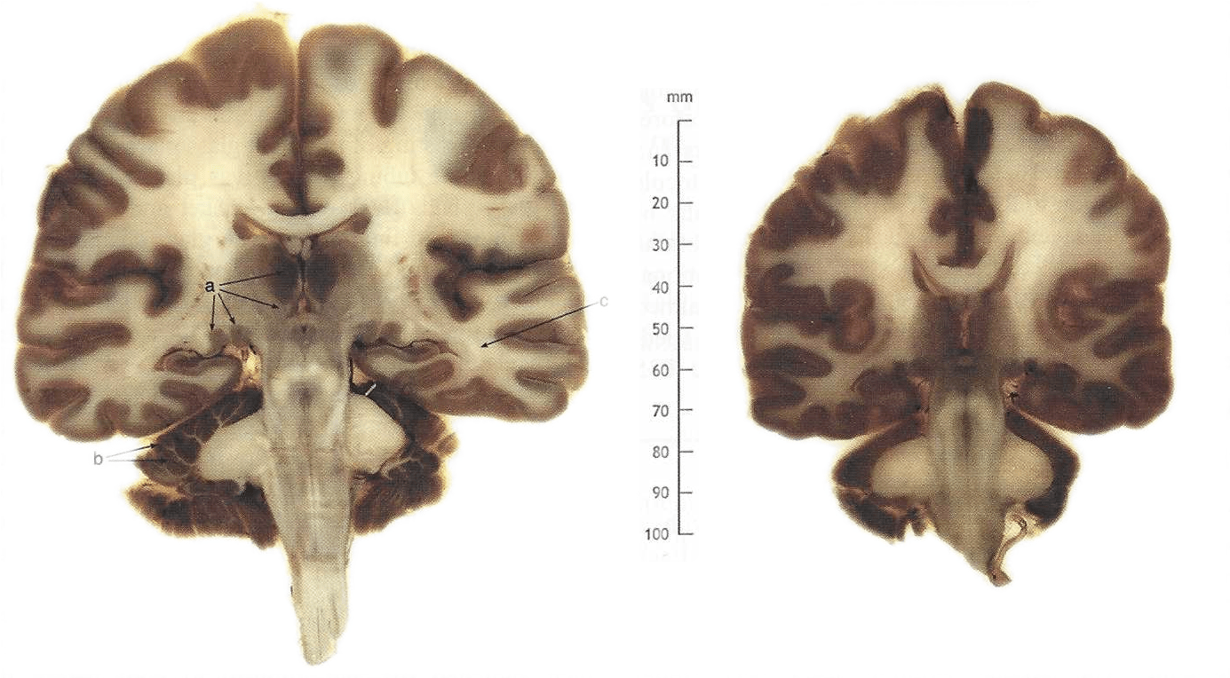
Figure 1. Human brain slices plastinated via the P35 process. Section on left embalmed with Dodge arterial mix. Section on right embalmed with Crosado mix. Thalamic nuclei (a), white matter of cerebellar folia (b), dense optic radiation (c).
demonstrated excellent definition between the grey and white matter, down to the finest details (Fig. 1). The individual nuclei of the thalamus are demarcated, as is the white matter of the cerebellar folia. Within the white matter, even the dense optic radiation is easily distinguished from the general cortical white matter. Crosado mix (60% alcohol base) yielded good gray/white definition but the finer details within both grey and white matter arc less apparent (Fig. 1). The Crosado fixed brain slices were smaller than the Dodge brain slices.
The P35 technique has previously shown to offer consistently good results for plastination of thin brain slices when used on fresh brain material fixed in formalin (Weber and Henry, 1992; Weber, 1994; Weiglein, 1996). This report demonstrates the successful use of P35 on brains which have been embalmed with either an alcohol-based (Crosado mix) or formalin-based (Dodge anatomical mix) embalming fluids.
Of the two embalming fluids tested, the formalin based mix resulted in better anatomical definition within the P35 plastinated slices than did the alcohol based mix. As well, the Dodge brain retained a more normal size.
The alcohol based Crosado embalming fluid was prepared and tested in our department in order to eliminate the exposure of students and staff to phenol and to reduce exposure to formalin. The Crosado mix has also been superior to the 10% formalin-based Dodge anatomical mix in controlling growth of mold on stored cadavers. However, for P35 slice-plastination purposes, it seems somewhat less successful in retaining the finer anatomical detail in the plastinated brains than is the Dodge Anatomical Mix.
We have stopped routine embalming with Dodge mix due to mold problems. However, we still embalm bodies with Dodge mix specifically for use in research and plastination and have found that it gives excellent results with most plastination techniques, including P35. The level of shrinkage was not determined in either of the brains used for this study. The brain fixed with Crosado mix was smaller upon removal than the Dodge arterial mix brain. This smaller size likely indicates that the Crosado mix embalmed brain, which contains 60% alcohol, shrunk prior to removal. Previous studies have shown shrinkage occurs during dehydration and impregnation (Tiedemann and Ivic-Matijas, 1988; Henry et al., 1998; Sora et al., 1999). Measurement of shrinkage before and after plastination would have been beneficial to note whether both embalming methodologies yielded similar shrinkage. There was no apparent visual distortion of anatomical structures due to shrinkage in either brain, thus providing us with a suitable resource for teaching neuroanatomy.
The ability to use brains from embalmed cadavers to produce P35 slices has now guaranteed a continued supply of brain slice sets for use in teaching neuroanatomy.
Acknowledgments
Crosado, B: Department of Anatomy and Structural Biology, University of Otago, Dunedin, New Zealand.
Barnett RJ. 1997: Plastination of coronal and horizontal brain slices using the P40 technique. J Int Soc Plastination 12(1):33-36.
https://doi.org/10.56507/YJVS5787
Henry R W, Brown A, Reed RB. 1998: Current topics on dehydration. Abstract presented at The 9th International Conference on Plastination, Trois-Rivieres, Quebec, Canada, July 5-10, 1998. J lnt Soc Plastination 13(2):27-28.
Jones GD, Barnett RJ. 1998: The contribution of plastination to the neuroanatomy teaching. Abstract presented at The 9th International Conference on Plastination, Trois-Rivieres, Quebec, Canada, July 5-10, 1998. J lnt Soc Plastination 13(2):41.
Jones GD, Galvin KA. 2002: Retention of body parts: Reflections from anatomy. New Zeal Med J 115:267- 269.
Sora MC, Brugger P, Traxler H. 1999: P40 plastination of human brain slices: comparison between different immersion and impregnation conditions. J Int Soc Plastination 14(1):22-24.
https://doi.org/10.56507/XLSJ5724
Tiedemann K, lvic-Matijas D. 1988: Dehydration of macroscopic specimens by freeze substitution m acetone. J Int Soc Plastination 2(2):2-12.
https://doi.org/10.56507/SCLL2742
von Hagens G. 1990: Preliminary leaflet for plastination of brain slices with Biodur™ P35. Unpublished Computer Printout: August.
von Hagens G. 1990: Plastination of brain slices according to the P40 procedure, a step-by-step description, December.
Weber W. 1994: Sheet plastination of brain slices. J Int Soc Plastination 8(1):23.
https://doi.org/10.56507/OWYV2878
Weber W, Henry RW. 1992: Sheet plastination of the brain - P35 technique, filling method. J Int Soc Plastination 6(1):29-32.
https://doi.org/10.56507/KWGD3312
Weiglein AH. 1996: Preparing and using SI0 and P35 brain slices. J Int Soc Plastination 10(1):22-25.
https://doi.org/10.56507/IXGV4189